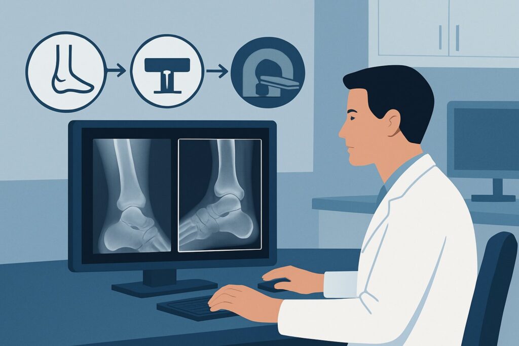
Chronic Ankle Pain with Normal X-Rays: Why Is MRI the Right Next Step?
It’s 4 PM on a busy clinic day. You’re seeing a 38-year-old patient with five months of persistent, nagging lateral ankle pain after a “tweak” while hiking. They’ve tried rest, ice, and physical therapy with minimal improvement. You ordered standard ankle radiographs a month ago, and the report was unequivocal: “unremarkable.” Now the patient is back, frustrated, and asking, “What’s next? Why does it still hurt?” This is a common and challenging clinical crossroads: chronic ankle pain of uncertain etiology despite normal initial imaging. The American College of Radiology (ACR) Appropriateness Criteria directly addresses this scenario, designating **MRI ankle without IV contrast** as *Usually Appropriate* for the next diagnostic step. This article details the clinical workflow for this specific situation, explaining the rationale behind the recommendation and outlining the downstream decision-making process.
Who Fits This Clinical Scenario?
This guidance applies to a specific patient population: adults or children with chronic ankle pain (typically defined as lasting more than three months) where the underlying cause is unclear after a clinical evaluation and normal initial radiographs. The key elements are the chronicity of symptoms and the diagnostic uncertainty. The initial X-rays have successfully ruled out obvious acute fracture, significant malalignment, or advanced degenerative joint disease, but the pain persists.
This workflow is distinct from scenarios where you have a strong clinical suspicion for a specific condition. If your examination points clearly toward a particular diagnosis, you should consult a different ACR variant. This guidance is NOT for patients with:
- A known history of degenerative joint disease seen on prior radiographs. That situation involves assessing the extent of arthritis, not finding an unknown cause of pain.
- High suspicion for a specific tendon abnormality. While MRI is also used, the pre-test probability is different, which can influence interpretation. (See: ACR variant for suspected tendon abnormality).
- Symptoms and signs strongly suggesting ankle instability. This may involve stress radiographs or a different MRI focus. (See: ACR variant for suspected ankle instability).
This article is for the true diagnostic dilemma: the patient with a “radiograph-negative” painful ankle where the differential remains broad.
What Diagnoses Are You Working Up in This Scenario?
When ankle radiographs are normal, the source of chronic pain is almost always in the soft tissues, cartilage, or bone marrow—structures that X-rays assess poorly. An advanced imaging study is necessary to investigate a differential that includes several common but radiographically occult conditions.
Osteochondral Lesion of the Talus (OCL): This is an injury to the cartilage and underlying bone of the talus, often post-traumatic. Patients may have deep, aching pain and mechanical symptoms like catching or locking. Radiographs are notoriously insensitive for non-displaced or early-stage OCLs, making this a primary consideration in this scenario.
Occult Tendon Pathology: Chronic microtrauma or an incompletely healed injury can lead to tendinosis, tenosynovitis, or small interstitial tears. The peroneal tendons (posterolateral pain) and the posterior tibial tendon (medial pain) are common culprits. These conditions are purely soft-tissue abnormalities and are invisible on X-ray.
Ligament Injury and Impingement Syndromes: A previous ankle sprain may result in chronic ligamentous laxity or scarring that leads to impingement. Anterolateral impingement syndrome, caused by synovial thickening and scar tissue in the anterolateral gutter, is a frequent cause of persistent pain after an inversion injury and is not visible on radiographs.
Sinus Tarsi Syndrome: This is a clinical syndrome of pain and instability localized to the sinus tarsi, a small cavity between the talus and calcaneus. It is often associated with subtalar joint instability and inflammation of the synovial recesses within the sinus, findings that require cross-sectional imaging to diagnose.
Bone Marrow Edema or Stress Fracture: Repetitive stress can cause a stress reaction or a non-displaced fracture that is not yet visible on radiographs. This is particularly common in runners or individuals who have recently increased their activity level. The resulting bone marrow edema is a hallmark finding on MRI.
Why Is MRI Ankle Without IV Contrast the Recommended Study?
The ACR designates **MRI ankle without IV contrast** as *Usually Appropriate* because it is the single most effective, non-invasive study for evaluating the wide range of potential pathologies in this scenario. Its superior soft-tissue contrast and ability to detect bone marrow edema make it uniquely suited to answer the clinical question.
Magnetic Resonance Imaging (MRI) provides exquisite detail of tendons, ligaments, cartilage, and bone marrow. It can directly visualize an osteochondral lesion, identify tendinosis or tears, reveal ligamentous thickening or scarring consistent with impingement, and detect subtle bone marrow edema from a stress injury. For the differential diagnoses listed above, MRI offers high sensitivity and specificity, allowing for a definitive diagnosis or exclusion in most cases. Importantly, this is achieved without any ionizing radiation (Adult RRL: O 0 mSv).
The recommendation specifically calls for a non-contrast study. For the initial workup of non-specific chronic pain, intravenous contrast is generally not required and is rated as *Usually Not Appropriate*. Contrast is typically reserved for cases where there is a specific concern for infection, inflammatory arthropathy, or a soft-tissue mass.
How Do Alternative Studies Compare?
- US ankle: Rated as *May be appropriate*, ultrasound is a valuable tool, particularly for evaluating superficial tendons like the peroneals or the Achilles. However, it is highly operator-dependent, has a limited field of view, and cannot adequately assess intra-articular structures, cartilage, or bone marrow. It is a reasonable alternative if the clinical suspicion is highly localized to a specific, accessible tendon, but it is less comprehensive than MRI for pain of uncertain etiology.
- CT ankle without IV contrast: Also rated as *May be appropriate*, CT provides excellent detail of the bone. It is superior to radiographs for detecting subtle fractures or mapping the osseous component of a known OCL. However, it provides very limited information about soft tissues and no information about bone marrow edema. Given that most of the differential is soft-tissue or marrow-based, CT is a suboptimal choice for the initial advanced imaging study in this scenario. It also involves a small amount of ionizing radiation (Adult RRL: ☢ <0.1 mSv).
What’s Next After MRI Ankle Without IV Contrast? Downstream Workflow
The results of the ankle MRI will guide the subsequent clinical pathway. The goal is to move from diagnostic uncertainty to a targeted treatment plan.
If the MRI is positive for a specific finding:
- Osteochondral Lesion: The next step is typically a referral to an orthopedic surgeon. Treatment depends on the size, location, and stability of the lesion and may range from conservative management to arthroscopic debridement, microfracture, or cartilage transfer procedures.
- Tendon Tear or Severe Tendinosis: This often leads to a more focused physical therapy regimen, bracing, or a sports medicine or orthopedic consultation. In some cases, particularly with full-thickness tears, surgical repair may be considered.
- Impingement Syndrome or Sinus Tarsi Syndrome: Management often begins with conservative measures, including physical therapy and activity modification. An image-guided corticosteroid injection, which is rated as *May be appropriate* by the ACR for diagnosis and therapy, can be both a diagnostic and therapeutic next step.
If the MRI is negative: A normal MRI is also a highly valuable result, as it effectively rules out most significant structural causes of pain. If the patient’s symptoms persist despite a normal MRI, the focus should shift. Consider a referral to sports medicine or physiatry for a functional assessment. An image-guided anesthetic injection into a specific joint or tendon sheath can help determine if that structure is the pain generator, even without an anatomic abnormality on MRI. Re-evaluation for non-orthopedic causes of pain may also be warranted.
Pitfalls to Avoid (and When to Get Help)
Navigating the workup of chronic ankle pain requires careful consideration to avoid common missteps.
- Stopping the Workup at a Normal Radiograph: The most significant pitfall is assuming a normal X-ray means “no pathology.” For chronic pain, radiographs are merely the first step to rule out gross osseous abnormalities.
- Ordering Contrast by Default: Unnecessary use of IV contrast for an initial MRI in this scenario increases cost and carries a small risk of adverse reaction without adding diagnostic value. Only add contrast if there is a specific question of tumor, infection, or inflammatory synovitis.
- Ignoring Clinical-Radiologic Correlation: MRIs can show incidental, asymptomatic findings (e.g., mild tendinosis). Always correlate the imaging findings with the precise location of the patient’s pain and their clinical history.
- Misapplying this Scenario: If your clinical exam strongly suggests a specific diagnosis like ankle instability or a peroneal tendon tear, consult the ACR variant for that specific indication, as the imaging recommendations may be more nuanced.
If the diagnosis remains elusive after a high-quality, non-contrast MRI, or if the findings are complex, consultation with a musculoskeletal radiologist or an orthopedic subspecialist is the appropriate next step.
Related ACR Topics and Tools
This article covers one specific scenario in depth. For a broader view of imaging for all chronic ankle pain presentations, or to explore the tools used to make these evidence-based decisions, please see the resources below.
- For breadth across all scenarios in Chronic Ankle Pain, see our parent guide: Chronic Ankle Pain: ACR Appropriateness Decoded.
- To explore other clinical situations, use the ACR Appropriateness Criteria Lookup.
- For technical details on performing the recommended study, see the Imaging Protocol Library.
- To discuss radiation exposure from alternative studies like CT with patients, use the Radiation Dose Calculator.
Frequently Asked Questions
Why not start with ultrasound if I suspect a tendon issue?
Ultrasound is rated as ‘May be appropriate’ and can be an excellent first choice if your clinical suspicion is highly localized to a single, superficial tendon (e.g., peroneal or Achilles). However, for pain of ‘uncertain etiology,’ where the differential includes deeper structures, intra-articular pathology like an OCL, or bone marrow edema, MRI is far more comprehensive. It provides a global assessment of the ankle that ultrasound cannot match.
Is an MR arthrogram ever useful for chronic ankle pain?
For this specific scenario of uncertain etiology, MR arthrography is rated ‘Usually not appropriate.’ Its primary use is in evaluating for subtle chondral injuries, loose bodies, or ligamentous tears related to instability. It is a more invasive procedure and is typically reserved for cases where a standard MRI is non-diagnostic but suspicion for these specific conditions remains high, often in a pre-operative context.
What if the patient has a contraindication to MRI, like a non-compatible pacemaker?
In cases of an absolute contraindication to MRI, you must rely on the next best alternatives. A CT scan (‘May be appropriate’) would be the best choice for evaluating for an occult fracture or the osseous component of an OCL. A high-quality ultrasound (‘May be appropriate’) could assess the tendons and ligaments. A combination of these may be needed. In complex cases, a bone scan with SPECT/CT (‘May be appropriate (Disagreement)’) could also be considered to localize metabolic activity, though it is less specific.
Does ‘chronic’ have a specific time definition in this context?
While there isn’t a universal, strict cutoff, ‘chronic’ in musculoskeletal medicine generally refers to pain lasting longer than the expected healing time for a typical soft-tissue injury, which is usually considered to be around 3 months. Pain persisting beyond this point suggests an underlying pathology that has failed to heal or a condition other than a simple sprain or strain.
If the MRI is completely normal but the pain is severe and localized, what’s the next step?
A normal MRI is a powerful finding that rules out most structural causes. If significant, localized pain persists, the next step is often a diagnostic/therapeutic injection. An image-guided injection of a local anesthetic into the suspected pain-generating structure (e.g., a specific joint, tendon sheath, or the sinus tarsi) can confirm the diagnosis if the patient experiences significant temporary relief. This is rated as ‘May be appropriate’ by the ACR and can be a crucial step in the diagnostic algorithm.
Reviewed by Pouyan Golshani, MD, Interventional Radiologist — May 21, 2026