Suspected Nerve Abnormality in Chronic Elbow Pain: Should You Order Ultrasound or MRI?
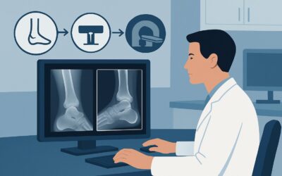
A 48-year-old software developer presents with six months of aching pain over his medial elbow and progressive tingling in his ring and little fingers, worsened by prolonged time at his desk. His physical exam is notable for a positive Tinel’s sign over the cubital tunnel, and initial radiographs of the elbow show only minor degenerative changes. You suspect an ulnar nerve entrapment, but you need to confirm the diagnosis and rule out a structural cause before referring him to a specialist. This article details the American College of Radiology (ACR) guided workflow for choosing the next imaging study in this specific scenario. For a patient with chronic elbow pain, suspected nerve abnormality, and normal or nonspecific radiographs, the ACR rates **US elbow** as *Usually Appropriate*.
Who Fits This Clinical Scenario for Suspected Nerve Abnormality?
This imaging workflow is designed for patients with chronic elbow pain (typically lasting more than three months) where the clinical picture points toward a peripheral nerve pathology. The key inclusion criteria are signs and symptoms of neuropathy, such as paresthesias, numbness, motor weakness in a specific nerve distribution (ulnar, median, or radial), or a positive provocative test like a Tinel’s sign or elbow flexion test. Critically, this guidance applies *after* initial radiographs have been performed and found to be normal or to show only nonspecific findings like mild osteoarthritis, which do not explain the patient’s primary symptoms.
This scenario is distinct from other causes of chronic elbow pain. This guidance does not apply if:
- The patient’s primary symptoms are mechanical, such as locking or clicking. This presentation suggests an intra-articular loose body or cartilage issue and routes to a different imaging pathway.
- The pain is highly localized to an epicondyle without radiating neuropathic symptoms. This is more consistent with chronic epicondylalgia or a tendon tear.
- There is a history of acute trauma or suspicion of an occult fracture. This requires a workup focused on bone and ligamentous injury.
Correctly identifying your patient’s presentation ensures the chosen imaging study has the highest diagnostic yield for the suspected pathology.
What Diagnoses Are You Working Up with Suspected Nerve Entrapment?
When ordering imaging for suspected nerve abnormality around the elbow, you are primarily investigating nerve compression or injury. The differential diagnosis is guided by the anatomical location of the symptoms.
The most common cause is Cubital Tunnel Syndrome, an entrapment neuropathy of the ulnar nerve as it passes through the fibro-osseous cubital tunnel at the medial elbow. Compression can result from fibrous bands, osteophytes, direct trauma, or repetitive flexion, leading to the classic “funny bone” tingling in the fourth and fifth digits.
A less frequent but important consideration is Pronator Teres Syndrome, which involves compression of the median nerve as it passes between the two heads of the pronator teres muscle in the proximal forearm. This can cause aching anterior elbow pain and sensory changes in the thumb, index, middle, and radial half of the ring finger, sometimes mimicking carpal tunnel syndrome.
Another potential diagnosis is Radial Tunnel Syndrome, involving compression of the posterior interosseous nerve (a deep branch of the radial nerve). This typically presents as deep, aching pain in the lateral elbow and dorsal forearm, often confused with lateral epicondylitis (“tennis elbow”), but without the hallmark focal tenderness directly over the epicondyle.
Finally, imaging can identify structural causes of nerve compression, such as a space-occupying lesion (e.g., a ganglion cyst), an anomalous muscle (like the anconeus epitrochlearis compressing the ulnar nerve), or dynamic instability, where the ulnar nerve subluxates out of its groove during elbow flexion.
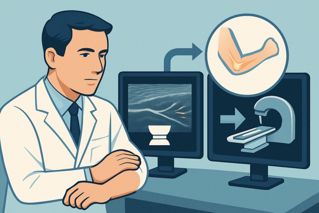
Why Is Ultrasound the Recommended First Study for Suspected Nerve Abnormality?
For a patient with suspected nerve pathology and unrevealing radiographs, both **US elbow** and **MRI elbow without IV contrast** are rated as *Usually Appropriate* by the ACR. However, ultrasound is often the preferred initial study for several key reasons.
The primary advantage of ultrasound is its exceptional spatial resolution for visualizing superficial nerves and its unique ability to perform dynamic assessment. A sonographer can watch the ulnar nerve in real-time as the patient flexes and extends their elbow, directly visualizing nerve subluxation or dislocation—a finding that is completely missed on static MRI. Ultrasound can precisely measure nerve cross-sectional area to identify swelling, detect focal constrictions, and characterize the nerve’s internal echotexture. It can also clearly identify compressive lesions like ganglion cysts or anomalous muscles.
While MRI is also highly effective for visualizing nerve morphology and detecting secondary signs like muscle denervation edema, it lacks dynamic capability and is generally more expensive and less accessible than ultrasound. For this specific clinical question, ultrasound often provides a more direct and functional answer.
Other imaging modalities are rated lower for this scenario:
- CT elbow without IV contrast is rated *May be appropriate*. It excels at showing bony anatomy, such as an osteophyte impinging on the cubital tunnel, but provides very poor visualization of the nerve itself.
- MR arthrography elbow is rated *Usually not appropriate*. The injection of contrast into the joint is designed to evaluate intra-articular structures like cartilage and ligaments, offering no benefit for assessing extra-articular nerve entrapment.
Both recommended studies, US and MRI, are radiation-free (0 mSv). When ordering, be specific: “High-resolution ultrasound of the medial elbow to evaluate the ulnar nerve, including dynamic assessment for subluxation during flexion.”
What’s Next After an Elbow Ultrasound? Downstream Workflow
The results of the elbow ultrasound will guide your next clinical steps. The workflow branches based on whether the findings are positive, negative, or indeterminate.
If the ultrasound is positive and clearly demonstrates nerve pathology (e.g., significant nerve swelling at a point of compression, a compressing ganglion cyst, or dynamic subluxation), the diagnosis is confirmed. This finding provides a clear target for treatment. The next step is typically a discussion with the patient about management options, which may include conservative measures like activity modification, splinting, and physical therapy, or a referral to a hand surgeon or orthopedic specialist for consideration of surgical decompression.
If the ultrasound is negative but your clinical suspicion for neuropathy remains high, the workup is not over. The next logical step is often to obtain electrodiagnostic studies (EMG/NCS). These tests assess nerve function directly and can confirm a physiologic neuropathy even when structural imaging is normal. Alternatively, if there is a strong suspicion of a deeper pathology not well-visualized by ultrasound, proceeding to an **MRI elbow without IV contrast** is a reasonable next step to look for intrinsic nerve signal changes or occult compressive sources.
If the ultrasound is indeterminate, showing borderline findings like mild nerve swelling without a clear compressive cause, correlation with EMG/NCS is highly valuable to determine the functional significance of the imaging findings before proceeding to more invasive treatments.
Pitfalls to Avoid (and When to Get Help)
In the workup of suspected elbow neuropathy, several common pitfalls can delay diagnosis. First, avoid ordering an MRI with contrast as the initial advanced imaging step; for primary nerve entrapment, contrast is rated *Usually not appropriate* and adds unnecessary cost and risk. Second, do not stop the workup after a negative ultrasound if clinical signs are compelling; a functional study like EMG/NCS is often the crucial next step. A third pitfall is failing to request a dynamic assessment when ordering an ultrasound for suspected ulnar neuropathy, as this can miss intermittent nerve subluxation. Finally, be wary of attributing neuropathic symptoms to incidental degenerative changes seen on radiographs. If a patient presents with progressive motor weakness or muscle atrophy, this is a red flag that warrants prompt escalation and referral to a specialist, regardless of imaging findings.
Related ACR Topics and Tools
This article focuses on a single clinical variant. For a comprehensive overview of all imaging scenarios related to chronic elbow pain, from epicondylalgia to occult fractures, please see our parent guide. For further exploration of imaging guidelines and techniques, the following GigHz resources are available:
- For breadth across all scenarios in Chronic Elbow Pain, see our parent guide: Chronic Elbow Pain: ACR Appropriateness Decoded.
- ACR Appropriateness Criteria Lookup — for adjacent scenarios
- Imaging Protocol Library — for technique on the recommended study
- Radiation Dose Calculator — for cumulative dose conversations
Frequently Asked Questions
Why is ultrasound preferred over MRI when both are ‘Usually Appropriate’?
Ultrasound is often preferred as the initial study because of its unique ability to perform dynamic assessment, which can visualize ulnar nerve subluxation during elbow flexion—a diagnosis MRI cannot make. It also tends to be more accessible, faster to schedule, and less expensive than MRI.
If the ultrasound is negative, what is the next step?
If clinical suspicion for a nerve abnormality remains high despite a negative ultrasound, the two most common next steps are 1) ordering electrodiagnostic studies (EMG/NCS) to assess nerve function, or 2) ordering an MRI without contrast to look for deeper pathology or intrinsic nerve signal changes that ultrasound may have missed.
Is contrast ever needed when imaging for suspected nerve entrapment in the elbow?
No, for the specific indication of chronic elbow pain with suspected nerve abnormality, both MRI with contrast and CT with contrast are rated as ‘Usually not appropriate’ by the ACR. Contrast does not improve visualization of the nerve or common compressive pathologies and is not recommended.
Can I skip radiographs and go straight to ultrasound or MRI?
The ACR workflow assumes that initial radiographs have already been performed. Radiographs are a crucial first step to rule out bony abnormalities like fractures, dislocations, or significant osteophytes that could be the primary cause of the patient’s symptoms. Advanced imaging is indicated when these initial radiographs are normal or nonspecific.
What if the patient’s symptoms are in the distribution of the median or radial nerve, not the ulnar nerve?
The same imaging principles apply. High-resolution ultrasound is an excellent modality for evaluating the median nerve (for pronator teres syndrome) and the radial nerve/posterior interosseous nerve (for radial tunnel syndrome). The sonographer will simply focus the examination on the relevant anatomical compartment based on the clinical suspicion.
Reviewed by Pouyan Golshani, MD, Interventional Radiologist — May 21, 2026