How Should You Image a Newborn with Bilious Vomiting and an Indeterminate Radiograph?
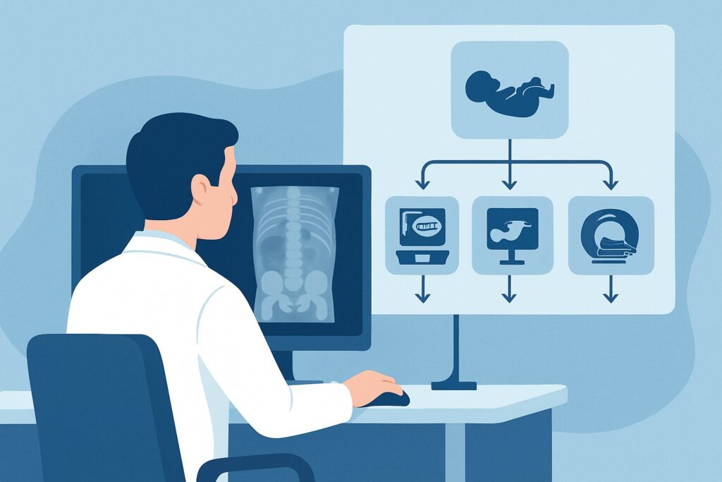
It’s 2 AM in the neonatal intensive care unit (NICU), and you are evaluating a 30-hour-old infant with new-onset, green-tinged emesis. The initial abdominal radiograph is equivocal—there are a few scattered, mildly distended loops of bowel, but no classic “double bubble” sign and a fair amount of gas distally. This presentation is a classic clinical crossroads. While the radiograph is not overtly alarming, bilious vomiting in a neonate is a potential surgical emergency until proven otherwise. The critical question is which imaging study will most reliably and rapidly differentiate a life-threatening midgut volvulus from other, less urgent causes of obstruction. According to the American College of Radiology (ACR) Appropriateness Criteria, for this specific scenario, a Fluoroscopy upper GI series is rated Usually appropriate.
Who Fits This Clinical Scenario for Neonatal Bilious Vomiting?
This clinical workflow is specifically for neonates presenting with bilious vomiting within the first 48 hours of life whose initial abdominal radiographs are indeterminate. This includes radiographic findings of a non-classic double bubble sign with gas present in the distal small bowel, a few non-specifically distended bowel loops, or even a completely normal bowel gas pattern. The key inclusion criteria are the combination of bilious emesis, age less than two days, and an initial radiograph that does not provide a definitive diagnosis.
It is crucial to distinguish this situation from other related presentations. This guidance does not apply if:
- The radiograph shows a classic “double bubble” sign with no gas distally. This finding is highly suggestive of duodenal atresia, often associated with malrotation, and follows a different diagnostic pathway.
- The radiograph clearly demonstrates a distal bowel obstruction. Findings like multiple, uniformly dilated bowel loops suggest a more distal process like jejunal/ileal atresia or meconium ileus, which may prompt a contrast enema as the next step.
- The infant is older than two days. While malrotation can present later, the initial workup and differential diagnosis can shift slightly in an older infant.
- The vomiting is nonbilious. Nonbilious emesis points toward a more proximal obstruction, such as hypertrophic pyloric stenosis or gastroesophageal reflux, which have their own distinct imaging algorithms.
What Diagnoses Are You Working Up in This Scenario?
With an equivocal radiograph, your primary goal is to urgently rule out conditions that require immediate surgical intervention. The differential diagnosis is focused on causes of proximal small bowel obstruction.
Midgut Volvulus: This is the most feared and time-sensitive diagnosis. It occurs when the small bowel twists on its own mesentery, compromising its blood supply. This is a direct consequence of intestinal malrotation. Without rapid diagnosis and surgical correction, this can lead to extensive bowel necrosis, short gut syndrome, or death. The presence of distal gas on a radiograph does not exclude this diagnosis, as the obstruction may be incomplete or intermittent early on.
Malrotation without Volvulus: This is the underlying congenital anomaly where the bowel does not rotate and fixate properly during fetal development. While not an emergency in itself, it creates the predisposition for a future volvulus. Identifying malrotation is critical because it typically requires elective surgery (a Ladd procedure) to prevent this life-threatening complication.
Duodenal Stenosis or Web: Unlike complete duodenal atresia, a stenosis (narrowing) or a web (a thin membrane across the lumen) causes a partial obstruction. This explains the clinical picture of bilious vomiting while still allowing some gas to pass distally, resulting in a non-classic radiographic appearance.
Jejunal Atresia or Stenosis: An obstruction in the proximal jejunum can also present with bilious vomiting. Depending on the level of the obstruction, the radiograph might show a few dilated proximal loops, which can be difficult to distinguish from other causes without further imaging.
Why Is a Fluoroscopy Upper GI Series the Recommended Study for This Presentation?
The American College of Radiology (ACR) rates a Fluoroscopy upper GI (UGI) series as Usually appropriate for this clinical scenario because it directly and reliably assesses for malrotation, the anatomic precursor to volvulus. The primary goal of the study is to identify the position of the duodenojejunal junction (DJJ), also known as the ligament of Treitz. A normally positioned DJJ is located to the left of the left-sided vertebral pedicle at the level of the duodenal bulb, effectively ruling out malrotation.
If malrotation is present, the DJJ will be inferior and to the right of its normal location. In the catastrophic event of a midgut volvulus, the UGI series will demonstrate a “corkscrew” or “pigtail” appearance of the twisted duodenum and proximal jejunum. The study can also identify partial obstructions from duodenal webs or stenosis. Its high sensitivity and specificity for these critical diagnoses make it the definitive examination in this setting.
Alternative imaging studies are rated lower for specific reasons:
- US abdomen (UGI tract) is rated May be appropriate. Ultrasound can be used to assess the relationship of the superior mesenteric artery (SMA) and superior mesenteric vein (SMV). Normally, the SMV is to the right of the SMA; an inverted relationship can suggest malrotation. In cases of active volvulus, a “whirlpool sign” may be seen. However, this study is highly operator-dependent, the SMA/SMV relationship can be normal in some cases of malrotation, and the DJJ is not as reliably visualized as with fluoroscopy. It is a reasonable alternative if fluoroscopy is not immediately available but is not considered the gold standard.
- Fluoroscopy contrast enema is rated Usually not appropriate for this specific question. This study evaluates the position of the colon and cecum. While a high-riding or malpositioned cecum can be associated with malrotation, the cecal position is notoriously variable in neonates. A normally positioned cecum does not exclude malrotation, making the contrast enema an unreliable study for this primary indication.
The recommended UGI series involves a pediatric radiation dose (ACR Pediatric Relative Radiation Level ☢☢☢, 0.3-3 mSv), a necessary trade-off for a rapid and definitive diagnosis of a potentially lethal condition.
What’s Next After Fluoroscopy Upper GI Series? Downstream Workflow
The results of the upper GI series will dictate your immediate next steps, creating a clear decision tree for managing the patient.
If the study is POSITIVE for malrotation (with or without volvulus): This is a surgical emergency. The immediate next step is an urgent consultation with pediatric surgery. The infant should be made NPO (nothing by mouth), have intravenous access established for fluid resuscitation, and a nasogastric tube placed for decompression. The surgical team will then prepare for an emergency exploratory laparotomy and Ladd procedure to untwist the bowel (detorsion) and prevent recurrence.
If the study is NEGATIVE for malrotation: A normally positioned duodenojejunal junction effectively rules out malrotation as the cause of the bilious vomiting. The workup must then continue to investigate other potential causes. If the UGI series also shows a partial duodenal obstruction (e.g., from a web or stenosis), surgical consultation is still warranted, though on a less emergent basis. If the UGI is entirely normal, the focus shifts to more distal or non-obstructive causes. Depending on the clinical picture, a contrast enema may be considered to evaluate for Hirschsprung disease or a meconium plug, which would now fall under a different clinical scenario.
If the study is INDETERMINATE: An equivocal UGI series is uncommon but can occur, for example, if the infant is uncooperative or has significant reflux preventing adequate filling of the duodenum. The first step is to discuss the findings immediately with the pediatric radiologist who performed the study. A repeat attempt at the UGI series may be necessary. Alternatively, an abdominal ultrasound could be performed as an adjunct to look for the whirlpool sign or an abnormal SMA/SMV relationship.
Pitfalls to Avoid (and When to Get Help)
In the high-stakes workup of neonatal bilious vomiting, several common pitfalls can lead to diagnostic delays and adverse outcomes. Be mindful of the following:
- Delaying the UGI Series: Time is bowel. Do not wait for the infant’s clinical condition to worsen before ordering the definitive study. Bilious vomiting is the primary indication, and the UGI should be performed urgently, regardless of the time of day.
- Being Falsely Reassured by a Normal Radiograph: A normal or non-specific abdominal radiograph is a common finding in early midgut volvulus. It does not exclude the diagnosis and should never be a reason to delay the UGI series.
- Over-reliance on Ultrasound: While a positive ultrasound for volvulus is helpful, a negative or indeterminate study does not rule out malrotation. The UGI series remains the diagnostic standard.
- Incomplete UGI Study: Ensure the radiologist visualizes the entire duodenal sweep and the definitive location of the DJJ. A study that only evaluates the stomach and duodenal bulb is incomplete and non-diagnostic for malrotation.
If there is any diagnostic uncertainty or if the infant’s clinical status is deteriorating despite a seemingly normal imaging workup, escalate immediately by consulting with pediatric radiology and pediatric surgery concurrently.
Related ACR Topics and Tools
This article covers one specific scenario within the broader topic of vomiting in infants. For a comprehensive overview of all related presentations, from esophageal reflux to distal bowel obstruction, please consult the parent guide. Additional GigHz tools can help you navigate adjacent scenarios and understand the technical aspects of the recommended imaging.
- For breadth across all scenarios in Vomiting in Infants, see our parent guide: Vomiting in Infants: ACR Appropriateness Decoded.
- To explore other clinical situations, use the ACR Appropriateness Criteria Lookup.
- For details on imaging techniques, visit the Imaging Protocol Library.
- To discuss radiation exposure with families, consult the Radiation Dose Calculator.
Frequently Asked Questions
Why is bilious vomiting in a newborn considered a surgical emergency?
Bilious (green) vomiting indicates that the obstruction is distal to the ampulla of Vater in the duodenum, where bile enters the gut. In a neonate, this symptom is the hallmark of a potential midgut volvulus, where the bowel twists on its blood supply. This can lead to rapid bowel ischemia and necrosis within hours. Therefore, it must be treated as a surgical emergency until malrotation and volvulus are definitively ruled out.
What if the initial radiograph shows a classic ‘double bubble’ with no distal gas?
That presentation points to a different clinical scenario. A classic ‘double bubble’ sign with no gas in the rest of the bowel is highly suggestive of complete duodenal atresia. While this also requires surgical correction, the immediate urgency is slightly less than for volvulus, as there isn’t typically an acute vascular compromise. The workup and management for that specific finding follow a separate pathway.
Can I order an abdominal ultrasound instead of a fluoroscopy upper GI series?
According to the ACR, an abdominal ultrasound is rated ‘May be appropriate’ but is not the primary recommended study. It can be useful for identifying the ‘whirlpool sign’ of active volvulus or an abnormal SMA/SMV vessel relationship suggestive of malrotation. However, it is highly dependent on the sonographer’s skill and may not detect malrotation if a volvulus is not present. The upper GI series is more reliable for definitively assessing the position of the duodenojejunal junction, which is the key to ruling out malrotation.
What specific findings on the upper GI series confirm or exclude malrotation?
To exclude malrotation, the study must show the duodenojejunal junction (DJJ) located to the left of the spine at the same horizontal level as the duodenal bulb. Malrotation is confirmed if the DJJ is positioned low and to the right of the spine. If a volvulus is also present, the contrast will show a ‘corkscrew’ or ‘pigtail’ appearance as it tries to pass through the twisted segment of bowel.
If the upper GI series is normal but the infant is still vomiting biliously, what’s next?
If the UGI series definitively excludes malrotation, the immediate surgical emergency is ruled out. However, persistent bilious vomiting requires further investigation. The differential diagnosis would then shift to other causes, such as severe dysmotility, sepsis-related ileus, or a more distal obstruction not apparent on the initial studies. The next step would likely involve a contrast enema to evaluate for conditions like Hirschsprung disease or meconium ileus, along with a broader clinical workup for non-surgical causes.
Reviewed by Pouyan Golshani, MD, Interventional Radiologist — May 29, 2026