What Is the Next Imaging Step for Suspected Osteomyelitis in a Child Over 5?
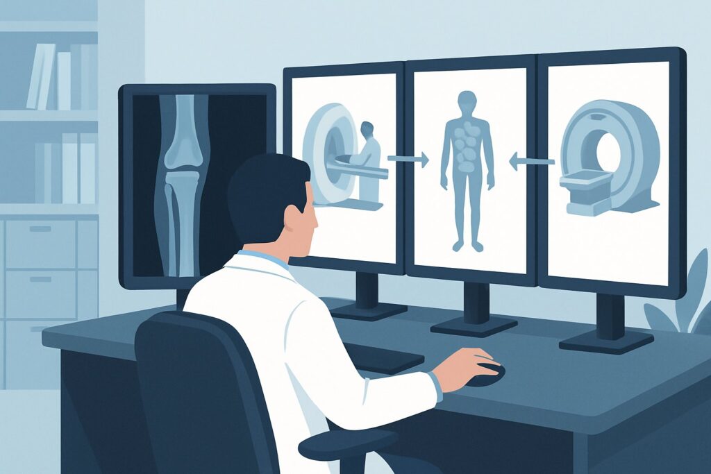
An 8-year-old boy presents to the emergency department with a three-day history of worsening left thigh pain, refusal to bear weight, and a low-grade fever. His C-reactive protein and erythrocyte sedimentation rate are significantly elevated. Initial radiographs of his femur are unremarkable, showing no fracture, periosteal reaction, or lytic lesions. You are now faced with a critical decision: which imaging study should you order next to evaluate for suspected acute osteomyelitis? This article provides a detailed workflow for this specific clinical scenario, grounded in the American College of Radiology (ACR) Appropriateness Criteria. For this presentation, the ACR rates `Image-guided aspiration area of interest` as ‘Usually Appropriate’, offering a direct path to diagnosis and treatment.
Who Fits This Clinical Scenario?
This guidance is specifically for children aged 5 years or older with a clinical suspicion for osteomyelitis or septic arthritis involving an extremity, where initial radiographs have already been performed and are either normal or show subtle, nonspecific findings like soft tissue swelling. The key inclusion criteria are:
- Age: 5 years or older.
- Location: An extremity (e.g., femur, tibia, humerus), excluding the axial skeleton (spine, pelvis).
- Clinical Picture: Signs and symptoms concerning for bone or joint infection, such as focal pain, tenderness, swelling, limp, or fever, often accompanied by elevated inflammatory markers (CRP, ESR).
- Prior Imaging: An initial radiograph has been completed and is non-diagnostic.
This workflow does not apply to several similar but distinct clinical situations. Exclusions include children younger than 5 years, where the differential diagnosis and imaging approach can differ significantly due to their unique skeletal anatomy and physiology. It also does not apply to the initial imaging workup (when no radiograph has been performed yet) or to patients where the clinical signs point overwhelmingly to septic arthritis as the primary diagnosis, as these represent separate ACR variants with tailored recommendations.
What Diagnoses Are You Working Up in This Scenario?
When a child over five presents with focal extremity pain and normal initial radiographs, your differential diagnosis is focused on identifying the source of inflammation and pain. The choice of the next imaging study is driven by the need to differentiate between these possibilities.
Acute Osteomyelitis: This is the primary concern. In children, it is most often a hematogenous infection that seeds in the highly vascular metaphyseal region of long bones. Early radiographic findings are absent, as it takes 10-14 days for bone changes like periosteal reaction or lytic lesions to become visible. The goal of advanced imaging is to detect early signs like bone marrow edema, abscess formation, or periosteal fluid collections.
Septic Arthritis: An infection within the joint space is a true orthopedic emergency requiring prompt drainage to prevent cartilage destruction. It can occur concurrently with osteomyelitis, especially when the metaphysis is intracapsular (e.g., hip, shoulder). Advanced imaging must be able to detect joint effusions and assess for synovial enhancement.
Cellulitis or Pyomyositis: A deep soft tissue infection can present with symptoms that closely mimic osteomyelitis, including fever, focal pain, and elevated inflammatory markers. Differentiating a soft tissue abscess from a subperiosteal abscess or intramedullary infection is a key role for cross-sectional imaging.
Trauma or Stress Fracture: An occult fracture or stress injury can also cause focal bone pain and swelling. While less likely to be associated with high fevers and systemic inflammatory response, it remains a crucial part of the differential, especially if the history is ambiguous.
Malignancy: Less common but critical to consider, primary bone tumors (like Ewing sarcoma or osteosarcoma) or hematologic malignancies (like leukemia) can manifest with bone pain and systemic symptoms. These conditions can have imaging features that overlap with infection, making definitive tissue sampling essential.
Why Is MRI or Image-Guided Aspiration the Recommended Next Step?
When initial radiographs are negative in a child over 5 with suspected osteomyelitis, the ACR designates both `MRI area of interest without and with IV contrast` and `Image-guided aspiration area of interest` as ‘Usually Appropriate’. The choice between them often depends on clinical stability, the presence of a suspected drainable fluid collection, and institutional resources.
Magnetic Resonance Imaging (MRI) is the premier non-invasive modality for this indication. Its high soft-tissue contrast resolution allows for the direct visualization of bone marrow edema, the earliest sign of osteomyelitis, often within 24-48 hours of symptom onset. MRI excels at defining the full extent of the infection, identifying intramedullary or subperiosteal abscesses, detecting concurrent septic arthritis or pyomyositis, and differentiating infection from tumor or trauma. The addition of intravenous (IV) contrast helps delineate non-enhancing abscesses from surrounding phlegmonous tissue and assess synovial enhancement in cases of septic arthritis. As an imaging modality with no ionizing radiation (O 0 mSv), it is particularly well-suited for pediatric patients.
Image-guided aspiration is also ‘Usually Appropriate’ because it is both diagnostic and potentially therapeutic. Under ultrasound or fluoroscopic guidance, a needle can be directed into a suspected fluid collection (e.g., a subperiosteal abscess or joint effusion). Obtaining fluid for Gram stain, culture, and cell count provides a definitive microbiological diagnosis, which is critical for tailoring antibiotic therapy. This procedure is particularly valuable when a drainable collection is suspected based on clinical exam or seen on another imaging study like ultrasound.
In contrast, other modalities are rated lower for this specific scenario:
- Ultrasound (US) area of interest is rated ‘May be appropriate’. US is excellent for detecting joint effusions and superficial soft-tissue fluid collections and can guide aspiration. However, it cannot directly visualize bone marrow and is limited in its ability to detect early osteomyelitis, making it a useful adjunct but not a comprehensive primary tool.
- 3-phase bone scan is also rated ‘May be appropriate’. While highly sensitive for detecting abnormal bone turnover, it is not specific for infection and can be positive in cases of trauma, inflammation, or tumor. Furthermore, it involves a significant radiation dose for children (☢☢☢☢ 3-10 mSv) and provides poor anatomic detail compared to MRI.
- Computed Tomography (CT) is ‘Usually not appropriate’. CT is inferior to MRI for detecting early bone marrow changes and evaluating soft tissues. While it can show cortical destruction or sequestra in more advanced disease, it exposes the child to ionizing radiation without offering the diagnostic advantages of MRI in the acute setting.
What’s Next After Imaging? Downstream Workflow
The results of your advanced imaging study will guide the subsequent management steps, forming a clear decision tree for the patient’s care.
If MRI or Aspiration is Positive for Osteomyelitis/Abscess: A definitive diagnosis triggers immediate consultation with pediatric orthopedic surgery and infectious disease specialists. The presence of a drainable abscess (subperiosteal or intramedullary) typically necessitates urgent surgical incision and drainage. Fluid and tissue samples obtained during surgery are sent for culture to guide long-term, targeted intravenous antibiotic therapy. The patient will be admitted for treatment and monitoring.
If MRI is Negative for Infection: A normal, high-quality MRI effectively rules out acute osteomyelitis and septic arthritis. The clinical focus should then shift to alternative diagnoses from the differential. This may involve re-evaluating for an occult fracture, considering rheumatologic causes of inflammation, or, if clinical suspicion for malignancy remains, pursuing further workup for neoplastic disease. The patient may be managed with supportive care and close outpatient follow-up.
If MRI is Indeterminate or Shows Only Soft Tissue Infection: Findings such as cellulitis or myositis without underlying bone or joint involvement can be managed with intravenous antibiotics alone, often without surgical intervention. If imaging findings are equivocal, a follow-up MRI or a switch to a more specific study (like a tagged white blood cell scan, though less common in children) might be considered in consultation with radiology and infectious disease experts.
Pitfalls to Avoid (and When to Get Help)
Navigating the workup for suspected pediatric osteomyelitis requires vigilance to avoid common diagnostic traps.
- Delaying Advanced Imaging: Relying solely on negative initial radiographs in a child with persistent, focal bone pain and elevated inflammatory markers is a significant pitfall. Early diagnosis and treatment are crucial to prevent bone destruction and chronic infection.
- Omitting IV Contrast with MRI: When ordering an MRI for infection, failing to request sequences both without and with IV contrast can limit the study’s diagnostic power. Contrast is essential for delineating abscesses and assessing soft tissue and synovial enhancement.
- Misinterpreting Normal Marrow Conversion: In children, hematopoietic (red) marrow is progressively replaced by fatty (yellow) marrow. This normal physiologic process can sometimes be mistaken for pathology. Consulting with a pediatric radiologist is key to accurate interpretation.
- Ignoring the Soft Tissues: The source of the child’s symptoms may be in the muscles or subcutaneous tissues (pyomyositis, cellulitis). Ensure the imaging report and your review thoroughly assess these structures, not just the bone.
If the clinical picture worsens despite a negative or equivocal workup, or if there is concern for a deep or extensive abscess, escalate care by ensuring prompt consultation with pediatric orthopedic surgery and infectious disease specialists.
Related ACR Topics and Tools
For a comprehensive overview of imaging recommendations across all pediatric age groups and presentations of suspected bone and joint infections, please consult the parent topic article. The following GigHz resources can also support your clinical decision-making:
- For breadth across all scenarios in Osteomyelitis or Septic Arthritis-Child (Excluding Axial Skeleton), see our parent guide: Osteomyelitis or Septic Arthritis-Child (Excluding Axial Skeleton): ACR Appropriateness Decoded.
- ACR Appropriateness Criteria Lookup — for adjacent scenarios
- Imaging Protocol Library — for technique on the recommended study
- Radiation Dose Calculator — for cumulative dose conversations
Frequently Asked Questions
Why is MRI preferred over a bone scan for a child over 5 with suspected osteomyelitis?
MRI is preferred because it offers superior anatomic detail, allowing it to precisely locate infection, identify abscesses, and evaluate adjacent soft tissues and joints. It does not use ionizing radiation, a key advantage in children. A bone scan is very sensitive but not specific (it can be positive for trauma or tumor) and involves a significant radiation dose (ACR rated ☢☢☢☢ 3-10 mSv for this scenario).
If we choose image-guided aspiration, do we still need an MRI?
Not always. If a drainable fluid collection is successfully aspirated and cultures are positive, this may be sufficient for diagnosis and to guide surgical planning. However, an MRI is often still valuable to define the full extent of bone and soft tissue involvement, which can influence the surgical approach and duration of therapy. The decision is often made in consultation with the orthopedic and radiology teams.
Is an MRI without contrast sufficient for this clinical scenario?
While an MRI without contrast can detect bone marrow edema, the ACR rates MRI both ‘without’ and ‘without and with IV contrast’ as ‘Usually Appropriate’. The addition of gadolinium-based contrast is highly recommended because it significantly improves the ability to identify and delineate abscesses (which appear as non-enhancing fluid collections with rim enhancement) from surrounding phlegmon or cellulitis. This distinction is critical for determining the need for surgical drainage.
What if the child is too unstable or uncooperative for a long MRI scan?
In cases where a child cannot tolerate an MRI due to clinical instability or motion, other options must be considered. A focused ultrasound can quickly identify a joint effusion or a superficial, drainable abscess that can be aspirated at the bedside or under light sedation. If sedation or general anesthesia is required for the MRI, it should be performed in a controlled setting with appropriate monitoring. Image-guided aspiration can sometimes be a faster procedure than a full MRI.
My patient has a metal implant from a previous fracture. Can they still get an MRI?
Most modern orthopedic implants are made of non-ferromagnetic materials (like titanium) and are safe for MRI. However, the metal will cause significant artifacts that can obscure the anatomy immediately surrounding the hardware. Special metal artifact reduction sequence (MARS) MRI protocols can help mitigate this, but diagnostic quality may still be limited. In these specific cases, a nuclear medicine study like a tagged white blood cell scan might be considered as an alternative.
Reviewed by Pouyan Golshani, MD, Interventional Radiologist — May 29, 2026