How Should You Restage Rectal Cancer After Neoadjuvant Therapy? An ACR-Guided Workflow
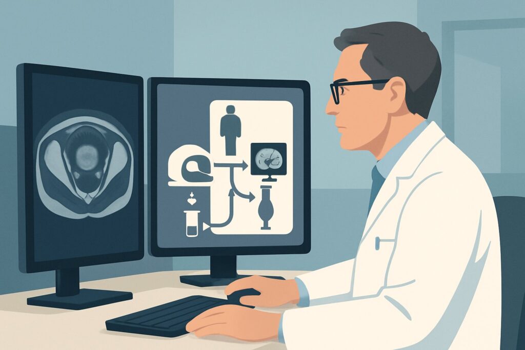
A 62-year-old male with locally advanced rectal adenocarcinoma has just completed eight weeks of neoadjuvant chemoradiation. He tolerated the treatment well, and his symptoms have improved. Now, in clinic, you are preparing him for his multidisciplinary tumor board review and subsequent surgical planning. The critical question is how to accurately assess the tumor’s response to therapy to determine the optimal surgical approach—or if, in a best-case scenario, a non-operative path is even possible. This requires precise locoregional restaging. According to the American College of Radiology (ACR) Appropriateness Criteria, an MRI of the pelvis without and with IV contrast is Usually Appropriate for this exact clinical scenario.
Who Fits This Clinical Scenario?
This guidance applies specifically to patients with biopsy-proven rectal cancer who have completed a full course of neoadjuvant therapy (typically chemoradiation) and now require locoregional restaging. The primary goal is to evaluate the degree of tumor response before a final decision is made about surgical resection. This assessment is crucial for determining the post-treatment tumor stage (ypTNM) and guiding the surgical strategy.
It is critical to distinguish this situation from other, similar-sounding clinical presentations that follow different imaging pathways:
- Initial Staging: This workflow is NOT for patients with a new diagnosis of rectal cancer who have not yet received any treatment. The initial workup follows the ACR guidelines for initial locoregional staging, which also relies on pelvic MRI but for a different clinical question.
- Evaluation for Distant Metastases: While locoregional staging is key, a separate evaluation for distant disease in the chest, abdomen, and pelvis (often with CT) is also part of the overall staging process. This article focuses only on the locoregional assessment of the primary rectal tumor post-treatment.
- Colon Cancer Staging: This guidance is specific to rectal cancer. The anatomical constraints of the pelvis, the importance of the mesorectal fascia, and the use of neoadjuvant therapy are unique to rectal cancer and dictate a different imaging approach than that for colon cancer.
What Are You Assessing After Neoadjuvant Therapy for Rectal Cancer?
In the post-treatment setting, the imaging goal shifts from initial diagnosis to assessing treatment response. The “differential” is less about identifying the disease and more about characterizing the extent of residual tumor versus treatment-related changes. The key questions you are trying to answer with imaging directly influence patient management.
Complete or Near-Complete Response: This is the optimal outcome, where neoadjuvant therapy has eradicated most or all of the viable tumor cells. Identifying this on imaging is paramount, as it may qualify certain patients for non-operative “watch-and-wait” management protocols. Imaging must confidently distinguish a complete response from minimal residual disease.
Partial Response with Residual Tumor: This is the most frequent outcome. The imaging must precisely delineate the size, morphology, and location of the remaining tumor. Critically, it must define the tumor’s relationship to key surgical landmarks, especially the mesorectal fascia, which serves as the circumferential resection margin (CRM). A threatened or involved CRM is a major prognostic indicator that the surgeon must know about preoperatively.
Post-Treatment Fibrosis vs. Viable Tumor: This is the central diagnostic challenge. Neoadjuvant therapy induces inflammation and fibrosis (scarring), which can look very similar to residual tumor on some imaging sequences. The chosen modality must have high soft-tissue resolution to differentiate between benign post-treatment changes and malignant tissue that requires resection.
Stable or Progressive Disease: A less common but crucial finding is the lack of response or, worse, tumor growth despite therapy. Identifying this signals a poor prognosis and necessitates an urgent re-evaluation of the treatment plan by the multidisciplinary team.
Why Is Pelvic MRI the Recommended Study for Restaging Rectal Cancer?
The ACR rates MRI of the pelvis without and with IV contrast as Usually Appropriate because its superior soft-tissue contrast resolution is unmatched for evaluating the rectal wall and surrounding mesorectal tissues after the anatomic distortion caused by chemoradiation. It is the most accurate non-invasive modality for differentiating residual tumor from fibrosis, which is the key to correct restaging.
A dedicated rectal cancer MRI protocol uses high-resolution, thin-slice T2-weighted images to visualize the layers of the rectal wall and the mesorectal fascia. This allows for an assessment of the radiologic tumor regression grade (mrTRG). Furthermore, functional sequences like Diffusion-Weighted Imaging (DWI) can help identify areas of high cellularity characteristic of viable tumor, which appear as areas of restricted diffusion. The addition of IV gadolinium contrast can further help by highlighting enhancing residual tumor, although post-radiation inflammation can also enhance, making interpretation complex.
Alternative studies are rated lower for this specific task:
- Transrectal Ultrasound (US): Rated May be appropriate (Disagreement), this modality is less accurate after neoadjuvant therapy. The inflammation and fibrosis induced by treatment obscure the normal tissue planes, making it very difficult to distinguish scar from tumor. Expert opinion is divided on its utility in this setting.
- CT of the Abdomen and Pelvis with IV Contrast: Rated May be appropriate, CT is excellent for assessing distant metastases and regional lymph nodes outside the mesorectum. However, its soft-tissue resolution within the pelvis is significantly inferior to MRI for delineating the primary tumor’s response and its relationship to the mesorectal fascia.
From a safety perspective, MRI is the ideal choice as it involves no ionizing radiation (0 mSv). This is particularly relevant for patients who have already received a significant radiation dose during therapy.
What’s Next After Pelvic MRI? Downstream Workflow for Rectal Cancer Restaging
The MRI report is not an endpoint; it is a critical input for the multidisciplinary tumor board (MDT) that directs the next phase of care. The downstream workflow depends directly on the imaging findings.
- If MRI suggests a complete response (mrCR): The findings will be correlated with the digital rectal exam and endoscopy with biopsies. If all signs point to a complete pathologic response, the patient may be offered entry into a non-operative management (NOM) or “watch-and-wait” program. This involves intensive surveillance with regular imaging and endoscopy rather than immediate surgery.
- If MRI shows residual tumor with clear margins: This is the most common finding. The MRI report acts as a detailed preoperative map for the surgeon, defining the extent of resection required. The patient proceeds to the planned surgery, such as a low anterior resection (LAR) or abdominoperineal resection (APR).
- If MRI shows a threatened or involved circumferential resection margin (CRM): This is a high-risk finding that signals a high likelihood of local recurrence. The MDT will discuss this urgently. The surgical plan may be altered to include a more extensive resection, or in some cases, additional therapy might be considered.
- If MRI findings are indeterminate: Sometimes, the distinction between fibrosis and a small amount of residual tumor is equivocal. In these cases, an FDG-PET/CT (rated May be appropriate) may be used as a problem-solving tool to look for metabolic activity suggestive of viable cancer. The final decision will rest on a consensus of the MDT, integrating all available clinical and imaging data.
Common Pitfalls in Post-Treatment Rectal Cancer Imaging (and When to Escalate)
Several pitfalls can compromise the accuracy of post-treatment staging. Awareness of these issues is key for the ordering clinician and the interpreting radiologist.
- Improper Timing of the Scan: Ordering the MRI too soon after the completion of chemoradiation is a common error. Post-treatment inflammation is at its peak immediately after therapy and can mimic residual tumor, leading to an overestimation of disease. The scan should be timed approximately 6-8 weeks after therapy completion to allow acute inflammation to subside.
- Using a Generic Pelvic MRI Protocol: A standard, large field-of-view pelvic MRI is inadequate. You must specifically order a dedicated, high-resolution rectal cancer protocol to obtain the necessary small field-of-view images of the tumor bed.
- Over-reliance on a Single Sequence: No single MRI sequence tells the whole story. Accurate interpretation requires synthesizing findings from T2-weighted images, DWI, and post-contrast sequences to distinguish scar from tumor.
If the MRI report is discordant with your clinical examination or endoscopic findings, it is crucial to escalate the case for review at the multidisciplinary tumor board before proceeding with a final treatment plan.
Related ACR Topics and Tools
Navigating imaging choices requires access to reliable, scenario-specific guidance. For breadth across all scenarios in Staging of Colorectal Cancer, see our parent guide: Staging of Colorectal Cancer: ACR Appropriateness Decoded.
For other clinical questions or to explore imaging protocols and radiation safety, the following GigHz resources are available:
Frequently Asked Questions
Why is MRI without contrast also rated ‘Usually Appropriate’ for post-treatment rectal cancer staging?
The ACR lists both ‘MRI pelvis without IV contrast’ and ‘MRI pelvis without and with IV contrast’ as ‘Usually Appropriate’. The core of the examination relies on high-resolution T2-weighted imaging and Diffusion-Weighted Imaging (DWI), which do not require contrast. While contrast can sometimes help identify enhancing residual tumor, post-radiation inflammation also enhances, which can complicate interpretation. Many institutions consider the non-contrast portion of the study to be the most critical component for locoregional restaging.
How long after completing neoadjuvant therapy should the restaging MRI be performed?
The optimal timing is a balance between allowing acute post-treatment inflammation to resolve and avoiding a significant delay to surgery. Most guidelines recommend performing the restaging pelvic MRI approximately 6 to 8 weeks after the completion of neoadjuvant chemoradiation.
Can FDG-PET/CT replace MRI for locoregional restaging of rectal cancer?
No. While FDG-PET/CT is rated ‘May be appropriate’, it is not a replacement for MRI. PET/CT assesses metabolic activity, which can be helpful, but it lacks the detailed anatomical resolution of MRI needed to evaluate the tumor’s relationship with the mesorectal fascia. It is best used as a problem-solving tool in equivocal cases or for assessing distant metastatic disease, not as the primary modality for locoregional restaging.
What if my patient has a contraindication to MRI, like an incompatible pacemaker?
In cases with a strong contraindication to MRI, you must rely on the next best available modalities. This would typically involve a high-quality CT of the abdomen and pelvis with IV contrast (‘May be appropriate’) for a broader view, combined with a detailed clinical exam and endoscopy with biopsies to assess the local tumor bed. The limitations of CT for soft-tissue delineation in the pelvis must be clearly communicated to the surgical team.
Does the MRI report give a definitive ‘yes’ or ‘no’ on whether there is a complete response?
Not definitively. The MRI provides a radiologic assessment of treatment response (mrTRG), but the gold standard for confirming a pathologic complete response (pCR) is histologic analysis of the resected specimen. MRI can strongly suggest a complete response, guiding a decision toward non-operative management, but it cannot prove it with 100% certainty. This is why such decisions are always made in a multidisciplinary context, weighing all available data.
Reviewed by Pouyan Golshani, MD, Interventional Radiologist — May 29, 2026