Post-Surgery Thoracic Back Pain: Should You Order Radiographs, CT, or MRI First?
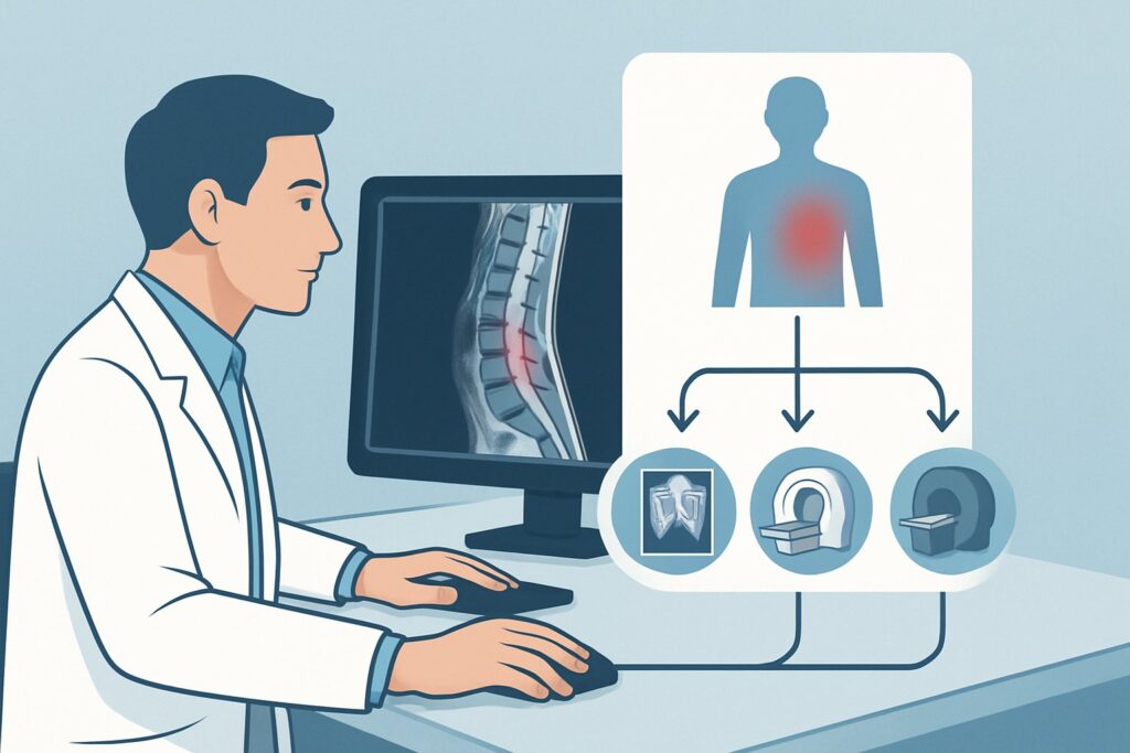
A 58-year-old patient is in your clinic for a six-month follow-up after a T7-T10 instrumented fusion for a traumatic fracture. For the past three weeks, they have reported a return of deep, aching mid-back pain, similar to their pre-operative symptoms, now with new, intermittent tingling in their legs. You perform a neurologic exam and find subtle hyperreflexia. The immediate question is how to evaluate the integrity of the surgical construct and rule out new pathology. This scenario—follow-up imaging for an adult with thoracic back pain after spine surgery—requires a deliberate, stepwise approach. According to the American College of Radiology (ACR) Appropriateness Criteria, the initial study, Radiography thoracic spine, is rated Usually Appropriate.
Who Fits This Clinical Scenario?
This guidance applies specifically to adult patients who have previously undergone thoracic spine surgery (e.g., decompression, fusion, instrumentation) and are now presenting for follow-up imaging due to persistent, recurrent, or new thoracic back pain. The presence of neurologic symptoms, such as radiculopathy (nerve root irritation) or myelopathy (spinal cord compression), is a key consideration within this scenario.
The defining characteristic is the post-operative context. The differential diagnosis and imaging challenges are fundamentally different from those in a surgery-naive patient. Hardware can obscure anatomy, and post-surgical changes can mimic pathology.
This workflow is distinct from several related clinical situations:
- Initial Imaging Pre-Surgery: A patient with thoracic back pain and myelopathy who has not yet had surgery follows a different diagnostic algorithm.
- Acute High-Velocity Trauma: A post-operative patient who sustains a new, significant traumatic injury may require immediate, advanced imaging like CT, bypassing initial radiographs.
- Suspected Cancer or Systemic Infection: If the primary concern is a new malignancy or the patient presents with systemic signs of infection (e.g., fever, rigors, markedly elevated inflammatory markers), MRI with contrast is often the most appropriate first-line study.
What Diagnoses Are You Working Up in This Scenario?
In the post-operative thoracic spine, pain can arise from several distinct mechanical or biological failures. Your imaging choice is designed to efficiently differentiate among these possibilities.
Hardware Complications: This is often the primary concern. Imaging is used to assess for hardware loosening, demonstrated by lucency around screws; hardware fracture, such as a broken pedicle screw or rod; or hardware malposition that may be impinging on neural elements. These are primarily mechanical issues that radiographs can often identify.
Pseudarthrosis (Failed Fusion): A common cause of persistent pain after fusion surgery is the failure of the bone to heal across the fused segments. This creates a non-union or “false joint” that allows for micromotion and subsequent mechanical pain. While subtle on radiographs, it is a key diagnosis to consider if hardware is intact.
Adjacent Segment Disease: Fusing a segment of the spine can transfer stress to the levels immediately above and below the construct. Over time, this can accelerate degenerative changes, leading to new stenosis, disc herniation, or instability at these adjacent levels, causing a return of symptoms.
Post-operative Infection or Fluid Collection: Though less common, spondylodiscitis (infection of the disc and vertebral bodies) or an epidural abscess can present with severe pain, with or without neurologic deficits. These are critical diagnoses to exclude, as they often require urgent intervention.
Why Is Thoracic Spine Radiography an Appropriate First Step?
While multiple advanced imaging modalities are also highly rated for this scenario, the ACR guidance supports starting with thoracic spine radiography because it is a fast, accessible, and low-dose method to assess the most immediate mechanical questions. It is rated Usually Appropriate.
Anteroposterior (AP) and lateral radiographs provide an excellent overview of spinal alignment and the integrity of surgical instrumentation. They are highly effective for detecting gross hardware failure like a fractured rod or a significantly backed-out screw. Flexion and extension views can sometimes be used to assess for instability or abnormal motion suggestive of pseudarthrosis. With a relative radiation level of ☢☢☢ (1-10 mSv), it provides crucial initial information while minimizing radiation exposure.
However, several other studies are also rated Usually Appropriate and serve as critical next steps or, in some cases, appropriate initial studies depending on the specific clinical question:
- MRI Thoracic Spine Without and With IV Contrast: This is the gold standard for evaluating the spinal cord, nerve roots, and soft tissues. It is superior for detecting epidural abscess, discitis, hematoma, or recurrent disc herniation. The “with contrast” portion is essential when infection or inflammation is suspected.
- CT Thoracic Spine Without IV Contrast: This modality provides the most detailed assessment of bony anatomy. It is the best test to definitively evaluate for pseudarthrosis, showing the presence or absence of a solid bony fusion mass. It is also excellent for clarifying suspected hardware-related issues seen equivocally on radiographs.
Conversely, a study like a Bone Scan with SPECT/CT is rated Usually not appropriate. While it is sensitive for metabolic activity, the expected post-surgical inflammatory and healing response makes it highly non-specific in this setting, leading to frequent false-positive results.
What’s Next After Radiography? Downstream Workflow
The initial radiograph is a triage tool that directs the subsequent diagnostic pathway. The next step depends entirely on its findings in the context of the patient’s symptoms.
- If radiographs show clear hardware failure (e.g., broken screw, rod displacement): The diagnosis is often made. The next step is typically a surgical consultation to plan for revision. A pre-operative CT may still be ordered to better define the bony anatomy for surgical planning.
- If radiographs are negative but neurologic symptoms are present: When a patient has signs of myelopathy or progressive radiculopathy, a negative radiograph is not reassuring. The workflow must escalate to an MRI of the thoracic spine to evaluate the spinal cord and nerve roots for compression from a soft-tissue cause like a hematoma, abscess, or disc herniation.
- If radiographs are negative and pain is primarily mechanical (without neurologic deficits): If the main suspicion is a failed fusion (pseudarthrosis), the next step is a CT of the thoracic spine without contrast. This will provide the definitive assessment of the fusion mass.
- If radiographs are equivocal or suggest infection (e.g., subtle lucency around a screw, endplate erosion): Proceed directly to an MRI of the thoracic spine with and without IV contrast and correlate with inflammatory markers (ESR, CRP). This is the most sensitive and specific non-invasive test for spondylodiscitis and epidural abscess.
Pitfalls to Avoid (and When to Get Help)
Navigating post-operative spine imaging requires careful correlation between imaging findings and the clinical picture. Here are common pitfalls to avoid:
- Stopping at a “Normal” Radiograph: Never dismiss significant neurologic symptoms like myelopathy just because the initial radiograph is unremarkable. The cause is likely in the soft tissues, which radiographs cannot visualize.
- Ignoring Comparison Films: Post-operative imaging is all about change over time. Always compare the new study to the immediate post-operative and subsequent follow-up films to detect subtle hardware migration or progressive degenerative changes.
- Artifact-Related Errors: Surgical hardware creates significant metallic artifact on both CT and MRI, which can obscure adjacent anatomy. Ensure your radiology department uses metal artifact reduction sequences (MARS) for MRI and appropriate reconstruction algorithms for CT.
- Over-interpreting Normal Post-op Changes: Some fluid collections and enhancement are normal in the early post-operative period. If imaging is performed too soon after surgery, these findings can be mistaken for infection.
If a patient develops rapidly progressive neurologic deficits or shows signs of sepsis, this constitutes a surgical emergency. Escalate immediately with an urgent MRI and consultation with the spine surgeon.
Related ACR Topics and Tools
This article covers one specific variant within the broader topic of Thoracic Back Pain. The optimal imaging strategy changes based on patient history, symptoms, and prior imaging findings. For a comprehensive overview of all clinical scenarios, from acute pain to suspected cancer, please consult our parent guide. Additional GigHz tools can help refine your imaging orders and patient conversations.
- For breadth across all scenarios in Thoracic Back Pain, see our parent guide: Thoracic Back Pain: ACR Appropriateness Decoded.
- To explore adjacent scenarios and their corresponding ACR ratings, use the ACR Appropriateness Criteria Lookup.
- For detailed technical specifications of the recommended studies, visit the Imaging Protocol Library.
- To discuss radiation exposure with your patients, consult the Radiation Dose Calculator.
Frequently Asked Questions
Why start with a radiograph if MRI and CT are also rated ‘Usually Appropriate’?
Starting with a radiograph is a pragmatic, high-yield approach. It is fast, inexpensive, widely available, and uses a lower radiation dose than CT. It can quickly answer the most common mechanical questions, such as gross hardware failure or malalignment. If the radiograph is negative or equivocal, it effectively triages the patient to the more specific problem-solving capabilities of MRI (for neural elements/infection) or CT (for bony fusion).
If a patient has new leg weakness after thoracic surgery, should I still get a radiograph first?
In the presence of significant or progressive neurologic deficits like myelopathy (e.g., leg weakness, gait instability, bowel/bladder changes), escalating directly to an MRI is often the most appropriate action. While a radiograph is not wrong, it cannot visualize the spinal cord. An MRI will directly assess for cord compression from causes like an epidural hematoma, abscess, or hardware impingement, which may require urgent intervention.
How long after surgery do I need to worry about metal artifact on MRI?
Metallic artifact is a permanent challenge related to the type of hardware implanted (e.g., titanium causes less artifact than stainless steel). It is not something that resolves over time. It is crucial to communicate with the radiologist that the patient has hardware, so they can employ specific metal artifact reduction sequences (MARS) to improve image quality around the instrumentation.
What is the role of CT myelography in this post-operative scenario?
CT myelography is rated ‘May be appropriate’. It is typically reserved for cases where MRI is contraindicated (e.g., patient has an incompatible implanted device) or when MRI results are inconclusive due to severe artifact. By introducing intrathecal contrast, it can delineate the contours of the spinal cord and nerve roots even in the presence of metal, helping to identify stenosis or compression.
Can I use a single MRI to evaluate for both infection and pseudarthrosis?
An MRI with and without contrast is the best test for infection. However, CT is superior to MRI for definitively evaluating bony fusion (pseudarthrosis). While MRI can show secondary signs of pseudarthrosis like fluid in the disc space or motion on kinetic studies, CT provides direct visualization of the bony bridging. Therefore, if both are a concern, you may need both studies, often starting with the one that addresses the most urgent clinical question.
Reviewed by Pouyan Golshani, MD, Interventional Radiologist — May 30, 2026