Should You Image a Classic Migraine? Why the ACR Says ‘Usually Not Appropriate
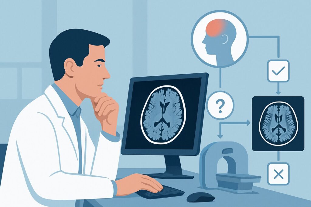
A 34-year-old woman presents to your clinic on a Tuesday afternoon with a familiar, throbbing, unilateral headache. The bright lights of the exam room make her wince, and she reports associated nausea. Her history is classic for migraine without aura, and her neurologic examination is, as always, completely normal. She mentions a news story about a celebrity’s brain tumor and asks, “Doctor, are you sure we shouldn’t just get an MRI to be safe?”
This scenario is one of the most common decision points in primary care and neurology. It pits clinical pattern recognition against patient anxiety and the temptation of defensive medicine. For this specific presentation—a primary migraine or tension-type headache with a normal neurologic exam—this article details the American College of Radiology (ACR) Appropriateness Criteria, which rates all forms of initial imaging, from MRI to CT to Arteriography, as Usually not appropriate.
Who Fits This Clinical Scenario?
This guidance applies to a well-defined patient population. The key is a confident clinical diagnosis of a primary headache disorder based on history and a normal physical examination.
Inclusion criteria for this workflow:
- Headache Pattern: The headache matches the typical characteristics of a primary headache, such as migraine (with or without aura) or tension-type headache. Features may include an episodic nature, a stable pattern over months or years, and a return to a normal baseline between attacks.
- Normal Neurologic Examination: This is a critical prerequisite. A thorough examination reveals no abnormalities in mental status, cranial nerves, motor function, sensation, coordination, or gait.
Exclusion criteria (these patients belong to different workflows):
- Sudden, Severe Onset: A “thunderclap” headache that reaches maximal intensity within an hour is a neurologic emergency and requires a different imaging pathway to rule out subarachnoid hemorrhage.
- Presence of “Red Flags”: Any of the following moves the patient to a higher-risk category where imaging is often warranted: a new or worsening headache in a patient with a history of cancer or immunosuppression, headache associated with fever or stiff neck, or a progressive headache that worsens over weeks or months.
- Focal Neurologic Deficits: New, persistent weakness, numbness, visual field loss, or other focal signs found on examination mandate imaging.
- Signs of Altered Intracranial Pressure: Headaches that are strictly positional (worse when standing, relieved by lying down) or associated with signs like papilledema on fundoscopy suggest intracranial hypotension or hypertension, respectively, and require dedicated imaging protocols.
What Diagnoses Are You Working Up in This Scenario?
In the case of a classic primary headache with a normal exam, the clinical goal is not to “work up” a long list of possibilities. Instead, it is to confidently conclude that the pre-test probability of a dangerous secondary cause is so low that the risks and costs of imaging outweigh any potential benefit. The strength of the history and normal exam effectively rules out the following sinister causes in this specific population.
Intracranial Mass (e.g., Tumor, Abscess): The most common patient fear. However, brain tumors typically cause progressive headaches that change in character or are associated with focal neurologic deficits, seizures, or altered mental status. In a patient with a stable, episodic headache pattern and a normal exam, the likelihood of finding a tumor is exceedingly low.
Subarachnoid Hemorrhage (SAH): A non-aneurysmal “sentinel bleed” can theoretically present as a less severe headache, but the classic presentation is a thunderclap headache. A typical migraine pattern is not consistent with SAH.
Cerebral Venous Sinus Thrombosis (CVST): While headache is a common symptom, CVST is often accompanied by other signs that would exclude a patient from this scenario, such as papilledema, seizures, or focal deficits that reflect venous infarction or congestion.
Cervical Artery Dissection: This condition typically presents with a combination of headache, neck pain, and often ischemic symptoms (like a transient ischemic attack or stroke) or a Horner syndrome. An isolated, classic migraine-phenotype headache would be a very atypical presentation.
Why Is Neuroimaging Usually Not Appropriate for This Presentation?
For a patient with a primary migraine or tension-type headache and a normal neurologic exam, the ACR rates all initial imaging modalities as Usually not appropriate. This includes non-contrast MRI of the head, CT of the head, and even advanced studies like CTA, MRA, and cervicocerebral arteriography. The rationale is based on a careful balance of benefit versus harm.
1. Extremely Low Diagnostic Yield Multiple large-scale studies have demonstrated that in patients who meet the clinical criteria for migraine and have a normal exam, the rate of finding a significant, causative abnormality on neuroimaging is virtually zero. The yield is no higher than that of finding an abnormality in an asymptomatic person of the same age. The clinical diagnosis is highly reliable on its own.
2. The High Risk of Incidental Findings The probability of finding an incidentaloma—an unrelated, asymptomatic abnormality—is far greater than finding a cause for the headache. Common incidental findings include arachnoid cysts, developmental venous anomalies, small meningiomas, or pineal cysts. Discovering these can trigger a cascade of patient anxiety, further specialist consultations, costly follow-up scans, and, in rare cases, unnecessary and risky interventions, causing iatrogenic harm.
3. The Inherent Risks and Costs of Imaging No imaging study is entirely without risk or cost.
- CT Head (without or with contrast): This study is rated Usually not appropriate. It exposes the patient to ionizing radiation (ACR Relative Radiation Level ☢☢☢, 1-10 mSv). While the risk from a single scan is small, it is not zero, and cumulative radiation exposure should be avoided, particularly in younger patients who may experience decades of headaches.
- MRI Head (without or with contrast): Also rated Usually not appropriate. While it avoids radiation, it is more expensive and less accessible than CT. If contrast is used, there is a very small risk associated with gadolinium deposition, especially in patients with renal impairment.
- Arteriography cervicocerebral: This invasive catheter-based study is definitively Usually not appropriate. It carries significant risks, including stroke, vessel dissection, and bleeding at the access site, in addition to a high dose of radiation (☢☢☢, 1-10 mSv) and iodinated contrast. It has no role in evaluating a primary headache disorder.
What’s Next After the Evaluation? Downstream Workflow
The recommended workflow for this scenario deliberately avoids imaging and pivots directly to clinical management.
If the Recommended Path (No Imaging) Is Followed:
- Next Step: Patient education and reassurance. Explain the diagnosis of migraine or tension-type headache and why imaging is not recommended by national guidelines. A confident explanation can be a powerful therapeutic tool.
- Action: Initiate appropriate evidence-based treatment. This may include abortive medications (e.g., triptans, NSAIDs), prophylactic medications for frequent headaches, and counseling on non-pharmacologic strategies like trigger identification and lifestyle adjustments.
- Safety Netting: Provide the patient with clear, explicit “red flag” symptoms that should prompt them to seek immediate medical attention. These include the sudden onset of the worst headache of their life, a headache accompanied by fever or confusion, or the development of any new neurologic symptoms.
If Imaging Is Performed and Is Negative:
- Next Step: Use the normal result to provide definitive reassurance. This can help break a cycle of anxiety for some patients.
- Action: Immediately pivot back to the clinical management plan for the primary headache disorder. The negative scan confirms the initial clinical diagnosis.
If Imaging Is Performed and Shows an Incidental Finding:
- Next Step: This creates a new, often complex, clinical pathway. It requires careful communication to explain that the finding is unrelated to the headaches.
- Action: Depending on the finding, this may necessitate a referral to a neurologist or neurosurgeon to determine if the finding requires surveillance imaging or has any clinical significance. This highlights the downstream cascade that non-indicated imaging can trigger.
Pitfalls to Avoid (and When to Get Help)
- Ordering for Reassurance Alone: Giving in to patient pressure or personal anxiety by ordering a low-yield test can lead to the incidentaloma cascade and reinforces the incorrect idea that a “cause” must be found on a scan.
- Misclassifying the Headache: The entire “no imaging” pathway depends on a correct clinical diagnosis. Be meticulous in screening for subtle red flags, such as a gradual change in headache character or frequency, that might suggest a secondary cause.
- Ignoring a Pattern Change: A patient with a 20-year history of stable migraine who presents with a new, different, and unrelenting headache no longer fits this scenario. A significant change in pattern is a red flag that warrants re-evaluation and likely imaging.
- When to Escalate: If at any point during the history or physical exam a red flag emerges (e.g., a new focal deficit, papilledema, suspicion of meningitis), the workflow must be abandoned. Escalate immediately to the appropriate next step, which is typically emergent neuroimaging and/or specialty consultation.
Related ACR Topics and Tools
This article covers a single, common scenario. For a broader view of imaging for all types of headache presentations, or to explore the tools and techniques involved, see the resources below.
- For breadth across all scenarios in Headache, see our parent guide: Headache: ACR Appropriateness Decoded.
- ACR Appropriateness Criteria Lookup — for adjacent scenarios
- Imaging Protocol Library — for technique on the recommended study
- Radiation Dose Calculator — for cumulative dose conversations
Frequently Asked Questions
My patient is convinced they have a brain tumor. How do I manage this without ordering a scan?
First, acknowledge and validate their concern. Then, provide clear education. Explain that the specific features of their headache (e.g., episodic, throbbing) and, most importantly, their completely normal neurologic exam make a dangerous underlying cause like a tumor extremely unlikely. Frame the decision not to image as adherence to best practices designed to protect them from unnecessary risks like radiation and the anxiety of incidental findings. Confident reassurance and a clear treatment plan for their migraine are key.
What if the patient has a family history of a brain aneurysm?
A significant family history (especially multiple first-degree relatives with aneurysms) can be considered a risk factor that may shift the patient out of this specific low-risk scenario. While a classic migraine pattern still makes a primary headache most likely, this history might lower the threshold for considering non-invasive vascular imaging (MRA). This is a nuanced decision, often best made in consultation with a neurologist, as the patient may no longer perfectly fit the ‘no red flags’ criterion.
Are there any exceptions where imaging is considered for a first-time classic migraine?
While the ACR guidance is ‘Usually not appropriate,’ some professional society guidelines suggest that imaging could be considered for an initial presentation of migraine with aura to rule out a mimic (like a seizure or transient ischemic attack). However, this is not considered mandatory, and the ACR’s rating reflects the extremely low diagnostic yield even in this situation, provided the neurologic exam is normal and there are no other red flags.
If I don’t order a scan, what documentation is most important?
Your documentation should clearly justify the clinical diagnosis. Note the classic features of the primary headache (e.g., using POUNDing criteria for migraine: Pulsatile, one-day duration, Unilateral, Nausea/vomiting, Disabling intensity). Most critically, document the absence of any red flag symptoms and the specific findings of your normal neurologic examination, covering mental status, cranial nerves, motor, sensory, cerebellar function, and gait. Conclude with a statement that the presentation is consistent with a primary headache disorder for which imaging is not indicated per established guidelines.
Does this guidance against routine imaging apply to children with primary headaches?
Yes, the principle is the same, and the imperative to avoid unnecessary ionizing radiation is even stronger in the pediatric population. The ACR data specifically notes pediatric radiation levels to emphasize this consideration. For children, a thorough history (often from parents as well) and a careful neurologic exam are paramount. If there is any diagnostic uncertainty, consultation with a pediatric neurologist is highly recommended before considering imaging.
Reviewed by Pouyan Golshani, MD, Interventional Radiologist — June 27, 2026