Which Imaging Study Is Best for Acquired Conductive Hearing Loss Without an Obvious Mass?
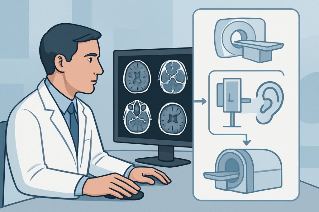
A 45-year-old male presents to your clinic with a six-month history of progressive, muffled hearing in his left ear. He denies pain, vertigo, or recent trauma. On physical examination, the external auditory canal is clear and the tympanic membrane appears normal, with no visible mass, effusion, or perforation. A Weber test lateralizes to the affected left ear, and the Rinne test confirms bone conduction is greater than air conduction, consistent with a conductive hearing loss. You suspect an issue with the ossicular chain or another middle ear structure not visible on otoscopy. The crucial next question is which imaging study will provide a definitive diagnosis without unnecessary radiation or cost. For this specific scenario, the American College of Radiology (ACR) finds that a CT temporal bone without IV contrast is Usually Appropriate.
Who Fits This Clinical Scenario?
This guidance applies to adult patients presenting with an acquired conductive hearing loss where the cause is not apparent on clinical examination. The key inclusion criteria are:
- Acquired Hearing Loss: The deficit is not congenital and developed after birth.
- Conductive Deficit: Confirmed by audiometry or tuning fork tests (e.g., Weber, Rinne), indicating a problem with sound transmission through the outer or middle ear.
- Clinically Unremarkable Middle Ear: Otoscopy reveals a normal-appearing tympanic membrane without an obvious mass, cholesteatoma, or effusion that would explain the hearing loss.
It is critical to distinguish this presentation from similar but distinct clinical situations that require different imaging pathways. This workflow does not apply if:
- A mass or cholesteatoma is visible on exam: If there is suspicion of intracranial or inner ear extension of a known lesion, a different ACR variant applies, often involving MRI.
- The hearing loss is sensorineural or mixed: Patients with sensorineural hearing loss (SNHL) or a mixed conductive/sensorineural picture are evaluated under a separate ACR guideline, as the differential diagnosis shifts toward the inner ear and retrocochlear structures, making MRI the primary modality.
- The patient has episodic or persistent vertigo: While hearing loss can accompany vertigo, the presence of significant vestibular symptoms points toward different ACR scenarios focused on evaluating the inner ear and central nervous system.
What Diagnoses Are You Working Up in This Scenario?
When a patient has conductive hearing loss with a normal-appearing eardrum, imaging is essential to visualize the middle ear structures that are hidden from direct view. The differential diagnosis in this scenario is focused on abnormalities of the ossicular chain and surrounding temporal bone.
Otosclerosis is a primary consideration. This is a metabolic bone disease of the otic capsule that causes abnormal bone remodeling. In its most common form (fenestral otosclerosis), it leads to fixation of the stapes footplate in the oval window, impeding sound transmission. While it often presents in younger adults, it can be diagnosed at any age. CT is highly sensitive for detecting the demineralized bone characteristic of otosclerotic foci.
Ossicular Chain Discontinuity or Fixation is another key diagnostic possibility. This can result from remote, forgotten trauma, chronic negative middle ear pressure, or congenital anomalies that manifest later in life. The incus is the most commonly affected ossicle in traumatic dislocations. CT provides exquisite detail of the ossicles and their articulations, allowing for direct visualization of dislocation, erosion, or fusion.
Superior Semicircular Canal Dehiscence (SSCD) is a less common but important cause. This condition involves a defect in the bone overlying the superior semicircular canal, creating a “third window” in the inner ear. While it can cause vestibular symptoms, it often presents with conductive hearing loss due to the abnormal transmission of acoustic energy. High-resolution CT is the definitive imaging modality for identifying the bony dehiscence.
Other less frequent causes include occult cholesteatoma (a non-neoplastic but destructive growth of squamous epithelium), glomus tympanicum (a small paraganglioma confined to the middle ear), or fibro-osseous lesions. CT is the first-line study to detect the subtle bone erosion or soft tissue density associated with these conditions.
Why Is CT Temporal Bone Without IV Contrast the Recommended Study?
The ACR designates CT temporal bone without IV contrast as Usually Appropriate for this clinical scenario because it directly and efficiently addresses the most likely differential diagnoses. The primary goal of imaging is to assess the bony anatomy of the middle ear, ossicular chain, and otic capsule, and CT is the superior modality for this purpose.
High-resolution, thin-section CT of the temporal bones provides unparalleled detail of the ossicles (malleus, incus, stapes), the oval and round windows, and the surrounding bony labyrinth. This allows for the confident diagnosis of otosclerosis, ossicular dislocation or erosion, and superior semicircular canal dehiscence. Because these are primarily bony pathologies, intravenous contrast is not necessary and adds no diagnostic value, while increasing cost and potential risk (e.g., contrast reaction, nephrotoxicity).
Alternative imaging studies are rated as Usually Not Appropriate for good reason:
- MRI head and internal auditory canal without and with IV contrast: While MRI is the gold standard for evaluating sensorineural hearing loss (assessing the vestibulocochlear nerve and brainstem), it provides very poor visualization of the fine bony structures of the middle ear. It cannot reliably diagnose otosclerosis or subtle ossicular chain abnormalities, making it the wrong initial test for a purely conductive deficit.
- CT head without IV contrast: A standard CT of the head is performed with much thicker image slices and a different reconstruction algorithm than a dedicated temporal bone CT. It lacks the spatial resolution needed to evaluate the millimeter-sized ossicles and would likely miss the key findings in this scenario. Ordering a “CT head” instead of a “CT temporal bone” is a common and critical error.
The recommended study, CT temporal bone, involves a moderate radiation dose (ACR Relative Radiation Level ☢☢☢, 1-10 mSv). However, this is a focused, high-yield examination where the diagnostic benefit of identifying a surgically correctable cause of hearing loss substantially outweighs the radiation risk.
What’s Next After CT Temporal Bone? Downstream Workflow
The results of the CT temporal bone will guide the subsequent clinical pathway, which is often surgical. The imaging findings create a clear branch point for patient management and counseling.
If the CT is positive for a clear diagnosis:
- Otosclerosis: The patient can be referred to an otologist for a discussion of treatment options, which may include hearing aids or surgical intervention such as a stapedotomy or stapedectomy. The CT confirms the diagnosis and helps in surgical planning.
- Ossicular Discontinuity: This finding also warrants referral to an otologist for surgical exploration and ossiculoplasty (reconstruction of the ossicular chain).
- Superior Semicircular Canal Dehiscence: Management depends on symptom severity. Options range from observation to surgical repair, such as canal plugging or resurfacing. The CT provides the definitive diagnosis needed for this discussion.
If the CT is negative or indeterminate:
A negative high-resolution CT in the setting of confirmed conductive hearing loss is uncommon but can occur. This may suggest a diagnosis not well-visualized by CT, such as ossicular fixation due to fibrous tissue (tympanosclerosis) that doesn’t calcify, or a subtle stapes footplate fixation below the resolution of CT. In this situation, the next step is typically a discussion with the patient about the risks and benefits of proceeding with surgical exploration of the middle ear based on the strong clinical and audiometric findings, even with a non-diagnostic scan.
Pitfalls to Avoid (and When to Get Help)
Navigating the workup for conductive hearing loss requires careful attention to clinical detail and precise imaging orders. Here are common pitfalls to avoid:
- Ordering a “CT Head” instead of “CT Temporal Bone”: This is the most frequent mistake. A standard head CT lacks the resolution to be diagnostic and results in a wasted scan and unnecessary radiation exposure. Be specific in your order.
- Ordering MRI as the first-line test: For a purely conductive hearing loss with a normal exam, MRI is a low-yield study. It should be reserved for cases with sensorineural, mixed, or pulsatile hearing loss, or when neurologic symptoms are present.
- Forgetting to obtain audiometry: While tuning forks are excellent screening tools, a formal audiogram is essential to confirm the type and severity of hearing loss and to provide a baseline for monitoring treatment efficacy.
- Dismissing minor trauma history: Patients may not recall a minor head injury from years ago that could have caused ossicular dislocation. A thorough history is key.
If the clinical picture is confusing, the imaging is equivocal, or the patient has mixed hearing loss, consultation with an otologist or neuroradiologist is the appropriate next step.
Related ACR Topics and Tools
This article covers one specific variant within the broader topic of hearing loss and vertigo. For a comprehensive overview of all related scenarios and to ensure you are applying the right criteria to your patient, please consult our parent guide. Additional GigHz resources can help you refine your imaging orders and discuss them with patients.
- For breadth across all scenarios in Hearing Loss and/or Vertigo, see our parent guide: Hearing Loss and/or Vertigo: ACR Appropriateness Decoded.
- To explore detailed techniques for studies like CT temporal bone, see our Imaging Protocol Library.
- For discussions about radiation exposure with your patients, our Radiation Dose Calculator can help quantify cumulative dose.
- For other clinical presentations, you can search the complete ACR Appropriateness Criteria Lookup.
Frequently Asked Questions
Why is IV contrast not recommended for the initial CT scan in this scenario?
Intravenous contrast is not recommended because the primary diagnostic targets—otosclerosis, ossicular chain abnormalities, and bony dehiscence—are all pathologies of bone. Contrast enhances soft tissues and blood vessels but provides no additional information for evaluating these specific conditions. Omitting contrast avoids potential risks and lowers the cost of the examination.
What if my patient also has tinnitus with their conductive hearing loss?
If the tinnitus is non-pulsatile, the workup remains the same, with CT temporal bone as the initial study. However, if the tinnitus is pulsatile (the patient hears a whooshing sound in sync with their heartbeat), the differential diagnosis expands to include vascular abnormalities. This represents a different clinical scenario, and CTA or MRA would become more appropriate.
Can a CT temporal bone miss otosclerosis?
Yes, although it is rare with modern high-resolution CT. Early or subtle cases of fenestral otosclerosis might not show obvious demineralization, leading to a false-negative scan. In cases with a strong clinical and audiometric suspicion for otosclerosis but a negative CT, the diagnosis may ultimately be made during surgical exploration of the middle ear.
My patient has a history of chronic ear infections. Does that change the imaging choice?
Yes, potentially. A history of chronic otitis media increases the suspicion for an occult cholesteatoma or significant tympanosclerosis, both of which can cause conductive hearing loss. CT temporal bone without contrast remains the best initial imaging test, as it is excellent for detecting the bone erosion characteristic of cholesteatoma and the calcifications of tympanosclerosis.
Is there any role for a plain radiograph (X-ray) of the mastoids?
No. Plain radiographs of the mastoids are considered obsolete for evaluating the cause of conductive hearing loss. They lack the spatial and contrast resolution to visualize the ossicles or detect the subtle bony changes of otosclerosis or canal dehiscence. CT is the required modality for detailed anatomical assessment.
Reviewed by Pouyan Golshani, MD, Interventional Radiologist — May 29, 2026