Should You Order a Head or Maxillofacial CT for a Suspected Frontal Bone Injury?
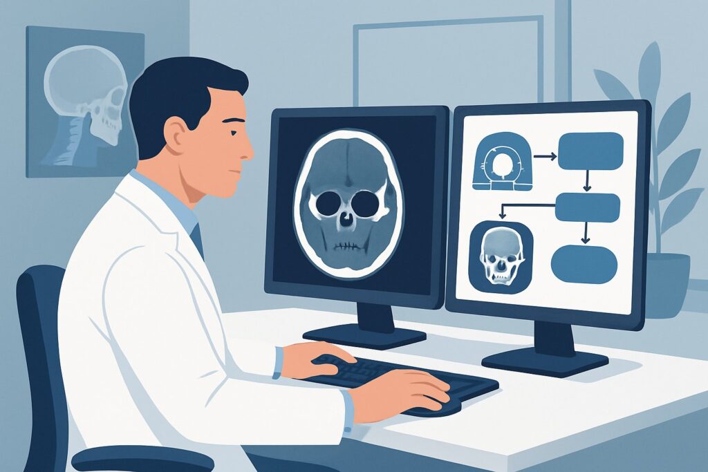
A 34-year-old male presents to the emergency department after a fall from a ladder, striking his forehead. He has no loss of consciousness and a GCS of 15, but there is significant swelling, a contusion, and marked tenderness to palpation directly over the frontal bone. The primary survey is complete and stable. Now, you must decide on the most appropriate initial imaging to evaluate for a potential frontal bone or frontal sinus fracture and to rule out associated complications. This article provides a detailed clinical workflow for this specific scenario, based on the American College of Radiology (ACR) Appropriateness Criteria. For a suspected frontal bone injury characterized by localized tenderness, contusion, or edema, the ACR rates `CT maxillofacial without IV contrast` as “Usually Appropriate.”
Who Fits This Clinical Scenario?
This guidance is intended for a specific patient presentation following a primary survey in a trauma setting. The key inclusion criteria are focal signs of injury limited to the frontal bone region.
Inclusion Criteria:
- Tenderness to palpation localized over the frontal bone.
- Visible contusion or hematoma over the forehead.
- Soft tissue edema or swelling confined to the frontal region.
- A clear clinical suspicion for an isolated or primary frontal bone injury.
This workflow is not appropriate for patients with more complex or widespread signs of facial trauma, which suggest different injury patterns. These patients require a different diagnostic approach.
Exclusion Criteria (These scenarios route to different guidelines):
- Suspected Midface or Zygomatic Injury: If the patient has pain with upper jaw manipulation, malocclusion, or a palpable deformity of the cheekbone (zygoma), the primary concern shifts to a Le Fort or zygomaticomaxillary complex fracture.
- Suspected Nasal Injury: If the primary findings are visible nasal deformity, localized nasal tenderness, or significant epistaxis, the workup should focus on a nasal bone fracture.
- Suspected Mandibular or Dental Injury: If the patient presents with trismus (difficulty opening the mouth), malocclusion, or loose/fractured teeth, the evaluation should be tailored to a mandibular fracture.
What Diagnoses Are You Working Up in This Scenario?
When you order imaging for a suspected frontal bone injury, you are evaluating for a spectrum of potential diagnoses, ranging from simple contusions to complex, high-risk fractures. The choice of imaging is driven by the need to differentiate these possibilities.
Isolated Anterior Table Frontal Sinus Fracture: This is a common injury involving only the anterior wall of the frontal sinus. While it can be a cosmetic issue and may require surgical intervention, it is generally less dangerous than fractures involving the posterior wall. CT is essential to confirm the diagnosis and assess the degree of displacement.
Combined Anterior and Posterior Table Frontal Sinus Fracture: This is a more significant injury. A fracture of the posterior table, which forms the anterior wall of the cranial vault, creates a direct communication between the contaminated sinus and the sterile intracranial space. This raises the risk of serious complications like meningitis, brain abscess, or cerebrospinal fluid (CSF) leak.
Frontal Bone Fracture with Intracranial Extension: A severely displaced posterior table fracture can be associated with direct intracranial injury. This includes pneumocephalus (air in the cranial cavity), epidural or subdural hematomas, or contusions of the frontal lobes. Identifying these findings is critical for neurosurgical consultation and management.
Naso-orbito-ethmoid (NOE) Complex Involvement: The frontal sinus outflow tract drains through the ethmoid sinuses. A fracture extending into this region can disrupt sinus drainage, leading to future complications like mucocele formation. CT is unparalleled in defining the anatomy of this delicate region.
Soft Tissue Injury Without Fracture: In many cases, the impact may only cause a soft tissue contusion or hematoma without an underlying fracture. Imaging confidently rules out a bony injury, allowing for conservative management and appropriate patient reassurance.
Why Is CT Maxillofacial without IV Contrast the Recommended Study?
The ACR designates both `CT maxillofacial without IV contrast` and `CT head without IV contrast` as “Usually Appropriate” for this clinical scenario. However, the maxillofacial CT is often the primary recommendation due to its specific advantages in evaluating the facial skeleton.
The primary rationale is the superior bony detail provided by computed tomography. CT offers high-resolution, cross-sectional images that eliminate the structural superimposition that makes plain radiographs inadequate. It is highly sensitive and specific for detecting cortical bone fractures, assessing fragment displacement, and evaluating the delicate walls of the paranasal sinuses.
A non-contrast study is sufficient because the primary goal is to assess bone and detect acute hemorrhage or pneumocephalus, neither of which requires intravenous contrast. Avoiding contrast also eliminates the risks of allergic reaction and contrast-induced nephropathy.
Comparing the “Usually Appropriate” Options:
- CT Maxillofacial without IV contrast (Adult RRL ☢☢): This study is optimized for facial bone imaging. It typically uses thinner image slices (e.g., 0.625-1.25 mm) and specific bone algorithms that provide exquisite detail of the anterior and posterior tables of the frontal sinus, the orbital rims, and the naso-orbito-ethmoid complex.
- CT Head without IV contrast (Adult RRL ☢☢☢): This study is optimized for evaluating the brain parenchyma and intracranial contents. While it provides excellent visualization of the cranial vault, including the frontal bone, its slice thickness may be greater than a dedicated maxillofacial protocol, potentially limiting the fine detail of sinus wall fractures. It remains an excellent and appropriate choice, especially if there is any concurrent concern for primary brain injury.
Why Other Studies Are “Usually Not Appropriate”:
- Radiography skull (Adult RRL ☢): While having a very low radiation dose, skull radiographs are rated “Usually Not Appropriate” due to their low sensitivity for complex facial fractures. The intricate anatomy of the frontal sinus is poorly visualized due to overlapping structures, and non-displaced fractures are frequently missed.
- MRI head without IV contrast (Adult RRL O): MRI provides no ionizing radiation but is poor at directly visualizing cortical bone. It is not the correct initial test for a suspected fracture. MRI may have a role later in the workup to evaluate for complications like CSF leaks or soft tissue injuries, but it is not the first-line imaging modality.
Once you’ve decided on the appropriate CT study, understanding the technical details is key. For a deeper dive into the technique, contrast considerations, and reading principles for a related study, see our protocol guide: CT Brain Without Contrast.
What’s Next After CT? Downstream Workflow
The results of the non-contrast CT will directly guide your next steps in management, including consultations and potential interventions.
If the CT is POSITIVE for a fracture:
- Isolated, non-displaced anterior table fracture: This may be managed conservatively. Consultation with Otolaryngology (ENT) or Plastic Surgery is often recommended for follow-up and to discuss potential cosmetic repair.
- Displaced anterior table fracture or posterior table fracture: This requires urgent consultation. A fracture involving the posterior table necessitates a Neurosurgery consult to evaluate for intracranial injury and risk of CSF leak. An ENT or Plastic Surgery consult is also critical for planning surgical repair.
- Evidence of frontal sinus outflow tract obstruction: This finding requires an ENT consultation, as it may necessitate surgical intervention to prevent long-term complications like mucocele formation.
- Pneumocephalus or intracranial hemorrhage: This is a neurosurgical emergency. The patient requires immediate neurosurgical evaluation, admission, and close monitoring.
If the CT is NEGATIVE for fracture or intracranial injury:
A negative CT scan effectively rules out a significant bony injury. The patient can be diagnosed with a soft tissue contusion or hematoma. Management typically involves symptomatic care with analgesics and ice. Discharge with clear head injury precautions and instructions for follow-up is appropriate.
If the CT is INDETERMINATE:
This is rare for bony fractures on modern CT scanners. However, if there is a question of a subtle CSF leak not clearly visualized, a downstream study like a CT cisternogram might be considered after specialist consultation. If there is concern for an associated vascular injury (e.g., a fracture line extending to a vascular canal), a CTA may be warranted, though this is uncommon for isolated frontal trauma.
Pitfalls to Avoid (and When to Get Help)
Navigating the workup for a suspected frontal bone injury requires attention to several key details to avoid common diagnostic and management errors.
- Underestimating Posterior Table Involvement: Always meticulously inspect the posterior table of the frontal sinus on every axial, coronal, and sagittal image. A missed posterior table fracture can lead to delayed diagnosis of a CSF leak or meningitis.
- Ignoring the Frontal Sinus Outflow Tract: Failure to assess the patency of the nasofrontal duct region can lead to chronic sinus complications. If a fracture extends into the naso-orbito-ethmoid complex, this should be explicitly mentioned in the report and clinical handoff.
- Dismissing “Minimal” Pneumocephalus: Any amount of intracranial air after trauma is abnormal and signifies a dural tear until proven otherwise. This finding mandates an urgent neurosurgical consultation, even if the patient appears neurologically intact.
- Ordering the Wrong CT: While both head and maxillofacial CTs are appropriate, ordering a standard head CT when the primary concern is a complex sinus fracture may result in suboptimal bony detail. Be specific with the ordering radiologist about the clinical question.
If you identify a displaced posterior table fracture, pneumocephalus, or any associated intracranial hemorrhage, escalate immediately to your institution’s neurosurgery service.
Related ACR Topics and Tools
This article covers one specific scenario within the broader topic of facial trauma imaging. For a comprehensive overview of all related clinical presentations and their recommended imaging pathways, please see our parent guide. Additional GigHz tools can help you apply these criteria in your daily practice.
- Parent Topic Hub: For breadth across all scenarios in Imaging of Facial Trauma Following Primary Survey, see our parent guide: Imaging of Facial Trauma Following Primary Survey: ACR Appropriateness Decoded.
- ACR Criteria Lookup: ACR Appropriateness Criteria Lookup — for adjacent scenarios and different clinical questions.
- Protocol Library: Imaging Protocol Library — for technical details on the recommended studies.
- Dose Calculator: Radiation Dose Calculator — for discussing cumulative radiation exposure with patients.
Frequently Asked Questions
Why is a CT maxillofacial preferred over a CT head if both are ‘Usually Appropriate’?
A CT maxillofacial is often preferred because its protocol is optimized for high-resolution bone imaging, using thinner slices and specific reconstruction algorithms. This provides superior detail of the delicate anterior and posterior walls of the frontal sinus and the naso-orbito-ethmoid complex. A CT head is still an excellent choice, especially if there’s a higher suspicion of primary intracranial injury, as it is optimized for brain parenchyma.
Is an MRI ever used for an acute frontal bone injury?
For the initial diagnosis of a bony fracture, MRI is ‘Usually Not Appropriate’ because it does not visualize cortical bone as well as CT. However, MRI may be used in a delayed or subacute setting to evaluate for complications, such as assessing for a suspected cerebrospinal fluid (CSF) leak or evaluating associated soft tissue or ligamentous injuries, but it is not the first-line test.
Do I need to order IV contrast for a suspected frontal bone fracture?
No, intravenous contrast is ‘Usually Not Appropriate’ for the initial evaluation. A non-contrast CT is sufficient to visualize bony anatomy, acute hemorrhage, and pneumocephalus. Contrast is generally reserved for cases where there is a secondary concern for vascular injury, infection, or tumor, which is not the primary question in this acute trauma scenario.
What is the most critical finding to look for on the CT scan?
The most critical finding is a fracture of the posterior table of the frontal sinus. This indicates a breach of the barrier between the sinus and the intracranial space, creating a high risk for complications like CSF leak, meningitis, and brain abscess. Any evidence of a posterior table fracture or associated pneumocephalus requires immediate neurosurgical consultation.
What if the patient also has a nasal deformity or epistaxis?
If the clinical signs point strongly to a nasal injury in addition to the frontal tenderness, the imaging workup may need to be adjusted. While a maxillofacial CT will cover the nasal bones well, the clinical focus shifts. This presentation fits a different ACR variant, which you can review in our parent topic guide on Imaging of Facial Trauma.
Reviewed by Pouyan Golshani, MD, Interventional Radiologist — May 29, 2026