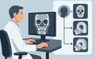
What Is the Best Initial Imaging for a Suspected Sinonasal Mass? An ACR-Guided Workflow
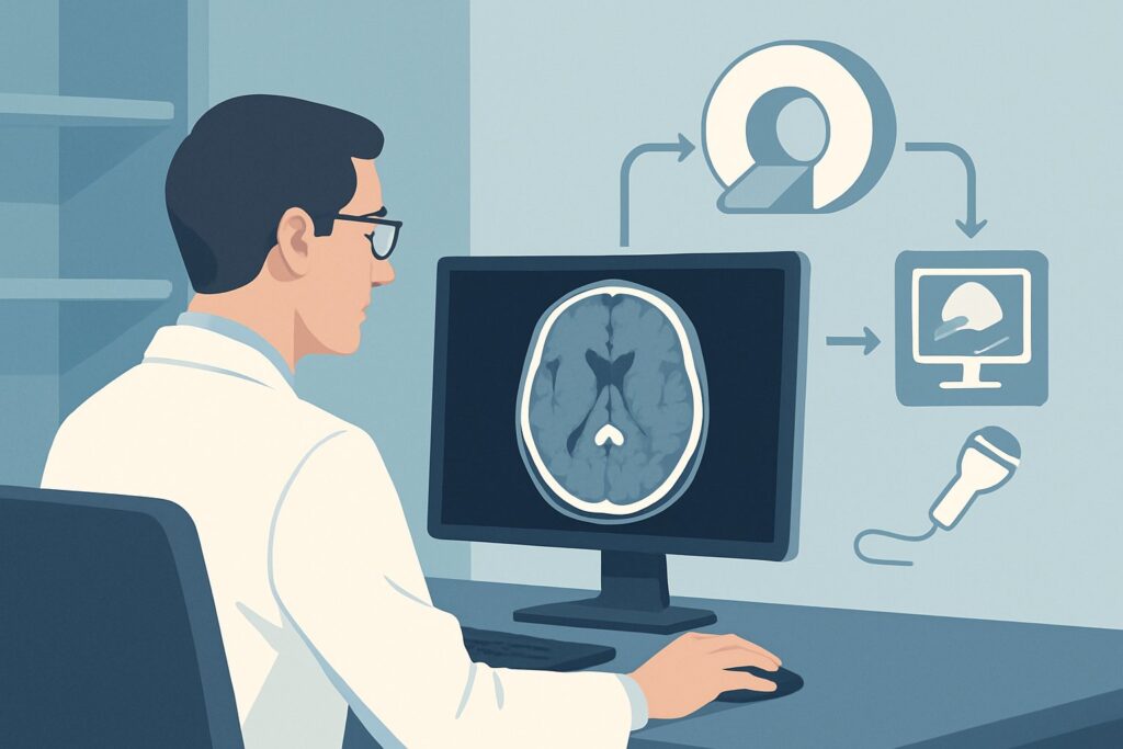
A 58-year-old patient presents to your clinic with three months of progressive right-sided nasal obstruction, intermittent epistaxis, and a new-onset sensation of facial fullness. An initial endoscopic exam is notable for a friable mass in the right nasal cavity, but the posterior extent is unclear. You suspect a sinonasal neoplasm and need to determine the optimal initial imaging study to characterize the lesion and define its extent before biopsy or referral. This critical first step informs the entire diagnostic and therapeutic pathway. According to the American College of Radiology (ACR) Appropriateness Criteria, for a suspected sinonasal mass, MRI of the orbits, face, and neck without and with IV contrast is rated Usually Appropriate as the initial imaging modality.
Who Fits the ‘Suspected Sinonasal Mass’ Scenario?
This clinical workflow is designed for patients presenting with signs and symptoms concerning for a neoplastic or other mass-like process within the nasal cavity or paranasal sinuses. The key is the suspicion of a mass, which differentiates this scenario from more common inflammatory conditions.
Inclusion criteria for this pathway typically involve:
- Unilateral sinonasal symptoms (e.g., obstruction, discharge, pain, or swelling) that are persistent or progressive.
- Recurrent or unexplained epistaxis, especially if unilateral.
- Visual changes such as proptosis, diplopia, or decreased visual acuity.
- Cranial nerve deficits, including facial numbness (V2 distribution), anosmia, or palsies.
- A palpable facial or intraoral mass.
- A mass visualized on physical examination or nasal endoscopy.
It is crucial to distinguish this presentation from other sinonasal pathologies that follow different imaging guidelines. This guidance does not apply to patients with:
- Acute uncomplicated rhinosinusitis: Patients with typical bilateral symptoms of a “common cold” lasting less than four weeks without red flags do not usually require imaging.
- Suspected invasive fungal sinusitis: This is an emergency scenario in an immunocompromised patient with rapid progression, often requiring urgent, specific imaging protocols to look for vascular invasion.
- Suspected Cerebrospinal Fluid (CSF) leak: Patients with a history of trauma or surgery presenting with clear, unilateral rhinorrhea require specialized high-resolution CT or cisternography protocols.
What Diagnoses Are You Working Up with Initial Imaging?
The differential diagnosis for a suspected sinonasal mass is broad, spanning from benign but locally aggressive lesions to highly malignant tumors. The primary goal of imaging is to characterize the lesion, define its anatomical extent, and identify features that narrow this differential.
A primary concern is Squamous Cell Carcinoma (SCC), the most common malignancy of the sinonasal tract. Imaging is critical for staging, as SCC can invade adjacent structures like the orbit, skull base, and cranial nerves. The pattern of enhancement and evidence of perineural tumor spread are key features sought on MRI.
Another important consideration is the Inverted Papilloma. While histologically benign, these tumors are locally aggressive, have a high rate of recurrence, and can harbor or degenerate into SCC. Imaging helps identify their characteristic origin from the lateral nasal wall and a specific convoluted cerebriform pattern on MRI.
In adolescent males presenting with recurrent, severe epistaxis, Juvenile Nasopharyngeal Angiofibroma (JNA) is a key differential. These are highly vascular, benign tumors that require careful preoperative planning, often including embolization. Their intense enhancement and characteristic location in the sphenopalatine foramen are classic imaging findings.
Less common but highly aggressive malignancies like Sinonasal Undifferentiated Carcinoma (SNUC), adenocarcinoma, or lymphoma are also on the differential. These often present as large, destructive masses, and imaging is essential to determine resectability and guide biopsy. Benign entities like an expansile mucocele can also present as a mass and must be differentiated from neoplastic processes.
Why Is MRI of the Orbits, Face, and Neck the Recommended First Study?
The ACR designates MRI of the orbits, face, and neck without and with IV contrast as Usually Appropriate because of its superior soft-tissue contrast, which is paramount for evaluating a suspected sinonasal mass. This modality excels at delineating the tumor’s relationship to critical adjacent structures.
The key advantages of MRI in this scenario include:
- Superior Soft-Tissue Resolution: MRI can distinguish tumor from adjacent inflamed mucosa, trapped secretions, and normal soft tissues far better than CT. This is crucial for determining the true extent of the mass.
- Detection of Perineural and Intracranial Spread: A primary route of spread for many sinonasal malignancies is along cranial nerves. Contrast-enhanced, fat-suppressed MRI is the most sensitive technique for detecting perineural tumor, a finding that significantly alters staging and treatment. It is also the best modality for assessing dural invasion or direct extension into the brain.
- Lack of Ionizing Radiation: MRI avoids radiation exposure (0 mSv), a significant benefit, particularly in younger patients or those who may require serial imaging.
While also rated Usually Appropriate, CT maxillofacial with or without IV contrast serves a complementary but distinct role. CT is superior for evaluating fine bony detail, making it excellent for assessing osseous erosion or destruction of the sinus walls, skull base, and orbital floor. However, its limited soft-tissue contrast makes it less ideal for defining the full extent of the tumor itself. Often, CT is performed as a second study for surgical planning after a mass is confirmed and characterized by MRI.
A CT of the head is rated lower (May be appropriate or Usually not appropriate depending on contrast) because its protocol is optimized for the brain parenchyma, providing suboptimal resolution and coverage of the sinonasal and facial structures. Similarly, plain radiographs are Usually not appropriate due to their extremely low sensitivity for detecting and characterizing sinonasal masses.
What’s Next After MRI? Downstream Workflow
The results of the initial MRI will guide the subsequent clinical pathway, including biopsy, further imaging, and multidisciplinary consultation.
- If the MRI is positive for a suspicious mass: The next step is typically a biopsy, often performed by an otolaryngologist via an endoscopic approach. The imaging findings are critical for guiding the biopsy to the most representative and safest area of the tumor, avoiding highly vascular regions. The patient should be referred to a multidisciplinary tumor board including ENT, radiation oncology, medical oncology, and neuroradiology. A staging CT of the chest is often performed for malignancies with a propensity for distant metastasis, and a CT of the face/sinuses may be added for surgical planning to better delineate bone anatomy.
- If the MRI is negative for a mass: If a high clinical suspicion remains despite a negative MRI, the patient’s symptoms should be re-evaluated. The findings may suggest an inflammatory process, which can be managed medically. If symptoms persist, a follow-up examination or repeat imaging in several months may be warranted. This may be an appropriate time to reconsider the diagnosis and explore other causes for the patient’s symptoms.
- If the MRI is indeterminate: In cases where the findings are ambiguous (e.g., distinguishing tumor from complex inflammation), a CT scan can provide complementary information about bone integrity. In rare, complex cases, a PET/CT scan (Usually not appropriate for initial workup) may be considered to assess for metabolic activity and nodal or distant metastatic disease, though its role in primary sinonasal tumor characterization is limited.
Pitfalls to Avoid (and When to Get Help)
Navigating the workup of a suspected sinonasal mass requires careful attention to detail to avoid common diagnostic errors.
- Pitfall 1: Ordering a non-contrast study. Intravenous contrast is essential for characterizing tumor vascularity, delineating tumor margins from adjacent inflammation, and detecting perineural or dural enhancement. Ordering an MRI or CT without contrast significantly limits its diagnostic value in this setting.
- Pitfall 2: Choosing the wrong study field of view. Ordering a “brain” or “head” MRI/CT instead of a dedicated “orbits, face, and neck” or “maxillofacial” protocol can result in poor resolution of the sinonasal cavity and inadequate assessment of the skull base and cranial nerves.
- Pitfall 3: Misinterpreting inflammation as tumor. Obstructed sinuses can fill with fluid and inflamed mucosa, which can be mistaken for tumor. MRI, particularly with diffusion-weighted imaging, is superior to CT in helping to differentiate trapped secretions from solid neoplastic tissue.
- Pitfall 4: Delaying biopsy for extensive imaging. While imaging is critical for staging, it should not unduly delay tissue diagnosis. Coordinate with the surgical or ENT team to ensure imaging and biopsy occur in a timely manner.
If you encounter a rapidly progressing mass, new-onset vision loss, or severe cranial nerve deficits, this represents a clinical emergency. Escalate immediately to an otolaryngologist and consider inpatient admission for expedited workup.
Related ACR Topics and Tools
This article focuses on a single clinical scenario. For a comprehensive overview of all variants within this topic, please consult our parent guide. For tools to help with ordering, protocoling, and patient communication, see the resources below.
- Parent Topic Hub: For breadth across all scenarios in Sinonasal Disease, see our parent guide: Sinonasal Disease: ACR Appropriateness Decoded.
- ACR Criteria Lookup: ACR Appropriateness Criteria Lookup
- Imaging Protocols: Imaging Protocol Library
- Dose Communication: Radiation Dose Calculator
Frequently Asked Questions
Why is MRI preferred over CT as the initial study for a suspected sinonasal mass?
MRI is preferred because of its superior soft-tissue contrast. It can better distinguish the tumor from surrounding inflamed tissue and trapped secretions, and it is the most sensitive modality for detecting subtle but critical findings like perineural tumor spread and intracranial extension, which are major factors in staging and treatment planning.
If I order an MRI, is a CT scan still necessary?
Often, yes. While MRI is the best initial study for characterization, a maxillofacial CT is superior for evaluating fine bony detail. It is frequently obtained after the MRI as a complementary study for preoperative planning, helping the surgeon understand the precise extent of bone erosion and identify key surgical landmarks.
What specific information should I include in the order for the MRI?
In your order, specify ‘MRI orbits, face, and neck without and with IV contrast.’ In the clinical history, clearly state the reason for the study, such as ‘unilateral nasal mass seen on endoscopy with epistaxis, rule out malignancy and assess for perineural or orbital invasion.’ This level of detail ensures the radiologist uses the correct protocol, including thin slices and fat-suppressed sequences through the skull base and cranial nerve foramina.
Should I order a PET/CT for the initial workup of a suspected sinonasal mass?
No. According to the ACR, FDG-PET/CT is ‘Usually not appropriate’ for the *initial* imaging of a suspected sinonasal mass. Its primary role is in staging for distant metastases or detecting recurrence *after* a diagnosis of malignancy has been established, not for initial characterization of the primary tumor.
What if the patient has a contraindication to MRI, like a non-compatible pacemaker?
If a patient cannot undergo an MRI, the next best study is a ‘CT maxillofacial with IV contrast,’ which is also rated as ‘Usually Appropriate’ by the ACR. While it has limitations in soft-tissue assessment, it is a robust alternative for defining the mass and assessing bone destruction. Be sure to document the contraindication to MRI in the order.
Reviewed by Pouyan Golshani, MD, Interventional Radiologist — May 29, 2026