Which Imaging Study Is Best for Suspected Complicated Otitis Media in Adults?
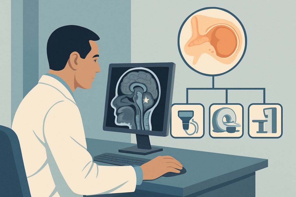
A 45-year-old patient presents to the emergency department with a three-week history of left-sided ear pain and purulent otorrhea, initially diagnosed as acute otitis media and treated with oral antibiotics. Over the past 48 hours, their headache has worsened, and they have developed a new, subtle facial droop on the left side. Otoscopy reveals a bulging, erythematous tympanic membrane. You are concerned this has progressed beyond a simple middle ear infection. The critical question is which imaging study to order first to evaluate for potentially life-threatening intracranial or intratemporal complications. This article details the American College of Radiology (ACR) evidence-based workflow for this specific scenario. For an adult with suspected complicated otitis media, the ACR designates MRI head and internal auditory canal without and with IV contrast as a Usually appropriate initial imaging study.
Who Fits This Clinical Scenario for Complicated Otitis Media?
This guidance applies specifically to adult patients with a clinical diagnosis of otitis media who present with new signs or symptoms suggesting the infection has spread beyond the middle ear cleft. These “red flag” features are the key differentiators that elevate a simple infection to a complicated one, mandating urgent imaging.
Inclusion criteria for this workflow:
- An adult patient with active or recent otitis media.
- Presence of new, concerning signs such as:
- Cranial nerve deficits (e.g., facial palsy, vertigo, diplopia)
- Signs of mastoiditis (postauricular pain, erythema, swelling, or fluctuance)
- Symptoms of intracranial pressure or infection (severe headache, altered mental status, seizures, nuchal rigidity)
- Failure to respond to appropriate antibiotic therapy, with worsening symptoms.
Exclusion criteria (patients who fit a different workflow):
- Uncomplicated Acute Otitis Media: Patients with typical ear pain and fever but no neurological deficits or signs of local spread do not require imaging. This is a clinical diagnosis managed medically.
- Chronic Otitis Media: Patients with persistent otorrhea and hearing loss, often being evaluated for cholesteatoma, follow a different imaging pathway. While CT is often primary, the clinical question is different.
- Complicated Otitis Externa: This typically refers to necrotizing or “malignant” otitis externa, an invasive infection of the external auditory canal and skull base, most common in diabetic or immunocompromised patients. While it requires imaging, the focus and differential are distinct.
What Diagnoses Are You Working Up in Complicated Otitis Media?
When ordering imaging for complicated otitis media, you are investigating the potential for the infection to have spread from the middle ear and mastoid air cells into adjacent critical structures. The differential diagnosis is a list of serious, and often emergent, conditions.
Coalescent Mastoiditis: This is the most common complication. The infection erodes the thin bony septa between the mastoid air cells, forming an abscess-like cavity within the mastoid bone. This can be a precursor to further intracranial spread and often requires surgical intervention.
Intracranial Abscess: A life-threatening extension of the infection into the cranial vault. This can occur via direct erosion of bone or through venous pathways. The abscess can be epidural (between the dura and the skull), subdural (beneath the dura), or within the brain parenchyma itself (cerebral or cerebellar abscess).
Dural Venous Sinus Thrombosis (DVST): Proximity of the mastoid process to the sigmoid and transverse sinuses makes them vulnerable. Inflammation can lead to the formation of a septic thrombus, obstructing venous outflow from the brain. This can cause venous infarction and a rapid increase in intracranial pressure.
Petrous Apicitis and Labyrinthitis: The infection can spread medially into the petrous apex of the temporal bone, causing Gradenigo’s syndrome (otorrhea, retro-orbital pain, and abducens nerve palsy). If it spreads into the inner ear, it causes labyrinthitis, resulting in severe vertigo, nystagmus, and sensorineural hearing loss.
Facial Nerve Palsy: The facial nerve (cranial nerve VII) travels through the temporal bone in close proximity to the middle ear. Inflammation can lead to neuropraxia or compression, resulting in ipsilateral facial paralysis.
Why Is MRI of the Head and IAC the Recommended First Study?
The ACR rates MRI head and internal auditory canal without and with IV contrast as Usually appropriate because of its superior ability to detect the soft tissue and vascular complications that define this clinical emergency. While bone erosion is a key feature, the immediate threat to the patient comes from abscesses, venous thrombosis, and neural inflammation—all best visualized with MRI.
The rationale for this choice involves several key factors:
- Superior Soft Tissue Contrast: MRI is exquisitely sensitive for detecting inflammation, pus, and edema. It can identify an early abscess, dural enhancement indicating meningitis or epidural inflammation, and abnormal signal within the brain parenchyma far earlier and more clearly than CT.
- Vascular Assessment: Post-contrast MRI sequences, combined with MR venography (MRV) which is often included in the protocol, are highly effective at identifying dural venous sinus thrombosis. The absence of flow and the presence of abnormal signal within the sinus are hallmark findings.
- Neural Evaluation: High-resolution sequences through the internal auditory canals (IAC) and temporal bone allow for direct visualization of the facial nerve and other cranial nerves, showing enhancement that indicates inflammation.
Why are other studies rated lower for this initial workup?
- CT temporal bone with IV contrast: While also rated Usually appropriate, it serves a complementary role. CT is superior for evaluating the fine osseous detail of the temporal bone, clearly showing erosion of the mastoid septa in coalescent mastoiditis or destruction of the tegmen tympani (the roof of the middle ear). However, it is less sensitive for early intracranial soft tissue complications. It is often a second-line or problem-solving study if MRI is unavailable or contraindicated, or if surgery is being planned. It also involves a moderate radiation dose (☢☢☢ 1-10 mSv).
- CT head without IV contrast: This study is rated Usually not appropriate. Without intravenous contrast, you cannot adequately assess for abscess wall enhancement, dural inflammation, or venous sinus thrombosis. A non-contrast CT can miss the most critical and life-threatening complications, providing false reassurance.
Ultimately, MRI with contrast provides the most comprehensive single examination to address the entire differential, from mastoiditis to intracranial abscess and venous thrombosis, all without the use of ionizing radiation (0 mSv).
What’s Next After the MRI? Interpreting Results and Planning Downstream Care
The results of the MRI will directly guide urgent management and consultations. The downstream workflow is a critical decision tree based on the specific findings.
- If the MRI confirms coalescent mastoiditis or a temporal bone abscess: This is a surgical emergency. An immediate consultation with Otolaryngology (ENT) is required for consideration of mastoidectomy and surgical drainage, alongside broad-spectrum IV antibiotics.
- If the MRI shows an intracranial abscess (epidural, subdural, or parenchymal): This requires an emergent Neurosurgery consultation. Management will involve a combination of surgical drainage and prolonged, targeted IV antibiotic therapy guided by an Infectious Disease specialist.
- If the MRI reveals dural venous sinus thrombosis: This is a neurologic emergency. Consultation with Neurology and potentially Hematology is critical. Treatment typically involves anticoagulation to prevent thrombus propagation and management of intracranial hypertension. A follow-up MRV or CTV may be needed to monitor treatment response.
- If the MRI is negative: If a high-quality MRI with contrast is unequivocally negative for complications, it provides strong evidence against the most serious diagnoses. The next step is to reconsider the primary diagnosis. This may involve a lumbar puncture to rule out meningitis if clinical suspicion remains high, or a focus on optimizing medical management of the otitis media itself with ENT consultation.
Pitfalls to Avoid (and When to Get Help)
Navigating this high-stakes scenario requires avoiding common diagnostic errors.
- Pitfall 1: Ordering a non-contrast study. Ordering a CT or MRI of the head without IV contrast is the most common and dangerous pitfall. It will not adequately evaluate for abscess or dural venous sinus thrombosis.
- Pitfall 2: Incomplete imaging protocol. Ordering a “routine” MRI of the brain may not include the high-resolution, thin-section images through the temporal bones and internal auditory canals needed to assess for petrous apicitis or facial nerve involvement. Specify the full protocol.
- Pitfall 3: Delaying imaging. The complications of otitis media can progress rapidly. If there is a high clinical suspicion based on red flag symptoms, imaging should be obtained emergently.
- Pitfall 4: Misinterpreting facial palsy. Attributing a new facial palsy in the setting of otitis media to a simple Bell’s palsy without imaging can lead to a missed diagnosis of an underlying abscess or cholesteatoma compressing the nerve.
If any red flag signs are present, especially altered mental status or focal neurologic deficits, escalate care immediately and consult the appropriate specialists (ENT, Neurology, Neurosurgery) concurrently with ordering imaging.
Related ACR Topics and Tools
For a comprehensive overview of imaging for all forms of ear infections and for tools to help with ordering and patient communication, the following resources are available. For breadth across all scenarios in Inflammatory Ear Disease, see our parent guide: Inflammatory Ear Disease: ACR Appropriateness Decoded.
- ACR Appropriateness Criteria Lookup: For exploring adjacent clinical scenarios or different patient presentations.
- Imaging Protocol Library: For detailed technical specifications on recommended imaging studies.
- Radiation Dose Calculator: For discussing cumulative radiation exposure with patients when considering CT.
Frequently Asked Questions
If my hospital’s MRI scanner is unavailable emergently, is CT an acceptable alternative?
Yes, in an emergency where MRI is not immediately available, CT temporal bone with IV contrast is rated ‘Usually appropriate’ by the ACR. It is excellent for assessing for coalescent mastoiditis and bone erosion. While less sensitive for early intracranial soft tissue changes, it is a reasonable first step. If the CT is negative but clinical suspicion for an intracranial complication remains high, the patient should be transferred or an MRI should be obtained as soon as possible.
Does the patient need a separate MRV or CTV study?
Not usually as a first step. A standard ‘MRI head and IAC with and without contrast’ protocol for this indication typically includes sequences that are sensitive for dural venous sinus thrombosis. Radiologists often add a formal MR Venogram (MRV) sequence if thrombosis is suspected during the scan. Similarly, a CT Angiography/Venography (CTA/CTV) can be performed, but MRI/MRV is preferred for its lack of radiation and superior soft tissue detail.
What if the patient has a contraindication to MRI, like a non-compatible pacemaker?
In cases of an absolute contraindication to MRI, CT temporal bone with IV contrast becomes the primary imaging modality. This would likely be paired with a CTV head with IV contrast to specifically evaluate the dural venous sinuses. This combination provides excellent osseous and vascular information, though it is less sensitive for early parenchymal brain infection and involves a significant radiation dose.
Is there a role for ultrasound in this scenario?
No, ultrasound has no role in the primary evaluation of complicated otitis media. It cannot penetrate the skull to visualize the middle ear, mastoid, or intracranial contents. Its use is limited to potentially identifying a superficial, palpable fluid collection over the mastoid process, but it cannot assess for deeper complications.
Should I order imaging for every patient whose otitis media doesn’t improve after one course of antibiotics?
Not necessarily. Failure to respond to a first-line antibiotic may warrant a change in antimicrobial therapy and close clinical follow-up. The key indication for imaging is the development of ‘red flag’ signs and symptoms of complication, such as new neurologic deficits, severe vertigo, signs of mastoiditis, or symptoms of increased intracranial pressure. Imaging is driven by suspicion of spread, not just by antibiotic failure alone.
Reviewed by Pouyan Golshani, MD, Interventional Radiologist — May 30, 2026