Should You Order an MRI for a Child’s First Primary Generalized Seizure?
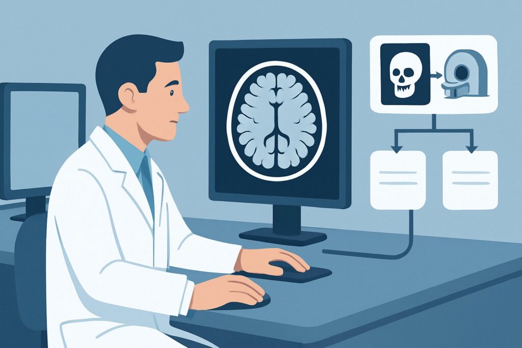
It’s a busy shift in the pediatric emergency department when you see a 9-year-old who had a witnessed generalized tonic-clonic seizure at school. This was their first-ever seizure. The event has resolved, the postictal period is over, and the child is back to their baseline. On examination, they are neurologically completely normal, with no focal deficits, a normal level of consciousness, and a non-focal history. The immediate question is whether to pursue neuroimaging. While the instinct may be to order a scan to rule out a serious underlying cause, the evidence for this specific presentation requires a nuanced approach. For a neurologically normal child with a first primary generalized seizure, the American College of Radiology (ACR) Appropriateness Criteria rate MRI head without IV contrast as `May be appropriate`, reflecting that imaging is not always required but is the best choice if a study is performed.
Who Fits This Clinical Scenario?
This guidance applies to a specific and common pediatric presentation: a child between 1 month and 17 years of age who has experienced their first seizure, which is clinically classified as a primary generalized seizure. A critical inclusion criterion is that the child has a completely normal neurologic examination after the postictal state has resolved. This implies no focal weakness, sensory changes, cranial nerve palsies, or altered mental status at their baseline.
It is crucial to distinguish this scenario from similar but distinct clinical situations that follow different diagnostic pathways:
- Focal Seizures: If the seizure had focal features at onset (e.g., involving only one side of the body or face), the likelihood of an underlying structural lesion is higher, and imaging is more strongly indicated. This is a separate ACR variant.
- Neurologically Abnormal Child: If the child has a known or newly discovered abnormality on their neurologic exam (e.g., hemiparesis, developmental regression, macrocephaly), this is a significant red flag. Imaging is more urgent and considered `Usually appropriate` in that scenario.
- Complex Febrile Seizures: While febrile seizures are common, a complex febrile seizure (defined as focal, prolonged >15 minutes, or recurrent within 24 hours) also warrants a different evaluation and has its own imaging recommendations.
- Post-Traumatic Seizures: A seizure occurring after head trauma is evaluated differently, with an initial focus on detecting acute injury like intracranial hemorrhage.
This article is exclusively for the workup of a first, unprovoked, primary generalized seizure in a child with a normal exam.
What Diagnoses Are You Working Up in This Scenario?
When considering imaging for a first generalized seizure in a neurologically normal child, the goal is to identify or exclude a structural cause that could alter management or prognosis. While the overall diagnostic yield of imaging in this population is low, a small but important subset of patients will have relevant findings.
Idiopathic or Genetic Generalized Epilepsy: This is the most common underlying diagnosis. In these syndromes (like childhood absence epilepsy or juvenile myoclonic epilepsy), the brain is structurally normal, and the seizures arise from network-level dysfunction. A normal MRI is the expected finding and helps confirm this diagnosis by exclusion. The primary diagnostic tool for these conditions is the electroencephalogram (EEG), not imaging.
Congenital or Developmental Malformations: These are the most common structural abnormalities found in this context. Conditions like cortical dysplasia, gray matter heterotopia, or polymicrogyria are subtle abnormalities of brain formation that can be highly epileptogenic. They are often invisible on CT scans but can be clearly delineated on high-resolution MRI, making it the modality of choice for their detection.
Low-Grade Neoplasms: Less commonly in this specific presentation, a slow-growing brain tumor (e.g., a ganglioglioma or dysembryoplastic neuroepithelial tumor) can present with a generalized seizure. While more typical with focal seizures, it remains a consequential diagnosis to exclude. MRI is far more sensitive than CT for detecting these types of tumors.
Vascular Malformations: Cavernous malformations or arteriovenous malformations (AVMs) can also be a source of seizures. While they can present with hemorrhage, a seizure may be the initial manifestation. MRI sequences like susceptibility-weighted imaging (SWI) are particularly sensitive for the blood products associated with these lesions.
Why Is MRI Head without IV Contrast the Recommended Study?
The ACR designates MRI head without IV contrast as `May be appropriate` for a child with a first primary generalized seizure and a normal neurologic exam. This rating reflects a careful balance: while the yield of finding a treatable structural cause is low, MRI is the superior modality if the clinical decision is made to proceed with imaging. The “May be appropriate” designation underscores that routine, automatic imaging for every child in this scenario is not the standard of care; the decision should be individualized based on clinical judgment, EEG findings, and family discussion.
The rationale for choosing a non-contrast MRI is driven by its high sensitivity for the most likely underlying structural pathologies without the need for ionizing radiation or intravenous contrast. It provides excellent gray-white matter differentiation, which is essential for identifying subtle cortical dysplasias or heterotopia that are the most common relevant findings. It is also highly effective at detecting low-grade tumors and evidence of vascular malformations.
Alternative studies are rated lower for specific reasons in this context:
- CT head without IV contrast is rated `Usually not appropriate`. While readily available and fast, it exposes the child to ionizing radiation (pediatric dose ☢☢☢ 0.3-3 mSv) and has very poor sensitivity for the key differential diagnoses like cortical dysplasia or small, non-calcified tumors. Its primary role is in emergent settings to rule out acute hemorrhage or large masses, which are not the main concern here.
- MRI head without and with IV contrast is also rated `Usually not appropriate`. The addition of gadolinium-based contrast does not typically increase the diagnostic yield for the most common etiologies in this scenario. Developmental malformations do not enhance, and most low-grade epilepsy-associated tumors are also non-enhancing. Avoiding contrast minimizes scan time, cost, and the rare risks associated with gadolinium administration.
Ultimately, if the decision is made to image, a non-contrast brain MRI provides the most relevant diagnostic information with the best safety profile (pediatric dose O 0 mSv).
What’s Next After MRI Head without IV Contrast? Downstream Workflow
The results of the brain MRI will significantly influence the subsequent clinical pathway. The workflow branches based on whether the study is normal or identifies an abnormality.
If the MRI is negative (normal): This is a reassuring result and the most common outcome. It strongly suggests a primary (idiopathic or genetic) epilepsy syndrome. The focus of management shifts entirely to clinical and electrographic data. The next steps typically involve a pediatric neurology consultation and an EEG to classify the epilepsy syndrome and determine the risk of seizure recurrence. The decision to start anti-seizure medication will be based on the EEG findings and clinical factors, not the imaging.
If the MRI is positive for a structural lesion: A positive finding, such as cortical dysplasia or a low-grade tumor, fundamentally changes the diagnosis from a primary epilepsy to a structural (or symptomatic) epilepsy. This has several downstream implications:
- Medication Management: The choice of anti-seizure medication may be influenced by the underlying cause.
- Prognosis: The prognosis for seizure control can be estimated more accurately.
- Surgical Evaluation: The patient is now a potential candidate for epilepsy surgery. A referral to a comprehensive epilepsy center for further evaluation, including video-EEG monitoring and advanced imaging, would be the appropriate next step.
If the MRI is indeterminate: Occasionally, an MRI may reveal non-specific findings, such as minor white matter signal abnormalities. These are often of unclear clinical significance. In such cases, correlation with a pediatric neuroradiologist is essential. Depending on the finding, a follow-up MRI or further advanced imaging may be recommended, but often these findings do not change immediate management and are simply monitored.
Pitfalls to Avoid (and When to Get Help)
Navigating the workup for a first pediatric seizure requires avoiding several common pitfalls to ensure optimal care and resource utilization.
- The Reflex CT Scan: Avoid ordering a CT head as the default imaging study in the emergency department for this specific scenario. It exposes the child to radiation with a very low likelihood of providing a definitive diagnosis for the most probable underlying causes.
- Ignoring the Need for Sedation: A high-quality MRI in a young child often requires sedation or general anesthesia to prevent motion artifact. Ordering an MRI without a plan for this can result in a non-diagnostic study. Coordinate with the radiology department and, if necessary, anesthesia services.
- Misinterpreting the “May be appropriate” Rating: This rating does not mean imaging should always be done. It signifies that the decision requires clinical judgment. A discussion with a pediatric neurologist before ordering imaging is often the best course of action.
If the clinical picture changes, such as the development of new focal neurologic deficits, a significant change in seizure type, or seizures that are refractory to initial medication, it is critical to escalate care and re-evaluate, which may include repeat or more advanced imaging.
Related ACR Topics and Tools
The ACR Appropriateness Criteria are extensive, covering numerous clinical variants. For a comprehensive overview of imaging recommendations across all pediatric seizure scenarios, from neonatal to post-traumatic, please consult our parent guide. For tools to help with ordering and patient communication, see the resources below.
- For breadth across all scenarios in Seizures-Child, see our parent guide: Seizures-Child: ACR Appropriateness Decoded.
- To explore other clinical presentations, use the Imaging Appropriateness Selector.
- For detailed procedural techniques, see the Imaging Protocol Library.
- To discuss radiation exposure with families, use the Radiation Dose Calculator.
Frequently Asked Questions
Why is MRI rated ‘May be appropriate’ instead of ‘Usually appropriate’ for this scenario?
The ‘May be appropriate’ rating reflects the low diagnostic yield of imaging in children with a first primary generalized seizure and a normal neurologic exam. Most of these children have a primary epilepsy syndrome where the brain is structurally normal. Imaging is not routinely required for every patient, and the decision should be individualized based on clinical factors, EEG results, and shared decision-making with the family.
If we decide to image, why is a non-contrast MRI preferred over one with contrast?
A non-contrast MRI is sufficient to identify the most common and important structural causes of seizures in this population, such as cortical dysplasia or low-grade tumors. These lesions typically do not enhance with contrast. Adding gadolinium increases scan time, cost, and introduces a small risk without providing additional diagnostic value for this specific clinical question.
In what situation would a CT scan be the right choice for a child with a first seizure?
A CT scan is primarily reserved for emergent situations where there is suspicion of an acute process like intracranial hemorrhage or skull fracture, such as a seizure immediately following significant head trauma. It is also used if MRI is unavailable or contraindicated. For an unprovoked seizure in a neurologically normal child, CT is considered ‘Usually not appropriate’ due to its radiation exposure and poor sensitivity for subtle epileptogenic lesions.
How does the recommendation change if the child’s neurologic exam is abnormal?
An abnormal neurologic exam is a major red flag that significantly increases the suspicion for an underlying structural brain lesion. In the ACR variant for a generalized seizure in a neurologically abnormal child, neuroimaging with MRI is more strongly recommended and is considered ‘Usually appropriate’. The clinical urgency is higher in that scenario.
Does a normal MRI guarantee that my patient won’t have more seizures?
No. A normal MRI is reassuring because it rules out a structural cause, but it does not predict the risk of seizure recurrence. That risk is determined by the underlying electrical function of the brain, which is assessed with an EEG, and other clinical factors. Many children with normal brain structure have epilepsy due to genetic or unknown causes.
Reviewed by Pouyan Golshani, MD, Interventional Radiologist — May 26, 2026