Should You Order an MRI for Suspected Cervicogenic Headache Without Neurologic Deficits?
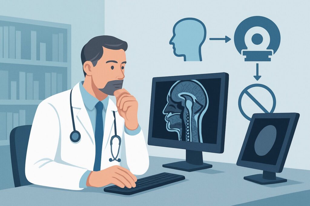
A 45-year-old patient presents to your clinic with a three-month history of a nagging, unilateral headache that starts in the back of their neck and radiates to the temporal region. The pain is a dull ache, worsened by prolonged sitting and specific neck movements, but they report no weakness, numbness, or changes in coordination. Your physical exam is unremarkable for any neurologic deficits. You suspect a cervicogenic headache and now face a common clinical question: is initial imaging necessary, and if so, which study is the most appropriate? This article provides a deep-dive into the American College of Radiology (ACR) Appropriateness Criteria for this exact scenario. For this presentation, an MRI of the cervical spine without IV contrast is rated as May be appropriate (Disagreement), reflecting the nuanced role of imaging in what is often a clinical diagnosis.
Who Fits This Clinical Scenario?
This guidance is specifically for an adult patient with a suspected cervicogenic headache and no neurologic deficits who is undergoing initial imaging. The key inclusion criteria are a headache pattern consistent with a cervical origin—typically unilateral pain referred from the neck, often provoked or aggravated by neck movement or sustained awkward postures—and a completely normal neurologic examination. This means the patient has full motor strength, intact sensation, and normal reflexes throughout the upper extremities.
It is critical to distinguish this scenario from similar but distinct clinical presentations that require a different diagnostic approach. This workflow does not apply if:
- Radiculopathy is present: If the patient reports radiating arm pain, numbness, tingling, or weakness, they fit the ACR variant for cervical pain with radiculopathy, which alters the imaging recommendations.
- “Red flags” are identified: Symptoms like fever, night sweats, unexplained weight loss, or a history of malignancy suggest a potential underlying infection or tumor. These cases fall under separate, more urgent ACR scenarios.
- There is a history of significant trauma: A traumatic mechanism of injury necessitates a different imaging algorithm focused on excluding fractures or ligamentous injury.
This guide is tailored for the common, non-emergent workup where the primary question is identifying a structural correlate for a suspected cervicogenic headache.
What Diagnoses Are You Working Up in This Scenario?
When ordering imaging for suspected cervicogenic headache, you are primarily investigating structural abnormalities in the upper cervical spine that can refer pain to the head. The differential diagnosis guides the choice of imaging modality.
Cervicogenic Headache (CGH): This is the primary diagnosis under consideration. It is a secondary headache caused by a disorder of the cervical spine and its component bony, disc, or soft tissue elements. Pain is typically referred from structures innervated by the C1, C2, and C3 cervical nerves. Imaging aims to identify a specific anatomical cause, though one is not always found.
Upper Cervical Facet Arthropathy: Degenerative osteoarthritis of the facet joints, particularly at the C2-C3 and C3-C4 levels, is a common cause of cervicogenic pain. The inflammation and mechanical dysfunction in these joints can directly refer pain into a headache pattern.
Upper Cervical Disc Herniation: While less common than in the lower cervical spine, a disc herniation or protrusion at C2-C3 or C3-C4 can impinge on the nerve roots or the thecal sac, generating referred pain. MRI is the only non-invasive modality that can directly visualize this pathology.
Occipital Neuralgia: This condition involves irritation or compression of the greater, lesser, or third occipital nerves. While often a clinical diagnosis, imaging can be used to rule out a structural cause of nerve compression, such as arthritic changes or other lesions along the nerve’s course.
Other Structural Lesions: Less commonly, other pathologies at the craniocervical junction, such as a Chiari malformation or rheumatoid pannus, can present with symptoms mimicking a cervicogenic headache. Imaging helps to exclude these consequential diagnoses.
Why Is MRI of the Cervical Spine Without Contrast Considered for This Presentation?
The ACR rates MRI of the cervical spine without IV contrast as May be appropriate (Disagreement) for an adult with suspected cervicogenic headache and no neurologic deficits. This rating is nuanced. It signifies that while imaging is not always required for this clinical diagnosis, if a study is performed, non-contrast MRI is the most suitable modality. The “Disagreement” qualifier indicates that there is variability in expert opinion on whether to image at all, often reserving it for cases that fail conservative therapy or have atypical features.
The rationale for choosing MRI centers on its superior soft-tissue contrast. It is highly sensitive for the key pathologies in the differential diagnosis, such as disc herniations, nerve root impingement, and spinal cord abnormalities, which cannot be visualized with other modalities. It also provides excellent detail of the facet joints and can identify inflammation or arthritic changes that may be the source of pain.
Alternative studies are rated lower for specific reasons:
- Radiography cervical spine is rated Usually not appropriate. While it can assess alignment and severe degenerative bony changes, it provides no information about the spinal cord, nerve roots, or intervertebral discs. It has a low diagnostic yield for this indication and exposes the patient to ionizing radiation (Relative Radiation Level ☢☢).
- CT cervical spine without IV contrast is also rated Usually not appropriate. Although it offers superior bone detail compared to radiography, it remains significantly inferior to MRI for evaluating soft tissues and involves a much higher dose of ionizing radiation (Relative Radiation Level ☢☢☢).
The recommended MRI is performed without IV contrast because contrast is not necessary to visualize the degenerative or mechanical causes of a typical cervicogenic headache. An MRI with contrast is rated Usually not appropriate for this scenario, as it is typically reserved for when there is a clinical suspicion of tumor, infection, or demyelinating disease—conditions outside the scope of this specific presentation. The non-contrast MRI approach avoids the costs and potential risks associated with gadolinium-based contrast agents while providing zero ionizing radiation (Relative Radiation Level O).
Once you’ve decided on MRI of the cervical spine without contrast, our protocol guide covers the technique, contrast, and reading principles: MRI Cervical Spine Without Contrast.
What Are the Next Steps After an MRI for Suspected Cervicogenic Headache?
The results of the cervical spine MRI will guide your downstream management plan. The workflow diverges based on whether a clear structural cause is identified.
If the MRI is positive for a likely pain generator: A finding such as significant C2-C3 facet arthropathy or a C3-C4 disc herniation that correlates with the patient’s symptoms provides a clear target for treatment. The next steps may include:
- Referral to physical therapy with a specific focus on the identified level.
- Consultation with a pain management specialist for diagnostic or therapeutic injections, such as a medial branch block or an epidural steroid injection.
- In rare, severe cases, a neurosurgical consultation may be warranted.
If the MRI is negative or shows only non-specific findings: A normal MRI is a very common outcome in this scenario. This result does not invalidate the patient’s symptoms or the diagnosis of cervicogenic headache. Instead, it effectively rules out a serious structural cause and reinforces that the pain is likely due to myofascial or functional issues not visible on imaging. Management should then focus on conservative, non-invasive therapies like manual therapy, physical therapy, posture correction, and medications.
If the MRI is indeterminate: The study may reveal mild, age-related degenerative changes whose clinical significance is unclear. In these cases, clinical correlation is paramount. A diagnostic nerve block can be a powerful tool to determine if the identified abnormality is the true source of the patient’s headache. If the pain is relieved by the block, it confirms the diagnosis and opens the door for targeted therapies like radiofrequency ablation.
Common Pitfalls to Avoid in This Workflow
Navigating the workup for suspected cervicogenic headache requires careful clinical judgment to avoid common missteps.
- Over-reliance on imaging: Remember that cervicogenic headache is a clinical diagnosis. A negative MRI does not rule it out, and a positive finding does not guarantee causation. Always correlate imaging with the patient’s history and physical exam.
- Attributing symptoms to incidental findings: Mild degenerative disc disease or spondylosis is nearly ubiquitous in adults. Be cautious about automatically blaming these common, often asymptomatic findings for the patient’s headache without strong clinical correlation.
- Starting with low-yield studies: Ordering plain radiographs as a first step is rated Usually not appropriate. This approach often fails to provide useful information, delays definitive imaging with MRI, and exposes the patient to unnecessary radiation.
If a patient with a suspected cervicogenic headache develops new or progressive neurologic deficits (e.g., weakness, gait instability, bowel/bladder changes), this constitutes a red flag. The clinical scenario has changed, and an urgent escalation, often involving a neurology or neurosurgery consult and potentially repeat imaging with contrast, is required.
Related ACR Topics and Tools
This article focuses on a single, specific clinical scenario. For a comprehensive overview of all variants related to neck pain and radiculopathy, or to explore the technical details of the recommended imaging study, please refer to the following resources.
- For breadth across all scenarios in Cervical Pain or Cervical Radiculopathy, see our parent guide: Cervical Pain or Cervical Radiculopathy: ACR Appropriateness Decoded.
- ACR Appropriateness Criteria Lookup — for adjacent scenarios
- Imaging Protocol Library — for technique on the recommended study
- Radiation Dose Calculator — for cumulative dose conversations
Frequently Asked Questions
Why not start with a simple X-ray of the neck for suspected cervicogenic headache?
According to the ACR, cervical spine radiography is rated ‘Usually not appropriate’ for this scenario. X-rays can only visualize bones and alignment, completely missing the soft tissue structures like intervertebral discs and nerve roots that are often the source of cervicogenic pain. An MRI is far more sensitive for the relevant pathologies.
My patient has a headache and neck pain, but also some tingling in their arm. Does this guide still apply?
No. The presence of radicular symptoms like tingling, numbness, or weakness in the arm moves the patient into a different clinical scenario: ‘Adult. Acute or increasing cervical pain with radiculopathy.’ That scenario has distinct imaging recommendations, and this guide for patients without neurologic deficits should not be used.
The MRI was negative. What does that mean for my diagnosis of cervicogenic headache?
A negative MRI does not rule out cervicogenic headache. In fact, it is a common finding. The diagnosis is primarily clinical, based on history and physical exam. A negative study is still valuable as it excludes serious structural causes, reinforcing a management plan focused on conservative therapies like physical therapy, manual therapy, and medication.
When would I ever need to add IV contrast to the cervical spine MRI for a headache?
IV contrast is reserved for specific clinical suspicions that fall outside of this initial workup scenario. You would consider an MRI with contrast if you were concerned about a tumor, infection (such as an epidural abscess), or an inflammatory or demyelinating condition like multiple sclerosis, all of which are ‘red flag’ conditions requiring a different diagnostic pathway.
What’s the significance of the ACR rating being ‘May be appropriate (Disagreement)’?
This rating reflects that while MRI is the best imaging test if one is chosen, the decision to perform imaging at all is not straightforward. There is a lack of high-level evidence and expert consensus on its routine use for this diagnosis. The decision to image often depends on factors like failure of an adequate trial of conservative therapy, atypical symptoms, or the need to rule out other conditions before proceeding with interventional treatments.
Reviewed by Pouyan Golshani, MD, Interventional Radiologist — May 29, 2026