What Is the Best Initial Imaging for Unilateral Facial Numbness or Trigeminal Neuralgia?
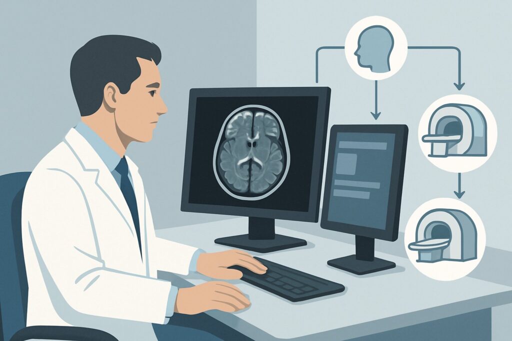
A 52-year-old patient presents to your clinic with a three-month history of progressive numbness over the right side of his face and new difficulty chewing, noting weakness when he bites down. He describes the sensation as a constant “pins and needles” feeling along his jawline and cheek. The clinical suspicion is a lesion affecting the trigeminal nerve (CN V), but the differential is broad, ranging from benign compression to malignancy. You need to decide on the most effective initial imaging study to delineate the cause without unnecessary radiation or delay. This article provides a clinical workflow for this specific scenario, explaining why the American College of Radiology (ACR) rates MRI head without and with IV contrast as Usually Appropriate.
Who Fits This Clinical Scenario for Trigeminal Nerve Imaging?
This guidance applies to patients presenting with new, isolated, and unilateral symptoms localized to the trigeminal nerve. This includes motor deficits, such as weakness or paralysis of the mastication muscles (masseter, temporalis, pterygoids), and sensory abnormalities, such as facial numbness, paresthesias, or pain consistent with trigeminal neuralgia. The key is that the symptoms are isolated to the distribution of CN V.
This workflow is NOT intended for patients with:
- Weakness of facial expression: If the patient cannot smile, raise an eyebrow, or close their eye tightly, the pathology likely involves the facial nerve (CN VII). This presentation falls under a different ACR scenario for Bell’s palsy or hemifacial spasm.
- Multiple cranial nerve palsies: If the trigeminal symptoms are accompanied by deficits in other cranial nerves (e.g., double vision from CN VI palsy or hearing loss from CN VIII involvement), the workup shifts. This suggests a more complex process at the skull base or within the cavernous sinus, routing to the ACR variant for multiple cranial neuropathies.
- Bilateral symptoms: While some systemic or central nervous system diseases can cause bilateral trigeminal symptoms, this specific ACR variant is tailored for a unilateral presentation, which has a distinct differential diagnosis.
What Diagnoses Are You Working Up in This Scenario?
When ordering imaging for suspected trigeminal neuropathy, the goal is to investigate the entire course of the nerve, from its nucleus in the brainstem to its peripheral branches. The differential diagnosis includes several key pathologies that imaging is well-suited to identify.
A primary concern is a schwannoma, a benign nerve sheath tumor that can arise from the trigeminal ganglion or any of its branches. These tumors are typically slow-growing but can cause significant symptoms through compression. Similarly, a meningioma arising from the dura near Meckel’s cave or the petrous apex can compress the trigeminal nerve root, leading to sensory or motor deficits.
For patients with classic trigeminal neuralgia—paroxysmal, lancinating facial pain—the most common cause is vascular compression of the nerve root by an adjacent artery or vein, most often the superior cerebellar artery. High-resolution imaging can directly visualize this neurovascular conflict at the root entry zone.
In younger patients or those with atypical features, multiple sclerosis (MS) must be considered. A demyelinating plaque located precisely at the trigeminal root entry zone in the pons can produce identical symptoms.
Finally, a less common but critical diagnosis to exclude is perineural tumor spread. Malignancies, particularly squamous cell carcinoma of the facial skin or adenoid cystic carcinoma of the salivary glands, can invade and track proximally along the trigeminal nerve branches into the skull base.
Why Is MRI of the Head With and Without Contrast Usually Appropriate for CN V Symptoms?
The ACR designates MRI head without and with IV contrast as Usually Appropriate because it provides the most comprehensive evaluation of the trigeminal nerve and the pathologies that affect it. Its superior soft-tissue contrast is unmatched for visualizing the nerve itself, the brainstem, and surrounding structures. The combination of pre- and post-contrast sequences is essential for a complete diagnostic picture.
The non-contrast sequences are excellent for assessing the anatomy of the cerebellopontine angle, identifying vascular loops that may be compressing the nerve root, and detecting the characteristic T2-hyperintense plaques of multiple sclerosis within the pons.
The administration of intravenous gadolinium-based contrast is critical for highlighting pathology. Schwannomas, meningiomas, and metastatic lesions typically demonstrate avid enhancement, making them conspicuous. Furthermore, contrast enhancement is the key to identifying perineural tumor spread, which often manifests as abnormal thickening and enhancement along the course of the nerve.
Why are other studies rated lower for this initial workup?
- CT maxillofacial with IV contrast is rated May be appropriate. While CT is excellent for evaluating the bony foramina of the skull base (e.g., foramen ovale, foramen rotundum) for erosive changes, its ability to visualize the nerve itself and non-calcified soft-tissue masses is significantly inferior to MRI. It also exposes the patient to ionizing radiation (ACR Relative Radiation Level ☢☢), making MRI the preferred initial test.
- MRI head without IV contrast is rated May be appropriate (Disagreement). Omitting contrast creates a significant diagnostic blind spot. A non-enhancing lesion could be missed, and more importantly, the presence and pattern of enhancement are crucial for differentiating between tumor types and identifying inflammatory or malignant processes. Relying on a non-contrast study alone is a potential pitfall that can lead to a missed or delayed diagnosis.
Ultimately, the combination of non-contrast and contrast-enhanced MRI sequences provides the highest diagnostic yield for the broad differential in this clinical scenario, all with a radiation dose of 0 mSv.
What Are the Next Steps After the Initial MRI?
The results of the MRI will guide the subsequent clinical pathway. The downstream workflow is highly dependent on the specific findings.
If the MRI identifies a schwannoma or meningioma, the next step is typically a referral to neurosurgery. Depending on the tumor’s size, location, and the severity of symptoms, management options may include observation with serial imaging, stereotactic radiosurgery, or surgical resection.
If the imaging confirms neurovascular compression consistent with the patient’s trigeminal neuralgia symptoms, the patient can be managed by neurology or a primary care physician with first-line medical therapies like carbamazepine or oxcarbazepine. If medical management fails, a neurosurgical consultation for microvascular decompression may be warranted.
A finding of a demyelinating plaque in the pons is highly suggestive of multiple sclerosis. This should prompt a referral to a neurologist for further workup, which typically includes an MRI of the cervical and thoracic spine to look for dissemination of lesions in space, and potentially a lumbar puncture.
In the case of a negative MRI, the management depends on the clinical picture. For classic trigeminal neuralgia, medical therapy can proceed. If symptoms are atypical or progressive despite a negative scan, particularly with concerns for perineural spread, a follow-up study or consultation with a head and neck surgeon or neurologist is advisable. In some cases, a dedicated high-resolution MRI with thin-slice sequences (e.g., FIESTA, CISS) may be requested to better scrutinize the nerve root.
Common Pitfalls to Avoid in Trigeminal Nerve Imaging
Navigating the workup for trigeminal neuropathy requires careful consideration to avoid common diagnostic errors.
First, avoid ordering a non-contrast MRI as the initial study. As noted, omitting contrast can miss key enhancing pathologies like small tumors or perineural spread, which are critical to identify. Always specify “without and with IV contrast.”
Second, provide specific clinical history on the imaging requisition. A generic request for “brain MRI” may result in a standard protocol that does not include the necessary thin-slice images through the skull base and posterior fossa. Clearly stating “unilateral facial numbness, rule out CN V lesion” helps the radiologist tailor the protocol to optimally visualize the entire trigeminal pathway.
Third, do not anchor on vascular compression too early. While it is the most common cause of classic trigeminal neuralgia, other serious pathologies can mimic its symptoms. A thorough review of the entire scan is necessary to exclude tumors or demyelination before attributing symptoms solely to a vascular loop.
If the initial MRI is negative but there are clinical red flags for malignancy—such as a history of facial skin cancer, progressive sensory loss rather than intermittent pain, or involvement of multiple trigeminal divisions—escalate care by consulting with neurology, neurosurgery, or an otolaryngologist for further evaluation.
Related ACR Topics and Tools
This article focuses on a single, specific clinical scenario. For a broader overview of imaging for all twelve cranial nerves, or to explore different patient presentations, the following resources are available.
- For breadth across all scenarios in Cranial Neuropathy, see our parent guide: Cranial Neuropathy: ACR Appropriateness Decoded.
- To look up other clinical scenarios, use the ACR Appropriateness Criteria Lookup tool.
- For technical details on the recommended study, consult the Imaging Protocol Library.
- To discuss radiation exposure from alternative studies like CT, use the Radiation Dose Calculator.
Frequently Asked Questions
Why is MRI so much better than CT for working up trigeminal neuralgia?
MRI is superior because of its excellent soft-tissue contrast, which allows direct visualization of the trigeminal nerve, adjacent blood vessels, and the brainstem. This is crucial for identifying the most common causes, such as vascular compression or a demyelinating plaque from multiple sclerosis. CT is poor at visualizing these structures and is primarily used to assess bone, making it a secondary choice. Furthermore, MRI does not use ionizing radiation.
Is intravenous contrast always necessary for an initial MRI for facial numbness?
Yes, for the initial diagnostic study, contrast is essential. The American College of Radiology rates MRI ‘without and with IV contrast’ as ‘Usually Appropriate,’ while MRI ‘without IV contrast’ is rated lower. This is because contrast is critical for detecting enhancing pathologies like schwannomas, meningiomas, and perineural tumor spread, which can present with facial numbness or pain. Omitting contrast can lead to a missed diagnosis.
What should I order if my patient has a contraindication to MRI, like an incompatible pacemaker?
If MRI is contraindicated, the next best option is ‘CT maxillofacial with IV contrast,’ which the ACR rates as ‘May be appropriate.’ This study can identify bony erosion at the skull base from a tumor and may show larger soft-tissue masses. However, it is important to recognize its limitations in sensitivity for nerve pathology, vascular compression, and demyelination compared to MRI.
The MRI report mentioned a vascular loop touching the trigeminal nerve, but the patient’s symptoms are atypical for classic trigeminal neuralgia. What does this mean?
An incidental finding of a vascular loop contacting the trigeminal nerve is common in asymptomatic individuals. The diagnosis of neurovascular compression as the cause of symptoms requires a strong clinical correlation with classic trigeminal neuralgia (i.e., paroxysmal, lancinating pain). If the symptoms are constant numbness or weakness, the vascular loop is likely an incidental finding, and other causes should be thoroughly investigated.
Should I order an MRA (Magnetic Resonance Angiography) instead of a standard MRI?
A standard ‘MRI head without and with IV contrast’ is the correct initial test. While MRA is rated as ‘May be appropriate,’ it is a specialized study focused only on the blood vessels. The standard MRI protocol provides a comprehensive evaluation of the nerve, brain parenchyma, and surrounding structures in addition to visualizing the vessels. An MRA alone would not be sufficient to evaluate for tumors, perineural spread, or demyelination.
Reviewed by Pouyan Golshani, MD, Interventional Radiologist — May 29, 2026