Should You Order Imaging for Unchanged Seizures in a Known Epilepsy Patient?
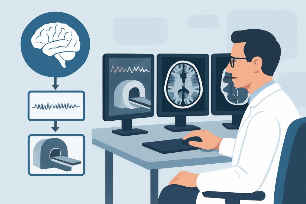
A 34-year-old patient with a five-year history of focal epilepsy, well-controlled on levetiracetam, presents for a follow-up. Last week, they had a single, brief breakthrough seizure while under significant stress and with poor sleep. The seizure’s characteristics—a rising epigastric sensation followed by right-hand twitching with preserved awareness—were identical to their typical seizures from before their medication was optimized. The patient has returned to their neurologic baseline. You are now considering whether repeat neuroimaging is warranted. This article provides a detailed clinical workflow for this specific scenario: a patient with a known seizure disorder and unchanged seizure semiology. For this presentation, the American College of Radiology (ACR) rates MRI head without and with IV contrast as May be appropriate, indicating that clinical judgment is the deciding factor.
Who Fits This Clinical Scenario?
This guidance applies to patients with an established diagnosis of a seizure disorder or epilepsy who experience a breakthrough seizure that is clinically identical to their previously characterized seizures. The core inclusion criteria are:
- Established Diagnosis: The patient has a known, previously evaluated seizure disorder.
- Unchanged Semiology: The clinical characteristics of the recent seizure(s)—including aura, motor, sensory, and autonomic features, as well as duration and postictal state—are consistent with the patient’s typical pattern.
- Return to Baseline: The patient has returned to their previous neurologic baseline without any new, persistent deficits.
It is critical to distinguish this situation from similar but distinct clinical presentations that require a different imaging approach. This workflow does not apply if the patient has:
- A change in seizure semiology: Any new or different seizure type, a significant increase in frequency or duration, or a change in the clinical pattern warrants a different workup, as detailed in the ACR variant for Known seizure disorder with a change in seizure semiology.
- A new neurologic deficit: The development of new, persistent weakness, numbness, vision changes, or cognitive decline suggests a new structural lesion and requires urgent evaluation.
- A known or suspected brain tumor: Patients with a history of a brain tumor have a lower threshold for imaging, as seizures can be the first sign of tumor recurrence or progression.
- A new-onset seizure: A patient presenting with their first-ever seizure requires a full diagnostic workup, which is a separate ACR scenario.
What Diagnoses Are You Working Up in This Scenario?
In a patient with stable epilepsy, a breakthrough seizure is most often triggered by non-structural factors. However, imaging is considered to ensure no new underlying pathology has developed that could lower the seizure threshold. The differential diagnosis in this context is focused on ruling out subtle or slowly evolving structural changes.
No New Structural Abnormality (Most Common) The most frequent cause of a breakthrough seizure in a well-controlled patient is a physiological stressor or a change in medication adherence. Common triggers include sleep deprivation, illness, metabolic disturbances (e.g., hypoglycemia, hyponatremia), alcohol or substance use, or missed doses of anti-epileptic drugs. In these cases, imaging is expected to be negative for any acute change and serves to confirm the absence of a new structural cause.
Slowly Progressive Lesion A previously undiagnosed, low-grade, or indolent lesion may be the underlying cause of the epilepsy. A subtle increase in the size of a low-grade glioma, dysembryoplastic neuroepithelial tumor (DNET), or cavernous malformation could be enough to provoke a breakthrough seizure, even without altering the seizure’s clinical manifestation. Repeat imaging after a significant time interval can help detect such slow changes.
Post-Treatment Changes For patients with a history of a treated brain lesion (e.g., post-surgical resection, post-radiation for a tumor or arteriovenous malformation), new seizures can be triggered by treatment-related effects. This includes radiation necrosis, gliosis, or encephalomalacia around a resection cavity, which can be epileptogenic. While less common, tumor recurrence must also be considered.
New Vascular or Inflammatory Process Less commonly, a new, small, and clinically subtle event like a small ischemic stroke, a microhemorrhage, or a focal inflammatory or infectious process (e.g., focal encephalitis) located within an already epileptogenic brain region could precipitate seizures without causing an obvious new neurologic deficit.
Why Is MRI Head Without and With Contrast Rated ‘May Be Appropriate’?
For a patient with a known seizure disorder and unchanged seizure semiology, routine repeat imaging after every breakthrough seizure is not necessary. The ACR rating of May be appropriate for MRI head without and with IV contrast reflects its role as a selective tool, reserved for situations where there is a clinical need to re-evaluate the underlying brain structure. The decision to image often depends on the time elapsed since the last scan, the specific epilepsy syndrome, and the overall clinical picture.
When imaging is pursued, MRI is the superior modality. Its high soft-tissue resolution is unmatched for visualizing the subtle structural abnormalities that can cause epilepsy.
- Non-contrast sequences (like FLAIR, T2-weighted, and susceptibility-weighted imaging) are essential for detecting cortical dysplasia, hippocampal sclerosis, gliosis, and cavernous malformations.
- IV contrast administration is crucial for identifying any breakdown of the blood-brain barrier, which can be seen in low-grade tumors that may not have been apparent on prior non-contrast studies, as well as in inflammatory, or infectious etiologies.
Why are other studies rated lower for this specific scenario?
- CT head without IV contrast: While also rated May be appropriate, its utility is limited. A non-contrast CT is primarily useful in an emergency setting to quickly rule out large, acute pathologies like a significant hemorrhage, hydrocephalus, or a large mass. However, it has very low sensitivity for the subtle epileptogenic lesions that MRI excels at detecting, such as cortical dysplasia or small, non-calcified tumors. It also involves ionizing radiation (1-10 mSv).
- MRI head without IV contrast: This study is rated May be appropriate (Disagreement). The panel’s disagreement highlights a key clinical debate. While a non-contrast MRI is powerful, forgoing contrast means potentially missing a small, enhancing lesion like a low-grade neoplasm or an inflammatory process. Given that the primary reason to re-image is to look for a new or evolved structural cause, many experts argue that the diagnostic value added by contrast administration justifies its use.
The choice to perform an MRI with and without contrast is a clinical one, balancing the low likelihood of finding a new lesion against the importance of not missing a treatable cause for worsening seizure control, even when the semiology is stable.
What’s Next After MRI Head Without and With Contrast? Downstream Workflow
The results of the MRI will guide the subsequent clinical pathway. The downstream workflow depends on whether the findings are negative, positive, or indeterminate.
- If the MRI is negative or unchanged from prior studies: This is the most common outcome. It provides reassurance that no new structural lesion has developed. The clinical focus should shift back to optimizing medical management. This includes reinforcing medication adherence, counseling on lifestyle modifications (e.g., sleep hygiene, stress management), and evaluating for and addressing any potential seizure triggers. No further immediate imaging is typically required.
- If the MRI is positive for a new or progressive lesion: The discovery of a new or enlarging mass (e.g., suspected low-grade glioma), a vascular malformation, or evidence of inflammation dictates the next steps. This typically involves:
- Referral to neurosurgery and/or neuro-oncology for further evaluation and management planning, which may include biopsy or resection.
- Adjustment of anti-epileptic drug therapy, as the underlying etiology may influence medication choice.
- Consideration for more advanced imaging or evaluation for epilepsy surgery if the lesion is identified as the clear seizure focus.
- If the MRI is indeterminate or shows non-specific findings: Ambiguous findings, such as a non-specific white matter signal abnormality or subtle questionable enhancement, may require further characterization. The next step could be a follow-up MRI in a shorter interval (e.g., 3-6 months) to assess for stability or change. In select cases, more advanced imaging techniques like FDG-PET, which is rated May be appropriate (Disagreement), could be considered to assess for metabolic activity in the suspicious area, though this is not a routine step.
Pitfalls to Avoid (and When to Get Help)
When managing a patient with stable epilepsy and a breakthrough seizure, several common pitfalls can occur.
- Pitfall 1: Routine, reflexive re-imaging. Avoid ordering imaging for every breakthrough seizure without a clear clinical question. The yield is low if the semiology is unchanged and known triggers are present.
- Pitfall 2: Ordering a non-contrast CT as the definitive study. While useful for emergent triage, a negative non-contrast CT does not adequately rule out the subtle structural causes of epilepsy. If there is a genuine concern for a new lesion, MRI is the appropriate test.
- Pitfall 3: Attributing all seizures to non-adherence. While common, automatically assuming non-compliance can lead to a missed opportunity to diagnose a new, underlying structural problem. A thorough history and, when indicated, selective imaging are key.
If a patient experiences a significant increase in seizure frequency, develops a new seizure type, or fails to return to their neurologic baseline, this represents a change in their clinical status. This situation requires escalation and should be managed according to the ACR Appropriateness Criteria for Known seizure disorder with a change in seizure semiology or new neurologic deficit.
Related ACR Topics and Tools
For a comprehensive overview of imaging recommendations across all common seizure-related presentations, and for tools to help in study selection and patient communication, the following resources are available:
- For breadth across all scenarios in Seizures and Epilepsy, see our parent guide: Seizures and Epilepsy: ACR Appropriateness Decoded.
- ACR Appropriateness Criteria Lookup — for adjacent scenarios
- Imaging Protocol Library — for technique on the recommended study
- Radiation Dose Calculator — for cumulative dose conversations
Frequently Asked Questions
How long after a previous ‘normal’ MRI should I consider re-imaging for unchanged seizures?
There is no strict timeline, and the decision is clinical. Many neurologists consider repeat imaging if it has been several years (e.g., >5 years) since the last scan, or if there is a subtle but persistent difficulty in achieving seizure control despite good medication adherence. The goal is to screen for very slowly progressive lesions like a low-grade glioma.
If my patient presents to the emergency department with an unchanged seizure, is a CT scan sufficient?
In the emergency setting, a non-contrast head CT is appropriate to rapidly exclude life-threatening conditions like a large hemorrhage or mass. However, if the patient is stable and the goal is to re-evaluate the underlying cause of their epilepsy, a negative CT is not sufficient. An outpatient MRI is the definitive study for detecting subtle epileptogenic lesions.
Does the type of epilepsy (e.g., focal vs. generalized) affect the decision to re-image?
Yes, to some extent. Patients with idiopathic generalized epilepsy (e.g., juvenile myoclonic epilepsy) are less likely to have a structural cause, and the yield of repeat imaging is extremely low. For patients with focal epilepsy, especially if the cause was not identified on initial imaging (non-lesional focal epilepsy), the rationale to re-image after a long interval is stronger to look for a previously occult lesion.
My patient is pregnant and had a breakthrough seizure. Is MRI safe?
Non-contrast MRI is considered safe at any stage of pregnancy. The use of gadolinium-based contrast agents during pregnancy is more controversial and generally avoided unless the potential benefit to the mother is deemed to outweigh any potential fetal risk. The decision should be made in consultation with the patient and a radiologist.
Why is MRI without contrast rated ‘May be appropriate (Disagreement)’?
The disagreement among the ACR panel reflects differing expert opinions. Some argue that a high-quality non-contrast MRI protocol is sufficient to detect most relevant structural changes. Others contend that since the primary reason for re-imaging is to look for a new or evolving lesion, the small risk and cost of IV contrast are justified to avoid missing a subtle enhancing abnormality, such as a low-grade tumor, which could significantly alter management.
Reviewed by Pouyan Golshani, MD, Interventional Radiologist — May 29, 2026