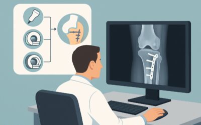
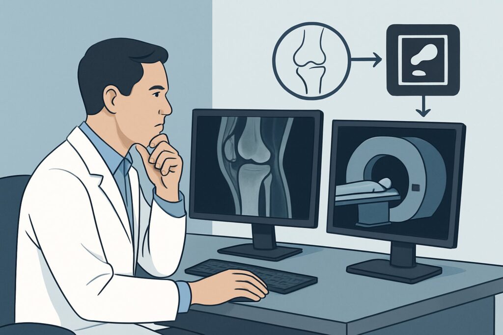
Should You Order MRI for Acute Knee Trauma with Negative Radiographs?
A 32-year-old patient presents to the urgent care clinic after a basketball game, describing a twisting fall and an audible “pop” from their right knee. They have a significant joint effusion and are unable to bear weight. Radiographs of the knee are obtained and show no acute fracture. Your clinical suspicion is high for an internal derangement, such as a meniscal tear or an anterior cruciate ligament (ACL) injury, but you also need to rule out an occult fracture not visible on the plain films. What is the next, most appropriate imaging study to order?
This exact clinical question is addressed by the American College of Radiology (ACR) Appropriateness Criteria. For an adult or skeletally mature child with acute knee trauma and negative radiographs where an occult fracture or internal derangement is suspected, the ACR rates MRI knee without IV contrast as Usually Appropriate. This article provides a detailed workflow for this specific scenario, outlining the differential diagnosis, the rationale for the recommended study, and the downstream clinical pathway.
Who Fits This Clinical Scenario for Post-Traumatic Knee Imaging?
This guidance is tailored for a very specific patient presentation. Correctly identifying if your patient fits this profile is the crucial first step to ensure you order the right test and avoid unnecessary imaging.
Inclusion criteria for this workflow:
- Patient: An adult or a skeletally mature adolescent.
- Mechanism: An acute traumatic event, typically a fall or a twisting injury.
- Initial Imaging: Radiographs (X-rays) of the knee have already been performed and are negative for a visible fracture.
- Clinical Suspicion: Despite negative radiographs, there is a persistent, strong clinical concern for a significant intra-articular injury. This is often based on physical exam findings like a large effusion, joint line tenderness, mechanical symptoms (locking or catching), or instability.
This workflow does NOT apply to several similar-appearing scenarios:
- Skeletally Immature Children: Patients with open growth plates have a different spectrum of injuries, particularly physeal (growth plate) fractures. Their evaluation follows a distinct diagnostic algorithm.
- Known Fracture on Radiographs: If the initial X-ray reveals a fracture, such as a tibial plateau fracture, the imaging goal shifts from diagnosis to characterization for surgical planning. In that case, CT is often the next step.
- Low-Risk Presentations: This guidance is not for patients who meet low-risk criteria (e.g., the Ottawa Knee Rules), such as those with no focal tenderness, no significant effusion, and the ability to bear weight. These patients may not require any advanced imaging.
What Diagnoses Are You Working Up When Knee Radiographs Are Negative?
When initial radiographs are negative, your differential diagnosis shifts from obvious bony injury to the complex soft tissue structures within the knee and subtle fractures that plain films can miss. The goal of advanced imaging is to interrogate these structures directly.
Ligamentous Injury
The most common and clinically significant injuries in this category involve the cruciate and collateral ligaments. An ACL tear is a classic example following a non-contact pivoting injury, often associated with a “pop” and rapid effusion. Posterior cruciate ligament (PCL), medial collateral ligament (MCL), and lateral collateral ligament (LCL) injuries are also highly prevalent and are best evaluated with a study that provides excellent soft-tissue resolution.
Meniscal Tears
The menisci are critical for shock absorption and stability. Twisting injuries can cause tears, leading to pain, locking, and catching sensations. These injuries are extremely common, especially in athletes and active adults, and are a primary target of investigation in this scenario.
Occult or Nondisplaced Fractures
Radiographs can miss subtle fractures, particularly non-displaced tibial plateau fractures, small avulsion fractures at ligamentous insertions, or patellar fractures. The hallmark of these injuries is bone marrow edema—an accumulation of fluid and blood within the bone—which is invisible on X-ray but vividly apparent on MRI.
Osteochondral Injury
Acute trauma can cause damage to the articular cartilage and the underlying subchondral bone, resulting in an osteochondral defect. These injuries can lead to long-term joint degeneration if missed. MRI is the only non-invasive modality that can directly visualize the articular cartilage to this degree of detail.
Why Is MRI Knee without Contrast Usually Appropriate for This Presentation?
The ACR’s recommendation for MRI knee without IV contrast is based on its superior diagnostic capability for the key differential diagnoses in this scenario, combined with a favorable safety profile.
The primary strength of Magnetic Resonance Imaging (MRI) is its unparalleled soft-tissue contrast. It provides detailed visualization of the menisci, cruciate and collateral ligaments, cartilage, and tendons, making it the definitive non-invasive study for diagnosing tears, sprains, and defects. Furthermore, MRI is exceptionally sensitive to bone marrow edema, allowing it to detect occult fractures that are invisible on both radiographs and often on CT scans.
Let’s compare MRI to the other imaging options considered by the ACR for this specific clinical problem:
- CT knee without IV contrast: This study is rated May be appropriate. While CT is excellent for delineating complex or subtle cortical bone fractures, it provides very limited information about ligaments, menisci, or cartilage. It is a reasonable alternative if MRI is contraindicated (e.g., incompatible implanted device) or unavailable in a timely manner, or if the pre-test suspicion for a subtle fracture is much higher than for a soft-tissue injury. However, it involves a small amount of ionizing radiation (adult relative radiation level ☢ <0.1 mSv).
- US knee: Ultrasound is rated Usually not appropriate for a global assessment of internal derangement. While it can be useful for evaluating specific superficial structures like the patellar tendon, quadriceps tendon, or fluid collections (e.g., a Baker’s cyst), it is operator-dependent and cannot adequately visualize intra-articular structures like the cruciate ligaments or the entirety of the menisci. It is not a comprehensive tool for this workup.
For this indication, intravenous contrast is not necessary. The joint effusion and inflammation from the acute injury create sufficient intrinsic fluid contrast on T2-weighted MRI sequences to highlight the relevant pathology. Therefore, MRI knee without IV contrast provides the optimal balance of diagnostic information and patient safety, with no ionizing radiation (0 mSv).
Once you’ve decided on MRI, our protocol guide covers the technique and reading principles in detail: MRI Knee Without Contrast.
What’s Next After the MRI? Interpreting Results and Planning Care
The results of the knee MRI will directly guide the next steps in management, branching into surgical, non-surgical, and further diagnostic pathways.
- Positive for a Surgically-Treated Injury: If the MRI confirms a diagnosis that often requires surgical intervention—such as a complete ACL tear in an active individual, a displaced meniscal bucket-handle tear causing a locked knee, or a significant osteochondral defect—the next step is a referral to an Orthopedic Surgeon. The MRI findings will be crucial for pre-operative planning.
- Positive for a Non-Surgical Injury: Many injuries identified on MRI are managed conservatively. Findings like a low-grade MCL sprain, a non-displaced stable meniscal tear, or a simple bone bruise typically call for a course of physical therapy, bracing, activity modification, and pain management. Follow-up is clinical, with repeat imaging rarely needed unless symptoms fail to improve.
- Negative or Indeterminate MRI: If the MRI is entirely negative but the patient remains highly symptomatic, the focus shifts. The differential may now include conditions not well-visualized on standard MRI, such as patellofemoral pain syndrome, plica syndrome, or complex regional pain syndrome. In these cases, a trial of physical therapy, a second opinion from a musculoskeletal specialist, or diagnostic/therapeutic injections may be considered. A truly negative MRI effectively rules out significant internal derangement, which is valuable information that allows the clinician to confidently pursue other diagnoses.
Pitfalls to Avoid (and When to Get Help)
Navigating this common clinical scenario has a few potential pitfalls that can delay diagnosis or lead to suboptimal care.
- Stopping at the Radiograph: The most common error is accepting a negative radiograph as the end of the workup in a patient with a compelling mechanism and persistent, significant symptoms (e.g., large effusion, mechanical locking, instability). Clinical suspicion should override a negative plain film.
- Ordering the Wrong Advanced Study: Defaulting to CT when the primary suspicion is meniscal or ligamentous injury is a frequent mistake. CT provides limited-to-no useful information for these structures and exposes the patient to unnecessary radiation.
- Delaying MRI Unnecessarily: While not always an emergency, a significant delay in obtaining an MRI for a locked knee or a suspected multi-ligamentous injury can complicate management. Timely diagnosis facilitates appropriate bracing and referral.
- Ignoring Skeletal Maturity: Applying this adult-focused workflow to a skeletally immature child can lead to missed Salter-Harris (physeal) fractures, which have different treatment and prognostic implications.
If the clinical picture is complex, such as a suspected knee dislocation (even if spontaneously reduced) or concern for a vascular injury, escalate immediately for an orthopedic and/or vascular surgery consultation.
Related ACR Topics and Tools
For a comprehensive overview of imaging guidelines for all knee trauma scenarios, and for tools to help with study selection and patient communication, the following resources are available:
- For breadth across all scenarios in Acute Trauma to the Knee, see our parent guide: Acute Trauma to the Knee: ACR Appropriateness Decoded.
- To explore other clinical scenarios and their corresponding ACR recommendations, use the ACR Appropriateness Criteria Lookup.
- For detailed technical specifications of imaging studies, consult the Imaging Protocol Library.
- To discuss radiation exposure from medical imaging with patients, the Radiation Dose Calculator can help frame the conversation.
Frequently Asked Questions
My patient has a knee effusion after a fall but negative X-rays. Is MRI always the next step?
Not always. If the patient meets low-risk criteria (e.g., per the Ottawa Knee Rules: age <55, no isolated patellar tenderness, no fibular head tenderness, able to flex to 90 degrees, and able to bear weight immediately and in the ED), they may not need any advanced imaging. MRI is indicated when there is high clinical suspicion for internal derangement (ligament/meniscus tear) or an occult fracture despite the negative radiographs.
Why is MRI without contrast recommended instead of with contrast?
In the setting of acute trauma, the joint typically fills with a bloody effusion. This fluid acts as a natural contrast agent on fluid-sensitive (T2-weighted) MRI sequences, outlining the internal structures of the knee. Intravenous gadolinium-based contrast adds little to no diagnostic information for acute meniscal or ligamentous tears and is therefore not necessary.
How soon should the MRI be performed after the injury?
The timing is situation-dependent. For a locked knee (suggesting a displaced bucket-handle meniscal tear) or a suspected multi-ligament injury, the MRI should be obtained urgently to facilitate timely orthopedic consultation. For less severe symptoms, the MRI can often be performed on an outpatient basis within a few days to a week.
What if my patient cannot get an MRI due to a pacemaker or severe claustrophobia?
If MRI is absolutely contraindicated, CT knee without contrast is rated as ‘May be appropriate’ by the ACR. It is an excellent alternative for detecting subtle or occult fractures but is very poor for evaluating the menisci, ligaments, and cartilage. The choice depends on whether the primary clinical suspicion is for a bony or soft-tissue injury. For severe claustrophobia, options include open MRI or sedation.
Does this guidance apply to a skeletally immature adolescent with the same injury?
No, this specific workflow is for adults or skeletally mature adolescents. A skeletally immature patient has open growth plates (physes), and their injury patterns are different, with a higher risk of physeal fractures (Salter-Harris injuries). While MRI is still often used, the clinical and radiographic evaluation follows a different pathway, and the ACR has a separate variant for that scenario.
Reviewed by Pouyan Golshani, MD, Interventional Radiologist — May 29, 2026