What Initial Imaging Should You Order for a Hemodynamically Unstable Adult After Major Blunt Trauma?
A 34-year-old male arrives by ambulance after a high-speed motor vehicle collision. It’s 2 AM in the emergency department, and he is hypotensive at 80/50 mmHg and tachycardic to 140 bpm. As the trauma team initiates resuscitation with fluids and blood products, you face a critical decision: what imaging is safe and effective to order right now to find the source of his instability? Transporting him to the CT scanner is a high-risk proposition. This article provides a detailed clinical workflow for this exact scenario, focusing on the initial imaging studies that can be performed rapidly at the bedside. For an adult patient with major blunt trauma who is hemodynamically unstable, the American College of Radiology (ACR) Appropriateness Criteria rate a Radiography trauma series as Usually Appropriate.
Who Fits the Scenario of an Unstable Adult After Major Blunt Trauma?
This guidance applies specifically to adult patients who have sustained major blunt force trauma and exhibit clear signs of hemodynamic instability.
Inclusion criteria for this workflow:
- Mechanism: Significant blunt trauma, such as from a motor vehicle accident, a fall from a significant height, or a crush injury.
- Patient Status: Evidence of hemodynamic instability, including hypotension (e.g., systolic blood pressure < 90 mmHg), persistent tachycardia, altered mental status consistent with shock, or the need for ongoing aggressive fluid and blood product resuscitation.
- Timing: The initial imaging evaluation phase, often occurring concurrently with primary survey and resuscitation in the trauma bay.
Exclusion criteria (patients who follow a different workflow):
- Hemodynamically Stable Patients: If the patient is normotensive and has a normal heart rate (or has quickly normalized with initial resuscitation), they are considered stable. This is the most critical distinction. Stable patients can typically proceed safely to more definitive imaging, such as whole-body CT, and follow a different diagnostic algorithm. This is covered in our guide for Major Blunt Trauma in stable adults.
- Penetrating Trauma: Gunshot wounds or stab wounds have different injury patterns and priorities, necessitating a distinct imaging and management pathway.
- Isolated, Minor Trauma: This workflow is for polytrauma. A patient with an isolated extremity fracture without systemic instability would not fit this scenario.
What Life-Threatening Diagnoses Are You Working Up in Unstable Trauma?
In the unstable trauma patient, the immediate goal of imaging is not a comprehensive diagnostic survey but a rapid search for life-threatening injuries causing massive hemorrhage. The differential is focused on conditions that can be quickly identified and temporized in the trauma bay or require immediate transfer to the operating room or interventional radiology suite.
Massive Hemothorax
A common and rapidly fatal injury, a massive hemothorax involves the accumulation of a large volume of blood in the pleural space. This can lead to hypovolemic shock from blood loss and obstructive shock by compressing the lung and mediastinal structures, impairing ventilation and cardiac function. A portable chest radiograph is the primary tool for its rapid diagnosis.
Unstable Pelvic Fracture with Hemorrhage
The pelvic ring, when disrupted, can create a massive potential space for retroperitoneal bleeding from the rich venous plexuses and arterial branches. Certain fracture patterns, like an “open-book” (anteroposterior compression) injury, are notoriously associated with life-threatening hemorrhage. An AP pelvis radiograph is critical for identifying these unstable patterns, which guides immediate intervention like pelvic binder placement.
Intra-abdominal Hemorrhage
Lacerations to solid organs, particularly the spleen and liver, are a frequent cause of exsanguination after blunt trauma. While plain radiographs cannot directly visualize these injuries, they are a key part of the initial trauma algorithm. The absence of findings on chest and pelvic radiographs in a persistently unstable patient strongly points toward an intra-abdominal or retroperitoneal source of bleeding, often prompting a bedside FAST exam and potentially an emergent laparotomy.
Major Thoracic Vascular or Cardiac Injury
Although less common, injuries like traumatic aortic disruption or cardiac tamponade must be considered. While definitive diagnosis requires CT angiography or echocardiography, the initial chest radiograph can provide crucial clues, such as a widened mediastinum, that raise suspicion and alter the diagnostic and therapeutic sequence.
Why Is a Radiography Trauma Series Usually Appropriate for the Unstable Patient?
The core principle guiding imaging in the unstable trauma patient is balancing diagnostic yield with patient safety. The primary advantage of radiography is its speed and portability, allowing for critical information to be obtained without moving a fragile patient away from the resuscitation area.
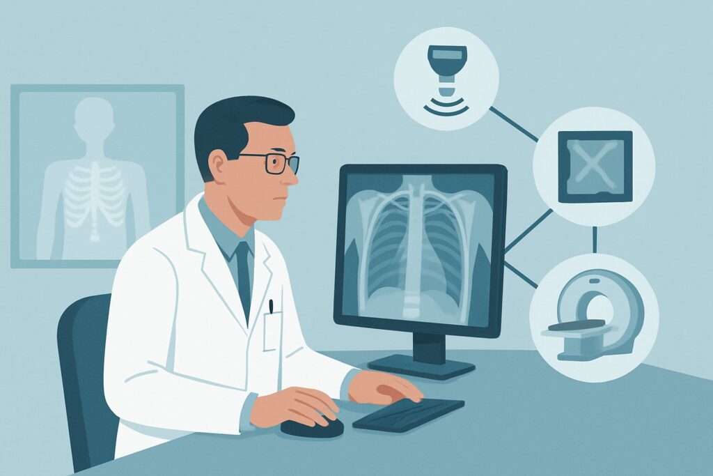
The standard trauma series typically consists of three portable views:
1. AP Chest Radiograph: This is the fastest way to diagnose a large pneumothorax, hemothorax, or signs suggestive of great vessel injury (e.g., widened mediastinum). These are actionable findings that can lead to immediate procedures like chest tube insertion.
2. AP Pelvis Radiograph: This view is essential for identifying mechanically unstable pelvic ring fractures. A positive finding prompts immediate application of a pelvic binder to reduce the pelvic volume and tamponade bleeding, a potentially life-saving maneuver.
3. Lateral C-Spine Radiograph: While CT has largely replaced radiography for definitive cervical spine clearance, a portable lateral view can quickly identify gross instability, such as a fracture-dislocation, that requires strict immobilization before any further patient movement.
The ACR rates Radiography trauma series as Usually Appropriate because it directly addresses the most immediate, mechanically correctable causes of shock in blunt trauma without the significant risk and time delay of transporting an unstable patient to a scanner.
Why Alternatives Are Rated Lower in This Specific Scenario
- CT whole body with IV contrast is rated May be appropriate. While CT is the gold standard for identifying and characterizing injuries in stable patients, its role in the actively unstable patient is more nuanced. The logistical challenge and physiological risk of moving a patient dependent on active resuscitation to the CT suite can lead to catastrophic decompensation. CT becomes appropriate only if the patient responds rapidly and durably to initial resuscitation, effectively transitioning them into the “hemodynamically stable” category.
- CT whole body without IV contrast is rated Usually not appropriate. Omitting intravenous contrast renders the study inadequate for evaluating the key concerns in trauma: solid organ injury, active arterial extravasation, and vascular dissection or transection. It provides little benefit over radiography while still carrying the risk of transport and a significant radiation dose.
- MRI is rated Usually not appropriate due to its long acquisition times, incompatibility with most resuscitation equipment, and limited utility in the acute hemorrhagic setting, making it entirely unsuitable for the initial evaluation of an unstable trauma patient.
The radiation dose for a trauma series (Relative Radiation Level ☢☢☢, 1-10 mSv) is considerably lower than that of a whole-body CT (☢☢☢☢, 10-30 mSv). In a life-threatening situation, radiation exposure is a secondary concern, but the lower dose is an ancillary benefit of the radiography-first approach.
What Is the Downstream Workflow After the Initial Trauma Series?
The results of the initial radiographs, combined with the patient’s response to resuscitation, dictate the next steps in a rapid, branching algorithm.
- If the Chest Radiograph is Positive: A large hemothorax or tension pneumothorax requires immediate tube thoracostomy (chest tube placement) in the trauma bay. A widened mediastinum raises high suspicion for aortic injury and prioritizes aggressive blood pressure control and plans for definitive imaging (CTA) or intervention once the patient is stabilized.
- If the Pelvis Radiograph is Positive: An unstable pelvic fracture pattern (e.g., open-book or vertical shear) necessitates the immediate application of a pelvic binder. This is a temporizing measure to control hemorrhage while arrangements are made for definitive care, which may include angiography with embolization by interventional radiology or surgical fixation.
- If Radiographs are Negative but the Patient Remains Unstable: This is a critical clinical scenario. Negative chest and pelvic films in a persistently hypotensive patient should dramatically increase suspicion for occult hemorrhage in the abdomen or retroperitoneum. The next diagnostic step is almost always a Focused Assessment with Sonography for Trauma (FAST) exam, performed at the bedside to detect free fluid (blood) in the pericardial, perihepatic, perisplenic, or pelvic spaces. A positive FAST exam in this context is often an indication for emergent exploratory laparotomy.
- If the Patient Stabilizes: If the patient responds well to initial fluid and blood administration and becomes hemodynamically stable, the management pathway changes. At this point, the patient may be safe for transport to the CT scanner for a CT whole body with IV contrast. This more comprehensive study is used to definitively identify all injuries and guide further, non-emergent treatment.
Pitfalls to Avoid (and When to Get Help)
- Delaying Imaging for Resuscitation: Initial radiographs should be obtained concurrently with the primary survey and resuscitation, not after. A chest x-ray can diagnose a tension pneumothorax that is causing the instability.
- Transporting an Unstable Patient: The decision to move a patient to the CT scanner is a major one. Avoid transporting a patient who is still receiving massive transfusions or has a labile blood pressure. Stabilize first.
- Over-reliance on Negative Radiographs: A normal trauma series does not rule out life-threatening injury. If the patient remains unstable, assume an occult source of bleeding (usually abdominal) and proceed immediately to the next step (FAST exam).
- Forgetting the Pelvic Binder: If an unstable pelvic fracture is identified, applying a pelvic binder is a critical, time-sensitive intervention that should not be delayed.
If the source of instability remains elusive despite initial radiographs and a FAST exam, or if complex interventions are required, immediate consultation with a trauma surgeon is mandatory.
Related ACR Topics and Tools
This article covers one specific scenario within the broader topic of Major Blunt Trauma. The clinical presentation and, most importantly, the patient’s hemodynamic stability, dictate the appropriate imaging pathway. For a comprehensive overview of all related scenarios, from stable patients to those with suspected specific organ injuries, please see our parent guide.
- For breadth across all scenarios in Major Blunt Trauma, see our parent guide: Major Blunt Trauma: ACR Appropriateness Decoded.
- To explore adjacent clinical scenarios and their ACR-recommended workups, use the ACR Appropriateness Criteria Lookup.
- For details on imaging techniques, consult the Imaging Protocol Library.
- To help in discussions about radiation exposure with patients or families, see the Radiation Dose Calculator.
Frequently Asked Questions
Why not just do a FAST exam first instead of radiographs?
The FAST exam and initial radiographs are complementary and often performed simultaneously. Radiographs are superior for diagnosing pneumothorax, hemothorax, and pelvic fractures—three critical and immediately treatable causes of instability. The FAST exam is superior for detecting intra-abdominal hemorrhage. In most trauma protocols (like ATLS), both are part of the initial adjuncts to the primary survey.
If the patient stabilizes after two units of blood, can I take them to CT?
A transient response to resuscitation is not the same as true hemodynamic stability. The decision to proceed to CT should be made in consultation with the trauma team. Generally, the patient should be considered stable enough for transport only if their vital signs have normalized and they are no longer requiring massive or escalating resuscitation efforts. If there is any doubt, it is safer to continue resuscitation and re-evaluate.
Is there any role for a portable head CT in this scenario?
A portable head CT may be considered in an unstable polytrauma patient with a severe head injury (e.g., GCS < 8, pupillary changes) where neurosurgical intervention is being contemplated. However, the priority remains identifying and controlling exsanguinating hemorrhage first, as hypotension will worsen any primary brain injury. The decision is complex and requires coordination between the trauma and neurosurgery teams.
What if my institution doesn’t have a portable X-ray machine in the trauma bay?
While uncommon in modern trauma centers, the absence of portable radiography capabilities presents a significant challenge. In this situation, the bedside FAST exam becomes even more critical as the primary imaging modality. If the FAST is negative or equivocal and the patient remains unstable, a diagnostic peritoneal lavage (DPL) may be considered, or the team may have to proceed to emergent laparotomy based on a high clinical suspicion of abdominal bleeding.
Does the radiography trauma series rule out a cervical spine injury?
No. A single lateral C-spine radiograph is a screening tool for gross instability only. It has a low sensitivity for detecting more subtle fractures. The definitive study for clearing the cervical spine in a major trauma patient is a non-contrast CT of the cervical spine. This is typically performed once the patient is stable enough to be transported to the CT scanner.
Reviewed by Pouyan Golshani, MD, Interventional Radiologist — May 29, 2026