Shoulder X-Ray Shows Calcific Tendinopathy: What Is the ACR-Recommended Next Step?

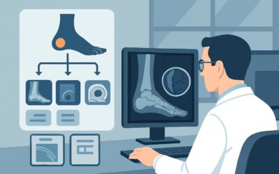
A 48-year-old patient presents with six months of worsening, atraumatic right shoulder pain, particularly at night and with overhead activity. Physical exam is notable for a painful arc of motion and impingement signs. You ordered initial radiographs, and the report is back: “Amorphous calcification is seen superior to the greater tuberosity, consistent with calcific tendinopathy of the supraspinatus tendon.” The diagnosis is clear, but the pain is persistent. The clinical question is no longer what is causing the pain, but rather confirming the calcification as the pain generator and initiating treatment. This article details the American College of Radiology (ACR) Appropriateness Criteria workflow for this specific scenario. For this presentation, the ACR rates an Image-guided anesthetic +/- corticosteroid injection as Usually Appropriate.
Who Fits This Clinical Scenario?
This guidance applies to adult patients with chronic shoulder pain (typically lasting more than 3-6 months) where initial two-view radiographs have already been performed and have positively identified calcific tendinopathy or calcific bursitis. The key inclusion criterion is the radiographic evidence of calcium deposits in or around the rotator cuff tendons or within the subacromial-subdeltoid bursa. The primary clinical goal has shifted from initial diagnosis to confirming the calcific deposit as the symptomatic source and planning the next therapeutic step.
This workflow is distinct from several related scenarios. It does not apply if:
- Initial imaging has not been performed. In that case, the workup starts with radiographs. See the guidance for initial imaging in chronic shoulder pain.
- Radiographs show significant glenohumeral osteoarthritis. This finding shifts the differential diagnosis and management priorities, routing to a different ACR workflow.
- The primary clinical suspicion is a labral tear or instability, often seen in younger, athletic patients with symptoms of clicking, locking, or apprehension. Even with normal radiographs, that presentation follows a separate imaging pathway.
This article is exclusively for the patient whose pain is attributed to calcifications already seen on an X-ray.
What Diagnoses Are You Working Up in This Scenario?
With calcifications identified on radiographs, the differential narrows significantly. The primary goal is to confirm that these deposits are the active source of the patient’s symptoms, as they can sometimes be incidental findings. The workup focuses on confirming symptomatic calcific tendinopathy and its associated inflammatory processes.
Symptomatic Calcific Tendinopathy: This is the leading diagnosis. The condition involves the deposition of calcium hydroxyapatite crystals, most commonly within the supraspinatus tendon. The natural history includes three phases: pre-calcific (metaplasia), calcific (with formative, resting, and resorptive sub-phases), and post-calcific. The resorptive phase is often the most acutely painful, as the body’s inflammatory response attempts to clear the deposits, increasing intratendinous pressure and chemical irritation.
Secondary Subacromial-Subdeltoid Bursitis: The calcific deposits can cause mechanical irritation and a secondary inflammatory response in the overlying subacromial-subdeltoid bursa. This bursitis can be a major contributor to the patient’s pain and impingement symptoms. The calcifications may even rupture from the tendon into the bursa, causing a severe, acute chemical bursitis.
Mechanical Impingement Syndrome: The enlarged, calcified tendon can physically crowd the subacromial space, leading to impingement against the acromion during arm elevation. While the underlying cause is the calcification, the clinical presentation is that of impingement, which is important to recognize and manage.
Why Is an Image-Guided Injection the Recommended Next Step?
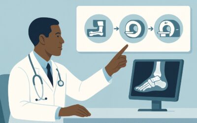
When radiographs have already confirmed the presence of calcific tendinopathy, the clinical priority shifts from anatomical diagnosis to confirming the pain generator and providing therapy. This is why the ACR designates an Image-guided anesthetic +/- corticosteroid injection as Usually Appropriate. This procedure is both diagnostic and therapeutic.
The diagnostic value comes from the immediate, albeit temporary, relief provided by the local anesthetic. If injecting around the calcific deposit and in the subacromial bursa significantly reduces the patient’s pain, it confirms this location as the primary source of symptoms. The therapeutic value comes from the corticosteroid, which provides potent, longer-lasting anti-inflammatory effects to manage the associated tendinitis and bursitis. Image guidance (typically ultrasound) is crucial for ensuring accurate needle placement, maximizing efficacy, and avoiding injury to adjacent structures like the rotator cuff tendon itself.
Why are other imaging studies rated lower for this specific scenario?
- Ultrasound (US) shoulder and MRI shoulder without IV contrast are rated May be appropriate. While these studies can provide more detailed anatomical information about the calcifications (e.g., consistency, exact location) and assess for concurrent rotator cuff tears, they do not offer a therapeutic benefit. Since the primary diagnosis is already known from the radiograph, these advanced imaging studies are often unnecessary as the next immediate step unless the injection fails or there is suspicion of a significant concurrent pathology like a full-thickness tear.
- Additional radiographs and all forms of CT imaging are rated Usually not appropriate. These studies add little to no new information beyond the initial radiograph and, in the case of CT, expose the patient to unnecessary ionizing radiation (CT shoulder without contrast carries a dose of ☢☢☢ 1-10 mSv).
In essence, the ACR guidance prioritizes a procedure that directly addresses the clinical question—”Is this calcification the source of pain?”—and simultaneously begins treatment. If further diagnostic imaging is later deemed necessary, an MRI is the preferred modality. For a detailed overview of the imaging technique, see our protocol guide: MRI Shoulder Without Contrast.
What’s Next After an Image-Guided Injection? Downstream Workflow
The patient’s response to the image-guided injection dictates the subsequent clinical pathway. The outcome provides crucial information for tailoring further management.
- Positive Response (Significant Pain Relief): If the patient experiences substantial and sustained pain relief, it confirms symptomatic calcific tendinopathy/bursitis. The next step is typically a course of formal physical therapy to address any underlying biomechanical issues, restore range of motion, and strengthen the rotator cuff and periscapular muscles. The injection breaks the pain cycle, creating a window of opportunity for effective rehabilitation.
- Partial or Temporary Response: If relief is incomplete or wears off quickly, several options exist. A repeat injection may be considered after an appropriate interval. Another option is a therapeutic ultrasound-guided procedure called barbotage (or lavage), which involves needling the calcific deposit to break it up and aspirating the calcium slurry. This is often followed by a corticosteroid injection into the bursa.
- Negative Response (No Pain Relief): If the patient has no significant pain relief from a technically successful, well-placed injection, it strongly suggests the calcific deposit is not the primary pain generator. This is a critical branch point. The diagnosis must be reconsidered. The next step is often to pursue further diagnostic imaging, such as an MRI of the shoulder (rated May be appropriate in the initial scenario) to evaluate for other pathologies like a rotator cuff tear, labral pathology, or adhesive capsulitis. One must also consider extrinsic sources of shoulder pain, such as cervical radiculopathy.
Pitfalls to Avoid (and When to Get Help)
Navigating this scenario requires careful attention to a few common pitfalls. First, avoid the temptation to order advanced imaging like MRI reflexively when the radiograph already provides the likely diagnosis; the injection is a more direct and efficient next step. Second, do not perform a “blind” (non-image-guided) injection, as accuracy is significantly lower and can lead to false-negative results (no pain relief despite the calcification being the true source) or iatrogenic tendon injury. Third, be sure to rule out a full-thickness rotator cuff tear on physical exam or with point-of-care ultrasound if available, as this may alter management. Finally, always consider referred pain from the cervical spine, especially if the pain distribution is atypical or associated with neurologic symptoms. If the patient has no relief after a well-placed injection or presents with red flags like weakness or sensory changes, referral to a musculoskeletal specialist or ordering an MRI of the cervical spine may be warranted.
Related ACR Topics and Tools
This article focuses on a single clinical variant. For a comprehensive overview of imaging for all common presentations of chronic shoulder pain, from initial workup to postoperative evaluation, please see our parent guide. The tools below can help you apply appropriateness criteria to other scenarios and understand the technical details of the recommended studies.
- For breadth across all scenarios in Chronic Shoulder Pain, see our parent guide: Chronic Shoulder Pain: ACR Appropriateness Decoded.
- ACR Appropriateness Criteria Lookup — for adjacent scenarios
- Imaging Protocol Library — for technique on the recommended study
- Radiation Dose Calculator — for cumulative dose conversations
Frequently Asked Questions
Why not just order an MRI first to see everything in the shoulder?
While an MRI provides excellent anatomical detail, it is rated ‘May be appropriate’ rather than ‘Usually Appropriate’ in this scenario because the radiograph has already identified a highly probable cause of pain (calcific tendinopathy). The most direct next step is to confirm this finding as the pain generator and initiate treatment simultaneously, which an image-guided injection accomplishes. An MRI is a purely diagnostic test that doesn’t offer therapy and is best reserved for cases where the injection fails or there is high suspicion of a different, coexisting pathology like a full-thickness rotator cuff tear.
What is the difference between calcific tendinopathy and calcific bursitis?
Calcific tendinopathy refers to calcium deposits forming within the substance of a tendon, most commonly the rotator cuff. Calcific bursitis occurs when those deposits rupture from the tendon into the overlying subacromial-subdeltoid bursa, causing a severe, acute inflammatory reaction. An injection can treat the inflammation in both scenarios.
How does an anesthetic and steroid injection help diagnostically?
The diagnostic component comes from the local anesthetic (e.g., lidocaine or bupivacaine). If the patient’s characteristic pain is significantly relieved within minutes of the injection, it provides strong evidence that the anatomical area injected is the source of the pain. This immediate feedback is invaluable for confirming the diagnosis before the longer-acting corticosteroid takes effect.
What is barbotage, and when is it considered?
Barbotage, also known as lavage or needling, is an ultrasound-guided procedure where one or two needles are used to break up the calcific deposit and aspirate the chalky calcium material. It is a more invasive therapeutic option than a simple steroid injection and is typically considered for patients with persistent, severe symptoms who have not responded adequately to more conservative measures like injections and physical therapy.
Are there alternatives if a patient cannot have a corticosteroid injection?
Yes. If a patient has a contraindication to corticosteroids (e.g., uncontrolled diabetes, allergy), an injection with local anesthetic alone can still be performed for its diagnostic value. If it provides temporary relief, confirming the diagnosis, then other treatments like physical therapy, oral anti-inflammatories, or barbotage (without a subsequent steroid injection) can be pursued.
Reviewed by Pouyan Golshani, MD, Interventional Radiologist — May 30, 2026