What Is the Best Initial Imaging for Suspected Osteomyelitis of the Foot in a Diabetic Patient?
A 64-year-old man with a 15-year history of type 2 diabetes mellitus presents to your clinic with a non-healing ulcer on the plantar aspect of his right foot, beneath the first metatarsal head. The area is erythematous, warm, and tender to palpation. You can probe the ulcer base with a sterile instrument and feel a hard, gritty surface, raising your suspicion for underlying bone involvement. The immediate clinical question is which imaging study to order first to evaluate for osteomyelitis. This is a common and high-stakes decision, as a missed diagnosis can lead to severe complications, including amputation.
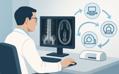
For this specific scenario—an adult with diabetes and suspected foot osteomyelitis requiring initial imaging—the American College of Radiology (ACR) Appropriateness Criteria rate Radiography foot as Usually appropriate. This article provides a detailed workflow for this initial diagnostic step.
Who Fits This Clinical Scenario?
This guidance is for clinicians evaluating an adult patient with diabetes mellitus who presents with new or worsening signs of a foot infection where osteomyelitis is a primary concern. The key inclusion criteria are:
- The patient is an adult with diagnosed diabetes mellitus.
- There is a clinical suspicion of osteomyelitis (e.g., a deep or chronic foot ulcer, positive probe-to-bone test, localized warmth, swelling, or pain).
- This is the initial imaging workup for this specific presentation; no prior imaging for this episode has been performed.
It is crucial to distinguish this situation from closely related but distinct clinical scenarios that require a different imaging pathway. This article does not apply if:
- Initial radiographs have already been performed and are negative or indeterminate. In that case, the workflow shifts to advanced imaging. This is addressed in the ACR variant for patients with negative or indeterminate initial radiographs.
- Radiographs are clearly positive for osteomyelitis. The imaging question then becomes about pre-operative planning, which may involve CT or MRI.
- The patient has known metallic instrumentation in the foot. Metal artifact significantly degrades CT and MRI, altering the choice and protocol of the imaging study.
Correctly identifying your patient’s scenario ensures the most efficient and clinically appropriate diagnostic pathway is chosen from the outset.
What Diagnoses Are You Working Up in This Scenario?
When ordering the initial radiograph for a diabetic foot infection, you are evaluating several potential diagnoses. The imaging findings, or lack thereof, will help differentiate among these possibilities.
Osteomyelitis is the primary concern. In patients with diabetes, peripheral neuropathy can mask the pain of a deep ulcer, allowing infection to penetrate from the skin and soft tissues directly into the underlying bone. The radiograph is assessed for classic signs of bone infection, such as cortical erosion, periosteal reaction, or the presence of a sequestrum (a piece of dead bone).
Cellulitis or a deep soft tissue abscess without bone involvement is another common diagnosis. While radiographs are not sensitive for directly visualizing soft tissue infection, they can reveal important secondary signs like soft tissue swelling, subcutaneous gas (indicating a gas-forming organism), or the presence of an occult foreign body that may be the source of infection.
Charcot neuroarthropathy is a critical mimic of osteomyelitis. This non-infectious, destructive process occurs in patients with neuropathy and can present acutely with erythema, warmth, and swelling, closely resembling infection. Early radiographs may show subtle fractures, joint subluxation, or bony fragmentation. Distinguishing acute Charcot from osteomyelitis is a notorious diagnostic challenge, as the two can also coexist.
Non-displaced fracture or foreign body can also be the underlying cause of pain, swelling, or a non-healing wound. A patient with severe neuropathy may not recall a traumatic event. A simple radiograph is an excellent tool for identifying radiopaque foreign bodies (like glass or metal) and assessing for fractures that could be a nidus for infection.
Why Is Foot Radiography the Recommended Initial Study?
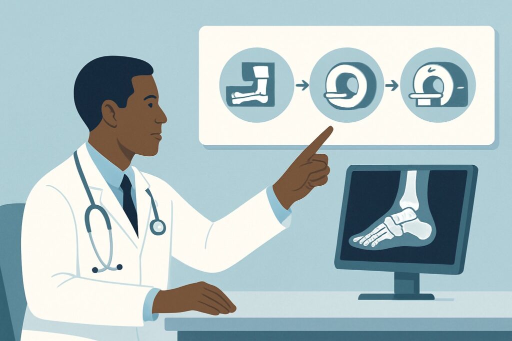
For the initial evaluation of suspected osteomyelitis in a diabetic foot, a standard multi-view radiograph is the cornerstone of the diagnostic process. The ACR designates Radiography foot as Usually appropriate because it provides a crucial, cost-effective baseline assessment that can guide all subsequent management, despite its known limitations in early disease.
The primary rationale is its high specificity when positive and its ability to identify alternative diagnoses. If classic radiographic signs of osteomyelitis—such as cortical destruction, periosteal reaction, or sequestrum—are present, the diagnosis is often confirmed, and the patient can proceed toward targeted antibiotic therapy or surgical planning. Furthermore, radiographs are invaluable for detecting fractures, joint dislocations, bony deformities associated with Charcot neuroarthropathy, soft tissue gas, and radiopaque foreign bodies.
It is important to acknowledge that the sensitivity of radiography for early osteomyelitis is low. Bony changes may not become visible for 10 to 14 days after the onset of infection. However, its role as a first-line test is justified by its wide availability, low cost, and minimal radiation dose (adult RRL=☢ <0.1 mSv).
In contrast, more advanced imaging modalities are rated as Usually not appropriate for the initial workup in this scenario:
- MRI foot without and with IV contrast: While MRI is the most sensitive and specific imaging test for osteomyelitis, it is not recommended as the first step. It is more expensive, less accessible, and often unnecessary if a radiograph provides a clear answer or suggests an alternative diagnosis. Its primary role is reserved for cases where the initial radiograph is negative or equivocal and clinical suspicion remains high.
- 3-phase bone scan: This nuclear medicine study is highly sensitive for detecting abnormal bone turnover but lacks specificity. It can be positive in osteomyelitis, but also in fractures, Charcot neuroarthropathy, and other inflammatory conditions, making it difficult to distinguish infection from its mimics. It also involves a significantly higher radiation dose (adult RRL=☢☢☢ 1-10 mSv).
Therefore, the clinical workflow begins with the simple, informative, and readily available radiograph.
What’s Next After Foot Radiography? Downstream Workflow
The results of the initial foot radiograph will direct your next steps. The clinical decision tree branches based on whether the findings are positive, negative, or indeterminate.
If the radiograph is positive for osteomyelitis: When findings like cortical erosion, periosteal reaction, or a sequestrum are clearly visible in the area of clinical concern, the diagnosis is strongly supported. The next step is typically to consult with infectious disease and podiatric or orthopedic surgery specialists. Further imaging may be needed for pre-operative planning, which falls under a different ACR scenario: Adult. Radiographs positive for osteomyelitis of the foot in patients with diabetes mellitus. Next imaging study for pre-operative planning.
If the radiograph is negative or indeterminate: This is a very common outcome, especially in early infection. A negative radiograph does not rule out osteomyelitis. If your clinical suspicion remains high (e.g., based on a positive probe-to-bone test or persistent signs of infection despite soft-tissue treatment), the next step is to proceed to advanced imaging. The appropriate follow-up is guided by the ACR scenario: Adult. Suspected osteomyelitis of the foot in patients with diabetes mellitus. Initial radiographs negative or indeterminate. This typically involves ordering an MRI with and without IV contrast.
If the radiograph suggests Charcot neuroarthropathy: Findings such as peri-articular fractures, joint subluxation, or bony fragmentation, especially in the midfoot, should raise suspicion for Charcot. This is a medical emergency requiring immediate and strict offloading (e.g., total contact casting) to prevent further joint destruction. Management should be coordinated with a podiatrist or endocrinologist specializing in diabetic foot care. Differentiating acute Charcot from osteomyelitis can be extremely difficult, and MRI is often required if superimposed infection is also suspected.
Pitfalls to Avoid (and When to Get Help)
Navigating the initial workup for a diabetic foot infection requires vigilance to avoid common diagnostic traps.
- Pitfall 1: Accepting a negative radiograph as the final word. The most significant error is to be falsely reassured by a normal radiograph in the first two weeks of infection. If clinical suspicion is high, a negative radiograph should prompt advanced imaging, not cessation of the workup.
- Pitfall 2: Misinterpreting acute Charcot as infection. Both can present with warmth, redness, and swelling. Mistaking Charcot for a simple infection and failing to enforce strict offloading can lead to catastrophic joint collapse and deformity.
- Pitfall 3: Not obtaining the correct views. Standard views include anteroposterior (AP), lateral, and oblique projections. Ensure all three are obtained to fully visualize the bones and joints of the foot. Weight-bearing views can be helpful if instability or Charcot deformity is the primary concern.
Escalate immediately if the patient shows signs of systemic illness (fever, leukocytosis, hemodynamic instability) or has evidence of deep-space infection or necrotizing fasciitis (e.g., crepitus, bullae, rapidly advancing erythema). These situations warrant emergency surgical consultation, often in parallel with any imaging workup.
Related ACR Topics and Tools
This article focuses on a single, common clinical scenario. For a comprehensive overview of all related variants, from post-operative evaluation to cases involving metallic hardware, please consult the parent topic guide. Additional GigHz tools can help you navigate adjacent scenarios and understand the technical aspects of imaging.
- For breadth across all scenarios in Suspected Osteomyelitis of the Foot in Patients with Diabetes Mellitus, see our parent guide: Suspected Osteomyelitis of the Foot in Patients with Diabetes Mellitus: ACR Appropriateness Decoded.
- ACR Appropriateness Criteria Lookup — for adjacent scenarios
- Imaging Protocol Library — for technique on the recommended study
- Radiation Dose Calculator — for cumulative dose conversations
Frequently Asked Questions
Why not just order an MRI first if it’s more sensitive for osteomyelitis?
While MRI is more sensitive, a plain radiograph is recommended as the initial study because it is fast, inexpensive, widely available, and uses very low radiation. It can definitively diagnose advanced osteomyelitis, identify important alternative diagnoses like fractures or Charcot foot, and provides a critical baseline for comparison. MRI is reserved for cases where the radiograph is negative or inconclusive but clinical suspicion remains high.
How long does it take for osteomyelitis to become visible on a radiograph?
Detectable bony changes from osteomyelitis, such as cortical erosion or periosteal reaction, typically take 10 to 14 days to develop. Therefore, a radiograph performed before this period may be falsely negative, even if an infection is present.
What specific radiographic views should I order for a suspected diabetic foot infection?
A standard foot series is appropriate. This includes three views: anteroposterior (AP), lateral, and medial oblique. These views together provide a comprehensive initial assessment of the bones and soft tissues of the foot.
If the radiograph is negative but my clinical suspicion is high, what is the next step?
If you still strongly suspect osteomyelitis despite a negative initial radiograph, the next step is to order advanced imaging. This falls under a different ACR clinical variant, and the most appropriate study is typically an MRI of the foot with and without IV contrast, which is highly sensitive and specific for detecting early bone marrow edema associated with infection.
Can a radiograph reliably distinguish between osteomyelitis and acute Charcot neuroarthropathy?
It can be very difficult, as the findings can overlap significantly, especially in the early stages. Classic Charcot involves midfoot collapse (‘rocker-bottom’ deformity) and peri-articular fragmentation, while classic osteomyelitis involves cortical destruction underlying an ulcer. However, many cases are ambiguous. If there is diagnostic uncertainty, an MRI is often necessary to differentiate the two conditions, as their management is drastically different.
Reviewed by Pouyan Golshani, MD, Interventional Radiologist — May 29, 2026