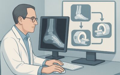
Should You Order MRI for Suspected Osteonecrosis After Inconclusive Radiographs?

A 45-year-old male with a history of long-term prednisone use for systemic lupus erythematosus presents to your clinic with six weeks of deep, throbbing, non-traumatic left hip pain. The pain is worse with weight-bearing and has begun to limit his daily activities. You obtain plain radiographs of the hip, which show no evidence of fracture, joint space narrowing, or other significant abnormality. Given his clinical risk factors and persistent symptoms, your suspicion for osteonecrosis remains high. This article details the American College of Radiology (ACR) recommended imaging workflow for this exact scenario: when you clinically suspect osteonecrosis, but initial radiographs are either normal or show only suspicious, non-diagnostic findings. For this presentation, an MRI of the area of interest without IV contrast is rated Usually Appropriate.
Who Fits This Clinical Scenario for Suspected Osteonecrosis?
This guidance applies to a specific and common clinical decision point: the patient in front of you has a compelling story for osteonecrosis (also known as avascular necrosis, or AVN), but the initial, readily available imaging—a plain radiograph—has not confirmed the diagnosis. This workflow is designed for patients who meet the following criteria:
- High clinical suspicion for osteonecrosis. This is typically based on the presence of significant risk factors (e.g., chronic corticosteroid use, excessive alcohol intake, sickle cell disease, Gaucher disease, history of trauma/dislocation) combined with characteristic symptoms like focal, deep, non-traumatic joint pain.
- An inconclusive initial radiograph. This includes radiographs that are interpreted as completely normal or those that show subtle, non-specific findings that are only suspicious for osteonecrosis, such as mild sclerosis or cystic changes, but are not definitive.
This workflow is not intended for:
- Patients with suspected osteonecrosis who have not yet had any imaging. That situation falls under the ACR variant for “Initial imaging,” which has a different set of considerations.
- Patients with known osteonecrosis and clear articular collapse on radiographs. If the diagnosis is established and imaging is being ordered for surgical planning, that constitutes a separate, pre-operative workup scenario.
- Patients with acute, high-energy trauma where an occult fracture is the primary concern. While osteonecrosis can be a long-term sequela of trauma, the immediate post-injury workup follows a different diagnostic pathway.
What Diagnoses Are You Working Up in This Scenario?
When radiographs are unrevealing in a patient with focal joint pain and risk factors for AVN, the differential diagnosis broadens. The goal of the next imaging study is to differentiate between osteonecrosis and its mimics, as the management for each can vary significantly.
Osteonecrosis (Avascular Necrosis)
This is the primary diagnosis of concern. It results from the disruption of blood supply to bone, leading to cellular death, subsequent structural weakening, and potential articular collapse. The femoral head is the most common site. Early diagnosis is critical because interventions may be more effective before the femoral head collapses. MRI is exceptionally sensitive for the early marrow changes of AVN that are invisible on plain films.
Subchondral Insufficiency Fracture
This condition can mimic osteonecrosis, presenting with acute joint pain, often in the absence of significant trauma, particularly in older individuals with osteoporosis. It represents a stress failure of weakened subchondral bone. On MRI, it typically appears as extensive bone marrow edema and a linear, serpentine low-signal fracture line parallel to the articular surface, which can be distinguished from the classic findings of AVN.
Transient Osteoporosis of the Hip
A less common, self-limiting condition characterized by acute pain and diffuse bone marrow edema on MRI. It typically resolves over several months with conservative management. Unlike osteonecrosis, it does not feature the focal, well-demarcated necrotic segment and is not associated with subchondral collapse. Differentiating it from AVN is crucial to avoid unnecessary interventions.
Stress Fracture
In patients with a history of new or repetitive physical activity, a stress fracture is a key consideration. Early stress fractures are often occult on radiographs but are readily visible on MRI as a fracture line surrounded by bone marrow edema. The location (e.g., femoral neck vs. femoral head) and patient history help distinguish this from osteonecrosis.
Why Is MRI Without Contrast the Recommended Study for Suspected Osteonecrosis?
When radiographs are normal or equivocal, the ACR designates MRI of the area of interest without IV contrast as Usually Appropriate. This recommendation is based on MRI’s superior diagnostic performance, safety profile, and ability to characterize the key pathologies in the differential diagnosis for this specific clinical scenario.
The primary strength of MRI is its unparalleled sensitivity and specificity for detecting the early stages of osteonecrosis. Long before radiographic changes like sclerosis, cystic formation, or the “crescent sign” of subchondral fracture appear, MRI can identify the hallmark findings. These include bone marrow edema on fluid-sensitive sequences (like STIR or T2-fat-suppressed) and, most characteristically, a focal, wedge-shaped area of abnormal signal in the subchondral bone on T1-weighted images. The presence of a “double-line sign”—a high-signal intensity line adjacent to a low-signal intensity line on T2-weighted images—is highly specific for osteonecrosis.
For the initial diagnosis, a non-contrast study is typically sufficient. The key morphological and signal characteristics of osteonecrosis and its mimics are well-delineated without the need for gadolinium. While an MRI with and without contrast is rated as May be appropriate, it is generally reserved for complex or equivocal cases where assessing the perfusion and viability of the femoral head is necessary.
Alternative studies are rated lower for valid reasons:
- Bone Scan: This is rated Usually not appropriate. A three-phase bone scan may show a photopenic (“cold”) area in the earliest stages, followed by increased uptake in later, reparative stages. However, these findings are highly non-specific and can be seen in fracture, infection, and arthritis, limiting the study’s diagnostic value in this context. It also involves significant radiation exposure (ACR RRL ☢☢☢, 1-10 mSv).
- CT without IV contrast: This is rated May be appropriate (Disagreement). CT is superior to radiographs for detecting subtle sclerosis and subchondral fractures but is far less sensitive than MRI for detecting the early bone marrow changes of osteonecrosis. It exposes the patient to ionizing radiation (ACR RRL Varies) and is not the preferred modality for initial detection when radiographs are negative.
Opting for a non-contrast MRI provides the highest diagnostic yield for the primary question at hand while avoiding the risks associated with IV contrast and eliminating any ionizing radiation exposure (ACR RRL O, 0 mSv).
What’s Next After the MRI? Downstream Workflow
The MRI report is the critical branch point that directs the subsequent clinical pathway. Your actions will depend on whether the findings are positive, negative, or indeterminate for osteonecrosis.
If the MRI is positive for osteonecrosis:
The next step is staging the disease (e.g., using the Ficat-Arlet or ARCO classification systems), which is typically done by the radiologist based on the MRI findings. Management is then dictated by the stage and location of the lesion, as well as patient factors. Early-stage, pre-collapse disease may be managed with protected weight-bearing or core decompression surgery. For later-stage disease with articular collapse, the patient should be referred to an orthopedic surgeon to discuss joint-preserving procedures or total joint arthroplasty. This corresponds to the ACR scenario, “Known osteonecrosis with articular collapse by radiographs. Surgery planned.”
If the MRI is negative for osteonecrosis but reveals an alternative diagnosis:
The workflow shifts to managing the identified condition. A finding of a subchondral insufficiency fracture or stress fracture will prompt a period of protected weight-bearing and a workup for underlying bone density issues. A diagnosis of transient osteoporosis of the hip leads to conservative management with analgesics and activity modification. If early degenerative joint disease is found, treatment will focus on arthritis management.
If the MRI is completely negative:
A normal MRI in the setting of persistent, localized joint pain is highly reassuring and effectively rules out osteonecrosis and other significant intra-articular bone pathology. The focus should shift to extra-articular causes of pain, such as soft tissue pathology (tendinopathy, bursitis), referred pain from the spine, or other systemic conditions. Further clinical evaluation and potentially physical therapy would be the logical next steps.
Pitfalls to Avoid (and When to Get Help)
Navigating this diagnostic pathway requires attention to a few common pitfalls to ensure timely and accurate diagnosis.
- Stopping the workup after a normal radiograph. In a patient with significant risk factors and persistent, focal pain, a normal X-ray should not be the end of the investigation. Delaying an MRI can allow pre-collapse osteonecrosis to progress to a more advanced stage.
- Ordering a bone scan or CT as the next step. While these modalities have roles in musculoskeletal imaging, they lack the sensitivity and specificity of MRI for early osteonecrosis detection and expose the patient to unnecessary radiation.
- Forgetting to image the contralateral joint. Osteonecrosis is bilateral in a high percentage of non-traumatic cases (e.g., steroid- or alcohol-induced). If the MRI of the symptomatic hip is positive, consider screening the asymptomatic contralateral hip, as early detection can impact management.
- Misinterpreting bone marrow edema. Diffuse edema can be seen in transient osteoporosis, while focal, subchondral edema is characteristic of AVN or an insufficiency fracture. Close collaboration with the interpreting radiologist is key to distinguishing these mimics.
If the diagnosis remains unclear after MRI or if the patient has advanced-stage disease, an early referral to an orthopedic surgeon with expertise in joint preservation is the appropriate escalation.
Related ACR Topics and Tools
For a comprehensive overview of imaging for osteonecrosis across all clinical presentations and for tools to help with ordering decisions, the following GigHz resources are available. For breadth across all scenarios in Osteonecrosis, see our parent guide: Osteonecrosis: ACR Appropriateness Decoded.
- ACR Appropriateness Criteria Lookup — For exploring adjacent clinical scenarios and official ratings.
- Imaging Protocol Library — For detailed technical specifications on MRI and other imaging protocols.
- Radiation Dose Calculator — For discussing cumulative radiation exposure with patients when considering CT or nuclear medicine studies.
Frequently Asked Questions
Why is a non-contrast MRI preferred over a contrast-enhanced MRI for initial diagnosis of osteonecrosis?
For the initial diagnosis of osteonecrosis, a non-contrast MRI is rated ‘Usually Appropriate’ because it is highly sensitive and specific for the key findings, such as changes in marrow signal and the characteristic ‘double-line sign.’ IV contrast is typically not needed to make the diagnosis and is reserved for equivocal cases or when assessing perfusion is critical, which is less common in the initial workup. A non-contrast study provides the necessary diagnostic information while avoiding the costs and potential risks of gadolinium.
If my patient has a contraindication to MRI (e.g., an incompatible pacemaker), what is the next best test?
In cases where MRI is contraindicated, the decision is more complex. A CT scan, rated ‘May be appropriate (Disagreement),’ would be the next logical step. While less sensitive than MRI for early marrow changes, it is more sensitive than plain radiographs for detecting sclerosis, microfractures, and early subchondral collapse. A nuclear medicine bone scan is ‘Usually not appropriate’ due to its low specificity but might be considered if CT is also inconclusive or contraindicated.
How soon after symptom onset can an MRI detect osteonecrosis?
MRI can detect the bone marrow edema and cellular changes of osteonecrosis within days to weeks of the vascular insult, long before any changes are visible on plain radiographs. Radiographic findings may not become apparent for months. This ability to provide an early diagnosis is the primary reason MRI is the recommended study in this scenario.
Should I order an MRI of the hip or the entire pelvis?
The ACR specifies ‘MRI area of interest.’ If the symptoms are clearly localized to one hip, a dedicated hip MRI is appropriate. However, given the high rate of bilateral disease in non-traumatic osteonecrosis, many radiologists will include screening sequences of the contralateral hip even on a unilateral order. If suspicion is high for bilateral involvement or if pain is less localized, ordering a bilateral hip or pelvic MRI may be more efficient.
What specific MRI sequences are most important for diagnosing osteonecrosis?
The most critical sequences are T1-weighted and fluid-sensitive, fat-suppressed sequences (like STIR or T2-weighted with fat suppression). T1-weighted images are excellent for delineating the focal, low-signal necrotic zone against the bright signal of normal fatty marrow. Fluid-sensitive sequences are highly sensitive for detecting the associated bone marrow edema.
Reviewed by Pouyan Golshani, MD, Interventional Radiologist — June 27, 2026