Should You Order MRI or CT for Chronic Foot Pain with Negative Radiographs?
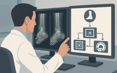
A 48-year-old marathon runner presents to your clinic with three months of worsening dorsal midfoot pain. The pain is localized over the navicular, exacerbated by running, and now bothers her even when walking. Initial radiographs ordered a month ago were interpreted as negative for acute fracture. Your clinical suspicion is high for a navicular stress fracture, but you also consider a symptomatic accessory navicular bone. This article provides a step-by-step clinical workflow for this exact scenario: an adult with chronic foot pain of suspected osseous origin, where initial radiographs are negative or indeterminate. For this presentation, the American College of Radiology (ACR) rates an MRI of the foot without IV contrast as Usually Appropriate.
Who Fits This Clinical Scenario?
This guidance is specifically for adult patients presenting with chronic foot pain (lasting several weeks to months) where the clinical history and physical exam point toward a bone-related issue. The key feature of this scenario is that initial radiographs have already been performed and were either negative or inconclusive, yet clinical suspicion remains high for an underlying osseous pathology.
Inclusion criteria for this workflow:
- Adult patient
- Chronic, localized foot pain
- Clinical suspicion for an occult fracture (e.g., stress fracture), a painful accessory ossicle, or another primary bone lesion (e.g., osteoid osteoma)
- Initial radiographs are negative, equivocal, or indeterminate
This workflow is NOT for:
- Initial Imaging: If this is the patient’s first presentation and no imaging has been done, the evaluation starts with radiographs. See the ACR variant for chronic foot pain of unknown etiology.
- Suspected Soft-Tissue Injury: If your primary concern is for a tendon, ligament, or fascia injury (like Achilles tendinopathy or plantar fasciitis), a different ACR workflow applies.
- Suspected Cartilage Abnormality: If the clinical picture suggests an osteochondral lesion or advanced degenerative joint disease, that follows a separate imaging pathway.
What Diagnoses Are You Working Up in This Scenario?
When radiographs are unrevealing, advanced imaging is used to investigate a specific differential diagnosis. The goal is to find subtle pathology that doesn’t show up on a standard X-ray.
Stress Fracture
This is a leading consideration, especially in athletes or individuals who have recently increased their activity levels. Bones like the navicular, calcaneus, and metatarsals are common sites. Radiographs are notoriously insensitive for detecting early-stage stress injuries, which initially manifest as bone marrow edema without a visible fracture line. Advanced imaging can detect these changes much earlier, guiding management and preventing progression to a complete fracture.
Symptomatic Accessory Ossicle
Accessory ossicles are common, normal anatomic variants, but they can become a source of pain. An os trigonum can cause posterior ankle impingement in dancers or soccer players. A painful os naviculare (prehallux) can be associated with posterior tibial tendon dysfunction. Imaging helps determine if the ossicle is simply present or if there is associated inflammation, bone marrow edema, or degenerative change indicating it is the pain generator.
Occult Traumatic Fracture
A non-displaced or minimally displaced fracture may not have been visible on the initial radiographs due to patient positioning, overlying structures, or the subtlety of the finding. Chronic, persistent pain after a minor trauma warrants investigation for a missed fracture, particularly in the tarsal bones.
Benign or Malignant Bone Lesions
Though less common, a primary bone lesion can present as chronic, localized pain. An osteoid osteoma, a benign tumor, classically causes night pain that is relieved by nonsteroidal anti-inflammatory drugs (NSAIDs). While often visible on radiographs, it can be subtle. Advanced imaging is definitive for diagnosis and localization before treatment.
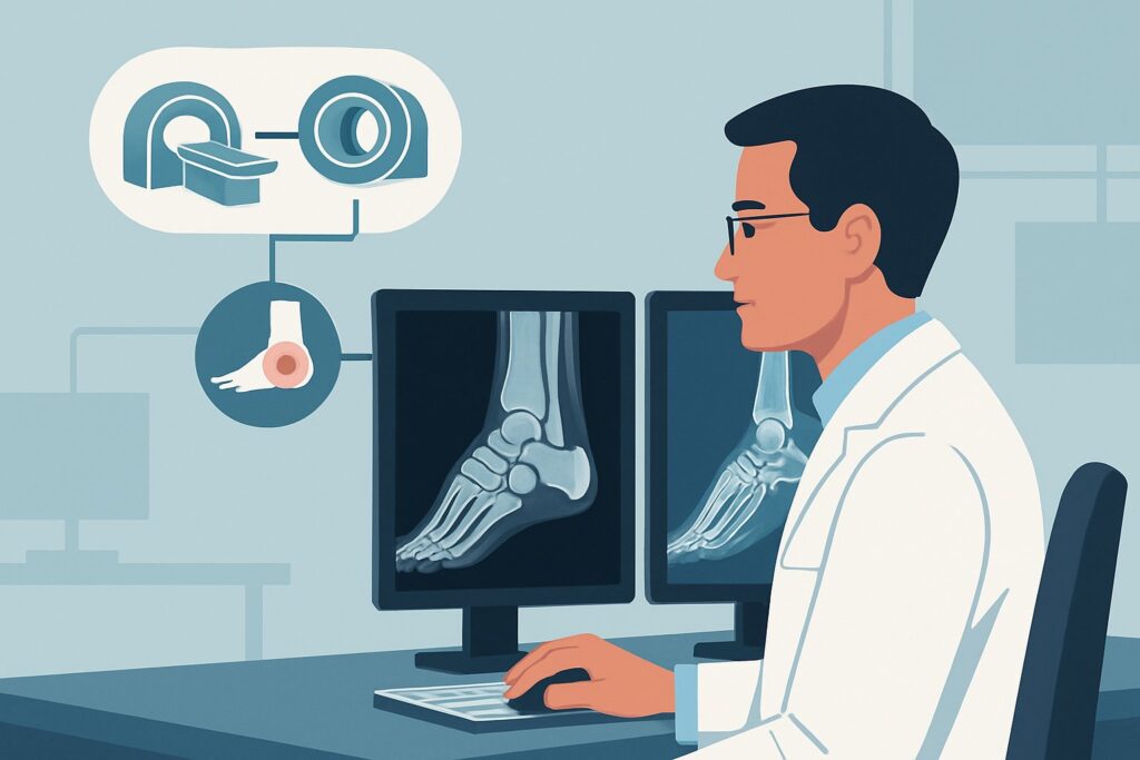
Why Is MRI Foot without IV Contrast the Recommended Study for This Presentation?
The ACR designates MRI of the foot without IV contrast as Usually Appropriate for this scenario because of its unparalleled ability to visualize bone marrow and surrounding soft tissues without using ionizing radiation.
The primary diagnostic strength of MRI is its high sensitivity for detecting bone marrow edema. This edema is the earliest sign of a stress reaction, occult fracture, or inflammation surrounding a painful ossicle. MRI can identify a stress injury weeks before a fracture line becomes visible on any other modality, including CT. It can also clearly depict associated soft-tissue abnormalities, such as synovitis or tendinosis, which can help confirm that an accessory ossicle is the source of symptoms.
How do alternative studies compare?
- CT foot without IV contrast: This study is also rated Usually Appropriate. CT provides superior spatial resolution and exquisite detail of cortical bone. It is excellent for precisely defining a fracture line, evaluating for healing, or assessing the morphology of an ossicle. However, it is less sensitive than MRI for detecting early stress reactions that only manifest as bone marrow edema. CT also involves a small dose of ionizing radiation (ACR RRL: ☢ <0.1 mSv). It is a strong choice if the primary question is the precise anatomy of a suspected fracture line seen equivocally on X-ray.
- Bone Scan (Technetium-99m): This study is rated Usually not appropriate. A triple-phase bone scan is highly sensitive for detecting areas of increased bone turnover. While it will likely be positive in the case of a stress fracture or painful ossicle, its findings are non-specific. A “hot spot” on a bone scan could represent a fracture, infection, arthritis, or tumor, almost always requiring follow-up with MRI or CT for definitive characterization. Given its non-specificity and higher radiation dose (ACR RRL: ☢☢☢ 1-10 mSv), it is rarely the best next step in this modern imaging era.
For this clinical question, intravenous contrast is typically unnecessary. Both MRI foot without and with IV contrast and CT foot with IV contrast are rated Usually not appropriate. The key diagnostic findings—bone marrow edema, fracture lines, and cortical integrity—are well-visualized on non-contrast sequences. Adding contrast increases cost, scan time, and introduces the small risk of an adverse reaction without providing significant additional diagnostic information for this specific differential.
What’s Next After MRI Foot without IV Contrast? Downstream Workflow
The MRI results will guide your next steps in management, whether the findings are positive, negative, or indeterminate.
If the MRI is positive for a stress fracture:
Management is dictated by the location and grade of the injury. For low-risk stress fractures (e.g., metatarsals 2-4), treatment typically involves activity modification and a period of protected weight-bearing. For high-risk fractures (e.g., navicular, fifth metatarsal base), management is more aggressive, often requiring strict non-weight-bearing and consultation with an orthopedic or sports medicine specialist, as these have a higher risk of nonunion or progression.
If the MRI is positive for a symptomatic accessory ossicle:
The report should describe findings like bone marrow edema within the ossicle, fluid in the synchondrosis (the cartilaginous joint between the ossicle and parent bone), and inflammation in the adjacent soft tissues. Initial management is typically conservative: rest, immobilization, and physical therapy. If symptoms persist, an image-guided corticosteroid injection (May be appropriate) can be both diagnostic and therapeutic. Surgical excision is reserved for refractory cases.
If the MRI is negative:
A high-quality negative MRI effectively rules out a significant osseous cause of pain. At this point, the diagnostic focus should shift. Re-evaluate the patient for a soft-tissue cause (tendon, ligament, nerve) or a referred pain pattern. This may lead you to a different ACR workflow, such as the one for a suspected soft tissue origin or neuropathic pain syndrome.
Pitfalls to Avoid (and When to Get Help)
Navigating the workup for chronic foot pain requires careful consideration to avoid common diagnostic errors.
- Stopping at the Radiograph: Do not dismiss persistent, focal bone tenderness in a high-risk patient just because the initial X-rays are negative. Radiographs have low sensitivity for early stress injuries.
- Ordering the Wrong Advanced Study: Choosing CT when the primary suspicion is an early stress reaction may lead to a false-negative result, as CT is less sensitive to bone marrow edema than MRI.
- Forgetting Anatomic Variants: Be aware of common accessory ossicles and other normal variants in the foot that can be mistaken for fractures. Correlating imaging findings with the precise location of the patient’s pain is critical.
- Ignoring High-Risk Locations: A delay in diagnosing a stress fracture of the navicular, anterior tibial cortex, or fifth metatarsal can lead to significant morbidity, including nonunion and need for surgery. If you suspect an injury in one of these locations, maintain a low threshold for advanced imaging and specialist consultation.
If the clinical picture and imaging findings are complex or discordant, or if a high-risk stress fracture is identified, consultation with an orthopedic surgeon or musculoskeletal radiologist is the appropriate next step.
Related ACR Topics and Tools
This article covers one specific scenario in depth. For a broader view of all clinical variants, or to explore the tools used to make these recommendations, see the resources below.
- For breadth across all scenarios in Chronic Foot Pain, see our parent guide: Chronic Foot Pain: ACR Appropriateness Decoded.
- To look up other clinical scenarios, use the ACR Appropriateness Criteria Lookup.
- To review technical details for the recommended study, visit the Imaging Protocol Library.
- To discuss radiation exposure with patients, consult the Radiation Dose Calculator.
Frequently Asked Questions
Why is MRI without contrast preferred over CT when both are ‘Usually Appropriate’?
MRI without contrast is often preferred as the first choice because of its superior ability to detect bone marrow edema, which is the earliest sign of a stress fracture. CT excels at showing cortical bone detail but may miss an early stress injury. MRI also avoids ionizing radiation. CT becomes the better choice if the goal is to precisely define a known or suspected fracture line.
If an MRI shows a symptomatic os naviculare, what is the next step?
The next step is typically conservative management, including rest, activity modification, orthotics, and physical therapy. If these measures fail, an image-guided injection with a corticosteroid and local anesthetic can be performed. This can be both diagnostic (confirming the ossicle as the pain source) and therapeutic. Surgical excision is reserved for cases that do not respond to conservative treatment.
Is there any role for ultrasound in this specific scenario?
The ACR rates ultrasound of the foot as ‘May be appropriate.’ While not the primary modality for evaluating bone, ultrasound can be useful for assessing the soft tissues immediately adjacent to a suspected painful ossicle or for guiding a diagnostic/therapeutic injection. It can sometimes visualize cortical irregularities from a stress fracture but is highly operator-dependent and less sensitive than MRI.
What if the patient has a contraindication to MRI, like a non-compatible pacemaker?
In cases where a patient cannot undergo an MRI, CT of the foot without IV contrast is an excellent alternative and is also rated ‘Usually Appropriate’ by the ACR. CT provides outstanding bone detail and is very effective for identifying fracture lines and assessing osseous anatomy. If CT is also contraindicated or non-diagnostic, a technetium-99m bone scan could be considered, though it is less specific.
How long should foot pain be present to be considered ‘chronic’ for this workflow?
While there is no strict cutoff, ‘chronic’ in this context generally refers to pain that has persisted for several weeks (e.g., more than 4-6 weeks) despite initial rest or treatment. This timeframe distinguishes it from acute traumatic injuries, where the initial imaging and management priorities are different.
Reviewed by Pouyan Golshani, MD, Interventional Radiologist — May 30, 2026