Should You Order Ultrasound or MRI for Chronic Foot Pain of Soft Tissue Origin?
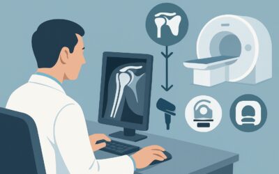
A 45-year-old avid runner presents to your clinic with four months of progressive right heel pain, worst with the first steps in the morning. Your physical exam localizes tenderness to the medial calcaneal tubercle and along the Achilles tendon insertion. You suspect either plantar fasciitis or insertional Achilles tendinopathy, but there’s some diagnostic ambiguity. Initial radiographs were unremarkable, showing no fracture or significant heel spur. You now face a common clinical decision: what is the most appropriate next imaging study to confirm the diagnosis and guide treatment? This article provides a detailed workflow for this specific scenario, grounded in the American College of Radiology (ACR) Appropriateness Criteria. For suspected soft tissue pathology in the foot after negative radiographs, US foot is rated Usually Appropriate.
Who Fits This Clinical Scenario for Chronic Foot Pain?
This guidance is specifically for an adult patient with chronic foot pain (typically lasting three months or longer) where the clinical history and physical examination strongly suggest a soft tissue origin. This includes conditions affecting tendons, ligaments, fascia, or muscles. A key prerequisite for this workflow is that initial radiographs have already been performed and were either negative or indeterminate, effectively ruling out an obvious osseous cause like a stress fracture or significant arthritis.
This workflow is intended for patients who fit these criteria:
- Adult patient
- Chronic symptoms (weeks to months, not acute trauma)
- Pain localized to a soft tissue structure on examination
- Negative or indeterminate initial radiographs
It is crucial to distinguish this presentation from similar but distinct clinical scenarios that require a different imaging approach. This guidance does not apply if you primarily suspect:
- An occult fracture or painful ossicle: This presentation, even with negative radiographs, often routes directly to MRI or CT for superior bone detail.
- An osteochondral lesion or advanced arthritis: While radiographs may be negative early on, MRI is the modality of choice for evaluating articular cartilage and subchondral bone.
- A neurologic cause: For suspected nerve entrapment (like Baxter neuropathy) or complex regional pain syndrome, the workup may involve specialized MRI neurography protocols or other diagnostic tests beyond standard imaging.
Applying this workflow to the wrong patient—for instance, using ultrasound to rule out a subtle navicular stress fracture—can lead to missed diagnoses and delayed care.
What Diagnoses Are You Working Up in This Soft Tissue Pain Scenario?
When a patient presents with chronic, localized soft tissue foot pain and negative X-rays, your differential diagnosis drives the imaging choice. The goal is to visualize the specific soft tissue structures implicated by the exam.
The most common diagnosis in this category is plantar fasciitis. This condition involves degenerative thickening and micro-tearing of the plantar fascia at its origin on the calcaneus. Patients classically report sharp, stabbing pain in the heel, especially with the first steps after a period of rest. Imaging is used to confirm fascial thickening, assess for tears, and rule out other causes of heel pain.
Another frequent consideration is tendinopathy, which can affect multiple tendons in the foot and ankle. Achilles tendinopathy is prevalent in active individuals and can be insertional (at the heel bone) or non-insertional (in the mid-portion of the tendon). Similarly, posterior tibial tendinopathy causes medial foot and ankle pain and can lead to progressive flatfoot deformity. On the other side of the ankle, peroneal tendinopathy is a common cause of lateral foot pain. Imaging helps characterize the extent of tendon degeneration, identify longitudinal splits or frank tears, and detect associated tenosynovitis.
Less common but important considerations include ligamentous injury or insufficiency. For example, chronic attenuation or tearing of the spring ligament complex is often associated with posterior tibial tendon dysfunction and acquired flatfoot. While acute sprains are a separate category, chronic ligamentous instability can be a source of persistent pain and is well-visualized with advanced imaging.
Why Is Ultrasound the Recommended Next Step for Suspected Soft Tissue Foot Pain?
For this specific clinical scenario, the ACR rates two studies as Usually Appropriate: US foot and MRI foot without IV contrast. While both are excellent, ultrasound often serves as the more practical and targeted initial choice.
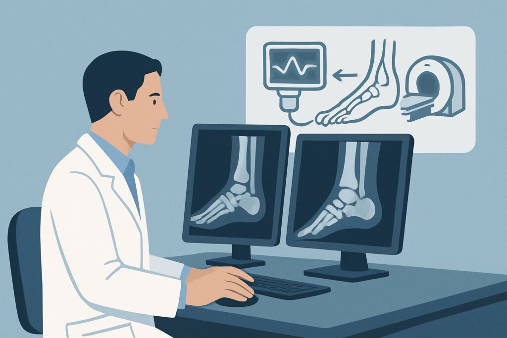
Ultrasound (US) of the foot is highly effective for this workup for several key reasons. First, it offers superb spatial resolution for superficial soft tissues, allowing for detailed evaluation of tendon fiber patterns, fascial thickness, and ligament integrity. Second, its dynamic capability is a unique advantage; a sonographer can assess a tendon as it glides during active or passive motion and can use the transducer to apply pressure to the exact site of the patient’s pain, correlating anatomy with symptoms in real-time. This can be invaluable for confirming the pain generator. Finally, ultrasound is cost-effective, widely available, and involves no ionizing radiation (0 mSv).
MRI of the foot without IV contrast is also Usually Appropriate and serves as an excellent alternative or a problem-solving tool after an inconclusive ultrasound. Its primary strength is providing a global, multi-planar view of all foot structures, including deep tendons, bone marrow, and intricate ligamentous complexes that may be difficult to access with ultrasound. It is superior for detecting bone marrow edema, which can be a clue to stress injuries or other pathologies.
The ACR rates other modalities lower for this specific presentation:
- MRI foot without and with IV contrast is rated Usually not appropriate. For the common degenerative conditions in the differential (tendinosis, plantar fasciitis), intravenous contrast adds little to no diagnostic information while increasing cost and procedural time. Contrast is typically reserved for cases where infection or a soft tissue mass is suspected.
- All variants of CT foot are rated Usually not appropriate. While CT excels at evaluating cortical bone, its soft tissue contrast resolution is far inferior to both US and MRI for assessing tendons, ligaments, and fascia. It also exposes the patient to ionizing radiation (☢ <0.1 mSv).
What to Do After the Foot Ultrasound: A Downstream Workflow
The results of the foot ultrasound will guide your next clinical steps. The workflow typically branches into one of three paths.
If the ultrasound is positive and confirms the suspected diagnosis (e.g., shows marked thickening of the plantar fascia consistent with plantar fasciitis, or tendinosis of the posterior tibial tendon), the diagnostic workup is often complete. This confirmation allows you to proceed confidently with targeted treatment, which may include physical therapy, activity modification, orthotics, or consideration of image-guided therapeutic injections. The ACR rates Image-guided anesthetic +/- corticosteroid injection as May be appropriate, and a positive ultrasound provides the clear target for such a procedure.
If the ultrasound is negative or equivocal but your clinical suspicion for significant pathology remains high, the next logical step is to order an MRI of the foot without IV contrast. As the other Usually Appropriate modality, MRI can overcome the limitations of ultrasound, such as evaluating deep structures, assessing the bone marrow, or providing a more comprehensive anatomical overview. A negative ultrasound with persistent, unexplained pain is a primary indication to escalate to MRI.
If the ultrasound identifies an unexpected finding that shifts the differential diagnosis (e.g., a soft tissue mass, signs of an abscess, or a complete tendon rupture), the downstream pathway changes accordingly. This may necessitate an MRI with contrast for better tissue characterization or an urgent referral to a subspecialist, such as an orthopedic surgeon or rheumatologist, depending on the finding.
Common Pitfalls to Avoid in This Imaging Workflow
Navigating the workup for chronic soft tissue foot pain requires careful consideration to avoid common errors that can delay diagnosis or lead to unnecessary testing.
First, avoid ordering a generic “foot ultrasound.” Be as specific as possible on the imaging requisition. Include the patient’s history and clearly state the area of maximal tenderness and the suspected pathology (e.g., “Rule out plantar fasciitis, pain at medial calcaneal tubercle” or “Evaluate peroneal tendons for tendinopathy”). This detail focuses the sonographer’s attention and ensures a high-quality, clinically relevant study.
Second, do not discount the value of dynamic assessment. Ultrasound’s ability to visualize tissues in motion is a key advantage over static imaging like MRI. If possible, communicate with the radiology department or sonographer to ensure a dynamic evaluation is performed, especially for suspected tendon subluxation or impingement.
Third, resist the reflex to order MRI as the first-line advanced study. While MRI is an outstanding imaging tool, ultrasound is often faster, more accessible, and more cost-effective for evaluating the superficial soft tissue structures that are the most common culprits in this scenario. Starting with ultrasound is a more judicious use of resources.
If you encounter clinical red flags such as fever, overlying erythema, or a palpable mass suggesting infection or neoplasm, escalate the workup promptly, which may involve MRI with contrast and an urgent subspecialty consultation.
Related ACR Topics and Tools
This article focuses on a single, common clinical scenario. For a comprehensive overview of all variants and imaging recommendations for chronic foot pain, please consult our parent guide. You can also use the tools below to explore adjacent scenarios, review imaging techniques, and discuss radiation exposure with patients.
- For breadth across all scenarios in Chronic Foot Pain, see our parent guide: Chronic Foot Pain: ACR Appropriateness Decoded.
- ACR Appropriateness Criteria Lookup — for adjacent scenarios
- Imaging Protocol Library — for technique on the recommended study
- Radiation Dose Calculator — for cumulative dose conversations
Frequently Asked Questions
Why is MRI also rated ‘Usually Appropriate’ if ultrasound is the preferred first choice?
Both ultrasound and non-contrast MRI are considered ‘Usually Appropriate’ because they are excellent modalities for evaluating soft tissues. Ultrasound is often preferred first due to its lower cost, wider availability, and unique ability to perform dynamic assessment and correlate findings with the patient’s exact point of pain. MRI is an equally valid choice and is particularly useful when a more global view of the foot is needed, when deeper structures are implicated, or when the ultrasound is negative or inconclusive despite high clinical suspicion.
Is there any role for CT scan in this specific scenario?
For a patient with suspected primary soft tissue pathology (tendon, fascia, ligament), a CT scan is rated ‘Usually not appropriate’ by the ACR. CT provides excellent detail of bone but has poor soft tissue contrast compared to ultrasound and MRI. It would not be the right test to diagnose plantar fasciitis or tendinopathy. Its use would be reserved for scenarios where an occult fracture or complex osseous anatomy is the primary concern.
When should I order an MRI with contrast for chronic foot pain?
An MRI with IV contrast is rated ‘Usually not appropriate’ for this specific scenario. The common differential diagnoses, like plantar fasciitis and degenerative tendinopathy, are not inflammatory or vascular processes that typically require contrast for diagnosis. Contrast is generally reserved for situations where there is a clinical suspicion of infection (abscess, osteomyelitis), a soft tissue tumor, or certain inflammatory arthropathies.
My patient’s radiograph mentioned a ‘heel spur.’ Does that change the imaging plan?
No. A calcaneal bone spur is a very common, often incidental finding on radiographs and is not considered the primary cause of pain in plantar fasciitis. The pain comes from the inflammation and degeneration of the fascia itself. The presence of a spur does not change the indication for further soft tissue imaging with ultrasound or MRI if clinically warranted to confirm the diagnosis or rule out other pathology.
What if my patient has bilateral chronic foot pain?
Bilateral symptoms should prompt consideration of systemic inflammatory conditions, such as seronegative spondyloarthropathies, which can present with enthesitis (inflammation where tendons and ligaments attach to bone). While the imaging choice (ultrasound or MRI) would still be appropriate to evaluate the soft tissues, the clinical context is different. You might order bilateral imaging and consider laboratory workup for inflammatory markers if the history is suggestive.
Reviewed by Pouyan Golshani, MD, Interventional Radiologist — May 30, 2026