What’s the Best Imaging for Chronic Shoulder Pain After Rotator Cuff Repair?
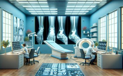
A 58-year-old patient, who underwent a rotator cuff repair 18 months ago, returns to your clinic. After a period of successful rehabilitation and good function, he now reports several months of progressive, deep shoulder pain and weakness, particularly with overhead activities. You perform a physical exam and obtain initial radiographs, which show surgically-placed anchors but are otherwise unremarkable for acute fracture, dislocation, or significant glenohumeral arthritis. Your primary concern is a recurrent rotator cuff tear or persistent subacromial bursitis. This article details the American College of Radiology (ACR) Appropriateness Criteria for this specific clinical scenario, explaining the recommended imaging workflow. For this presentation, the ACR rates US shoulder as Usually Appropriate.
Who Fits This Clinical Scenario for Post-Surgical Shoulder Pain?
This guidance is tailored for a specific patient population. The key inclusion criteria are:
- Chronic Shoulder Pain: The patient’s symptoms are persistent or recurrent over weeks to months, not the result of acute, high-energy trauma.
- History of Prior Rotator Cuff Repair: There is a documented surgical history of rotator cuff repair. The time since surgery can vary, but the symptoms represent a new or worsening problem after an initial recovery period.
- Suspected Rotator Cuff Disorder or Bursitis: The clinical picture, including pain patterns and physical exam findings (e.g., positive impingement signs, weakness with resisted strength testing), points toward a retear, tendinopathy, or inflammation of the subacromial-subdeltoid bursa.
- Normal or Inconclusive Radiographs: Initial X-rays have already been performed and have not identified a definitive cause for the patient’s pain, such as hardware failure, significant arthritis, or fracture.
This workflow is not intended for patients with different presentations, even if they have chronic shoulder pain. For instance, a patient with no prior surgery would fall under a different ACR variant. Similarly, if your primary clinical suspicion is for labral pathology or shoulder instability (often suggested by clicking, popping, or a sense of apprehension), the imaging strategy would shift, as that scenario requires a different workup. Finally, a patient presenting with global, profound stiffness in all planes of motion may be better evaluated under the adhesive capsulitis (frozen shoulder) scenario.
What Diagnoses Are You Working Up in This Post-Repair Scenario?
When a patient presents with recurrent pain after rotator cuff surgery, the differential diagnosis is focused on complications of the repair and other common sources of shoulder pathology. The choice of imaging is designed to differentiate among these possibilities.
Rotator Cuff Retear: This is the primary concern. A retear can occur at the tendon-suture interface, through the tendon substance adjacent to the repair, or at the bone-anchor interface. Tears can be partial-thickness or full-thickness. Imaging must be able to clearly visualize the repaired tendon, assess its integrity, and characterize the size and location of any new defect.
Subacromial-Subdeltoid Bursitis: Inflammation of the bursa is a frequent cause of shoulder pain and impingement symptoms. It can occur as a primary inflammatory process or, more commonly, as a secondary reaction to an underlying mechanical issue like a small retear, tendinosis, or hardware prominence. Imaging can identify bursal fluid and thickening, confirming this as a pain generator.
Tendonitis or Tendinosis: The repaired tendon or adjacent cuff tendons (e.g., infraspinatus, subscapularis) can develop degenerative or inflammatory changes. The long head of the biceps tendon is another common source of pain and can be evaluated concurrently during the imaging study.
Suture Granuloma or Hardware Complications: Though less common, a foreign body reaction to suture material can create a painful, inflammatory mass (granuloma). Similarly, suture anchors can migrate or cause local irritation. Advanced imaging can help identify these less frequent causes of post-operative pain.
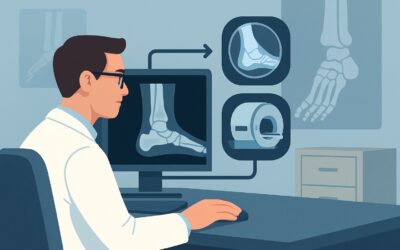
Why Is Ultrasound the Recommended Study for Suspected Rotator Cuff Retear?
For a patient with chronic shoulder pain after a rotator cuff repair and inconclusive radiographs, the ACR designates US shoulder as Usually Appropriate. This recommendation is based on a balance of diagnostic accuracy, safety, and accessibility.
Ultrasound offers high spatial resolution and provides a dynamic, real-time assessment of the shoulder. An experienced sonographer can directly visualize the rotator cuff tendons, identify fluid in the subacromial-subdeltoid bursa, and assess for tendon defects that indicate a retear. The ability to move the patient’s arm during the scan is a unique advantage, allowing for the direct visualization of tissues that may be causing mechanical impingement. Furthermore, ultrasound involves no ionizing radiation (0 mSv) and does not require intravenous contrast, making it a very safe procedure.
Several other imaging studies are also rated for this scenario, providing alternatives based on clinical context and local resources:
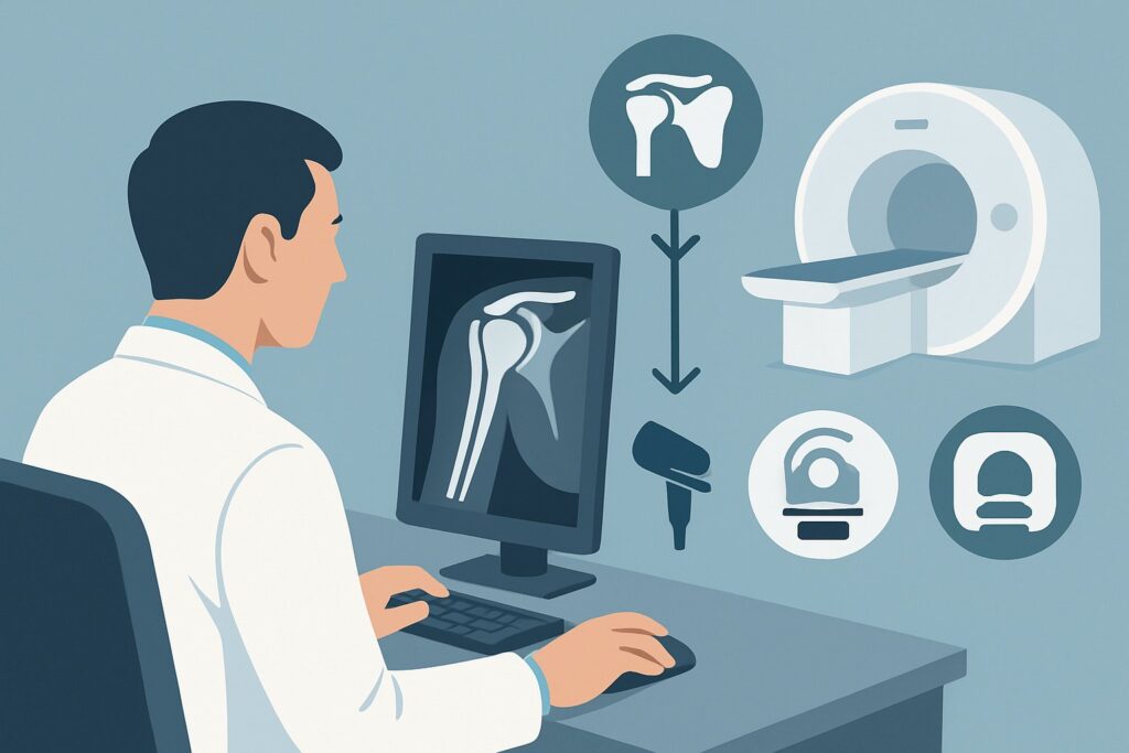
- MRI shoulder without IV contrast and MR arthrography shoulder are also rated as Usually Appropriate. MRI provides excellent soft tissue contrast and is highly sensitive for detecting retears, evaluating muscle fatty atrophy (a sign of chronicity), and identifying bone marrow edema. MR arthrography, which involves injecting contrast into the joint, is particularly sensitive for small partial-thickness tears but is more invasive. The choice between high-quality ultrasound and MRI often comes down to institutional expertise, cost, and patient factors like claustrophobia or MRI contraindications.
- CT arthrography shoulder is also Usually Appropriate but is typically reserved for patients who cannot undergo an MRI. While it provides excellent detail of the cuff and bone, it involves a significant radiation dose (☢☢☢☢ 10-30 mSv).
- MRI shoulder without and with IV contrast is rated as Usually not appropriate. The addition of intravenous gadolinium does not typically add significant diagnostic value for evaluating a rotator cuff retear compared to a non-contrast MRI, and it adds unnecessary cost and potential risk.
When ordering, be specific about the clinical question. A request for “US shoulder to evaluate for rotator cuff retear and bursitis in a post-operative patient” provides the radiologist with the necessary context to perform a targeted, high-yield examination.
What’s Next After the Shoulder Ultrasound? Downstream Workflow
The results of the shoulder ultrasound will guide your next clinical steps. The post-imaging workflow is a decision tree based on whether the findings confirm your suspicion, are negative, or are inconclusive.
If the ultrasound is positive for a significant rotator cuff retear: The next step is typically a consultation with an orthopedic surgeon. The surgeon will correlate the imaging findings with the patient’s functional demands, symptoms, and physical exam to determine if revision surgery is indicated. For some patients, particularly those with lower functional demands or significant medical comorbidities, non-operative management with physical therapy and injections may still be the preferred course.
If the ultrasound is negative for a retear but shows bursitis or tendinosis: This finding directs management toward non-operative treatments. Options include a course of physical therapy focused on strengthening and mechanics, anti-inflammatory medications, or an image-guided corticosteroid injection. The ACR rates Image-guided anesthetic +/- corticosteroid injection as May be appropriate, serving as both a diagnostic and therapeutic maneuver.
If the ultrasound is negative or inconclusive and the patient remains highly symptomatic: If there is a strong clinical suspicion for a tear despite a negative or technically limited ultrasound, the next logical step is to proceed with an MRI. An MRI shoulder without IV contrast is an excellent problem-solving tool in this situation. It can provide a more global assessment of the joint, including muscle quality and subtle intra-articular pathology that may not be well-visualized on ultrasound.
Pitfalls to Avoid (and When to Get Help)
Navigating the workup for post-operative shoulder pain requires careful consideration to avoid common missteps. One major pitfall is failing to correlate imaging findings with the patient’s clinical presentation; an imaging-identified small tear may not be the primary source of a patient’s functional disability. Another error is not providing a clear clinical history on the imaging requisition, which can lead to a non-diagnostic or non-specific report. Also, be aware that post-surgical anatomy can be complex; relying on an imaging center with musculoskeletal expertise is crucial for an accurate interpretation of ultrasound or MRI. If the clinical picture and imaging results are discordant, or if the patient has red flag symptoms like fever, night sweats, or unexplained weight loss, escalate care with an urgent referral to an orthopedic specialist to rule out deeper infection or other serious pathology.
Related ACR Topics and Tools
The ACR Appropriateness Criteria are extensive. For a comprehensive overview of all clinical variants related to chronic shoulder pain, from initial workup to specific pathologies like adhesive capsulitis or labral tears, please refer to our parent guide. Additional tools can help you apply these criteria in your daily practice.
- For breadth across all scenarios in Chronic Shoulder Pain, see our parent guide: Chronic Shoulder Pain: ACR Appropriateness Decoded.
- ACR Appropriateness Criteria Lookup — for adjacent scenarios
- Imaging Protocol Library — for technique on the recommended study
- Radiation Dose Calculator — for cumulative dose conversations
Frequently Asked Questions
Why is ultrasound recommended over MRI when both are ‘Usually Appropriate’?
While both are excellent options, ultrasound is often recommended first due to its lower cost, wider availability, lack of ionizing radiation, and ability to perform dynamic imaging (assessing tissues during movement). It is particularly effective for answering the primary questions of a full-thickness retear or significant bursitis. MRI is an outstanding alternative, especially if ultrasound is inconclusive or if there is a need to evaluate muscle atrophy or bone marrow changes.
Should I order an MR arthrogram for every patient with a prior cuff repair?
No. While MR arthrography is also rated ‘Usually Appropriate’ and is highly sensitive, it is an invasive procedure that involves injecting contrast directly into the joint. It is typically reserved for cases where there is a high suspicion of a small partial-thickness retear or concomitant labral pathology, which may be more difficult to visualize on non-arthrographic MRI or ultrasound.
What if the patient has surgical hardware that could cause artifact on an MRI?
Most modern suture anchors used in rotator cuff repair are made of MRI-compatible materials (like PEEK or biocomposite materials) that produce minimal artifact. However, if the patient has older metallic hardware or a device of unknown composition, artifact can be a concern. In these cases, CT arthrography, which is also ‘Usually Appropriate’, can be an excellent alternative as it is less susceptible to metallic artifact.
Is a corticosteroid injection safe after a rotator cuff repair?
An image-guided corticosteroid injection into the subacromial-subdeltoid bursa can be a safe and effective treatment for bursitis in a post-operative shoulder. However, injections directly into the repaired tendon substance are generally avoided due to a theoretical risk of weakening the repair. The decision to inject should be made by weighing the potential benefits for pain relief against any risks, and it is often performed under ultrasound or fluoroscopic guidance to ensure accurate placement.
My patient’s pain started immediately after the surgery and never improved. Does this workflow still apply?
This workflow is designed for chronic pain that develops or recurs after an initial period of recovery. If a patient has had persistent, severe pain from day one post-op, the differential diagnosis broadens to include early hardware failure, infection, or complex regional pain syndrome. While the imaging choice might be similar (US or MRI), the clinical urgency is higher, and an early consultation with the operating surgeon is critical.
Reviewed by Pouyan Golshani, MD, Interventional Radiologist — May 30, 2026