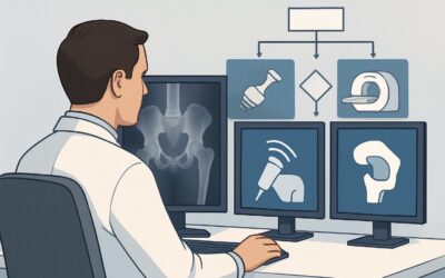
What Imaging Is Best for a Stable Patient with a Ballistic Abdominal Injury?
A 28-year-old male arrives in the trauma bay after a single gunshot wound to the right lower quadrant. He is awake, alert, and his blood pressure is 120/80 mmHg with a heart rate of 95 bpm. The entry wound is clear, but there is no exit wound, and the projectile’s path is uncertain. As the primary survey is completed, the trauma surgeon and emergency physician must decide on the initial imaging strategy to evaluate for internal injury without delaying potential intervention. This article details the American College of Radiology (ACR) Appropriateness Criteria workflow for this specific scenario: a normotensive adult with ballistic penetrating trauma limited to the abdomen and pelvis. For this presentation, the ACR rates a Radiography trauma series as Usually Appropriate.
Who Fits This Clinical Scenario?
This guidance applies to a specific subset of trauma patients. The inclusion criteria are precise: an adult patient who has sustained ballistic penetrating trauma (e.g., a gunshot wound) where the suspected trajectory is confined to the abdomen and/or pelvis. Critically, the patient must be hemodynamically stable, typically defined as being normotensive and not exhibiting signs of shock. This stability is a key decision point, as it affords the clinical team time for diagnostic imaging rather than proceeding directly to emergency surgery.
This workflow should not be applied to patients who fall outside these parameters. Key exclusion criteria include:
- Hemodynamic Instability: A hypotensive patient with penetrating abdominal trauma is a surgical emergency. Imaging should not delay transfer to the operating room. This presentation is covered in a different ACR variant for hypotensive penetrating torso trauma.
- Unknown Trajectory: If the projectile’s path is unknown or could involve the chest, the imaging workup must be expanded to include the thorax.
- Nonballistic Trauma: Stab wounds or impalement injuries have different injury patterns and may be managed differently, often with local wound exploration or focused imaging.
Correctly identifying the patient as normotensive and the injury as a ballistic wound confined to the abdominopelvic cavity is essential for applying this imaging pathway.
What Diagnoses Are You Working Up in This Scenario?
In a stable patient with a ballistic abdominal injury, imaging aims to identify or exclude several life-threatening conditions to determine whether the patient can be managed non-operatively or requires surgical or endovascular intervention. The differential diagnosis is broad and drives the choice of imaging modality.
Vascular Injury and Active Hemorrhage: This is a primary concern. High-velocity projectiles can cause direct laceration or secondary blast injury to major vessels like the aorta, inferior vena cava, iliac arteries, or mesenteric vessels. Identifying active contrast extravasation is a critical finding that signals ongoing bleeding and necessitates urgent intervention.
Solid Organ Injury: The liver, spleen, and kidneys are highly vascular organs and are commonly injured. Imaging is used to grade the severity of lacerations, identify hematomas (subcapsular or intraparenchymal), and detect active bleeding. The grade of injury helps guide non-operative versus operative management.
Hollow Viscus Perforation: The stomach, small bowel, and colon can be perforated by a projectile. A missed bowel injury can lead to peritonitis and sepsis. The key imaging finding is often extraluminal (“free”) air within the peritoneal cavity, though direct signs of bowel wall discontinuity or mesenteric stranding are also important.
Retroperitoneal Injury: Injuries to the retroperitoneum, including the pancreas, duodenum, ascending/descending colon, and major vessels, can be clinically occult. A retroperitoneal hematoma on imaging is a significant finding that requires careful evaluation of the underlying structures.
Why Is a Radiography Trauma Series Considered? The Role of CT
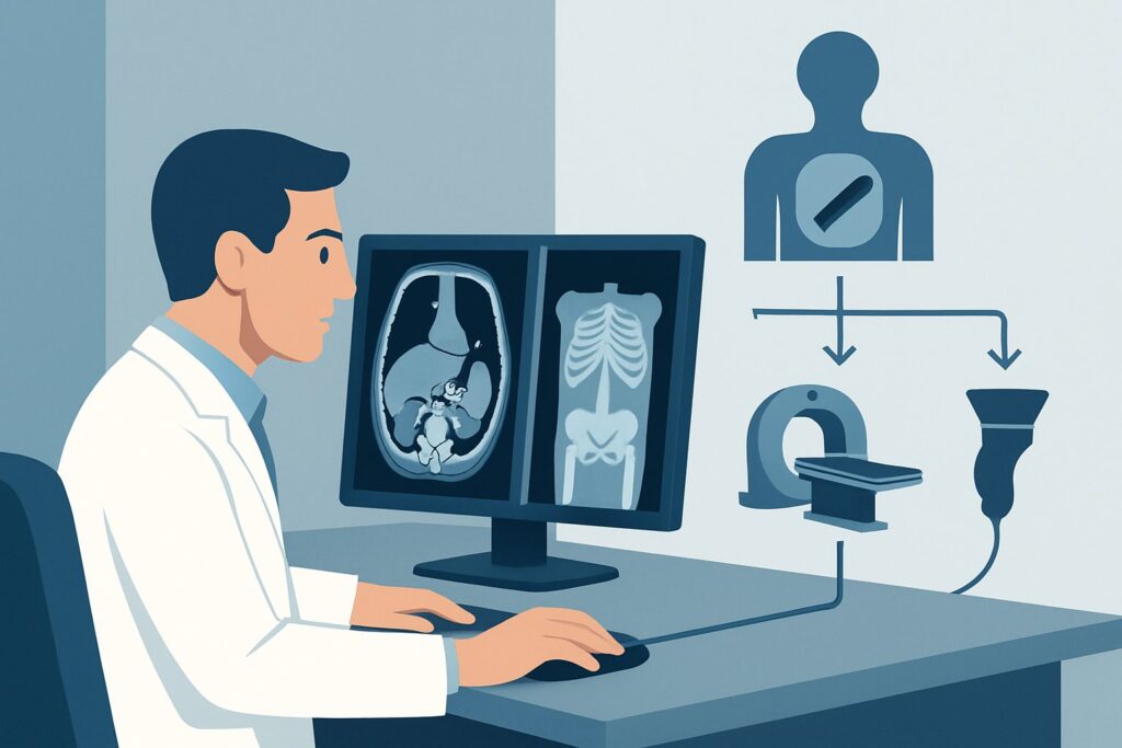
The ACR designates a Radiography trauma series as Usually Appropriate for this scenario. This typically includes anteroposterior (AP) views of the chest and abdomen, and a pelvic radiograph. The primary utility of radiography is to quickly identify the number and location of retained projectiles and significant bony fractures. Knowing the bullet’s final location can help infer its trajectory, which is crucial for surgical planning or for determining if a transperitoneal path was likely. Radiographs are fast, can be performed with a portable machine in the trauma bay, and involve a moderate radiation dose (☢☢☢ 1-10 mSv).
However, in modern trauma care, Computed Tomography (CT) of the abdomen and pelvis with intravenous (IV) contrast has become the mainstay for evaluating stable patients with penetrating abdominal trauma. The ACR also rates this study as Usually Appropriate (☢☢☢ 1-10 mSv). CT provides a comprehensive assessment that radiographs cannot, directly visualizing solid organ injury, active vascular extravasation, free fluid, free air from a bowel perforation, and retroperitoneal hematomas. For suspected arterial injury, Computed Tomography Angiography (CTA) of the abdomen and pelvis is also Usually Appropriate and provides superior detail of the vasculature, albeit with a higher radiation dose (☢☢☢☢ 10-30 mSv).
The combination of these ratings reflects a common real-world workflow: initial radiographs in the trauma bay to localize fragments, followed by a definitive CT scan to guide management. The CT is the primary tool for deciding between operative and non-operative pathways.
Alternative studies are rated lower for specific reasons:
- CT abdomen and pelvis without IV contrast is Usually Not Appropriate. Without IV contrast, solid organ injuries are poorly characterized, and active hemorrhage cannot be detected, defeating the primary purpose of the scan.
- MRI of the abdomen and pelvis is Usually Not Appropriate. It is too slow for the acute trauma setting, is susceptible to motion artifact, and poses a significant safety risk due to the potential for movement of metallic ballistic fragments within the patient.
What’s Next After Imaging? Downstream Workflow
The imaging results directly dictate the subsequent management steps in a highly structured decision tree. The findings on CT are the primary driver for determining the need for intervention.
Positive for Significant Injury:
- If the CT scan demonstrates active arterial contrast extravasation, the patient may be taken for angiography and embolization by Interventional Radiology or directly to the operating room (OR), depending on the location of the injury and institutional protocols.
- Findings of hollow viscus perforation (e.g., free intraperitoneal air) or a projectile trajectory that clearly crosses the peritoneal cavity are typically indications for an exploratory laparotomy in the OR.
- A high-grade solid organ injury (e.g., shattered spleen or liver laceration with hemodynamic instability) will also prompt surgical intervention.
Negative or Non-Operative Findings:
- If the CT scan is negative for any significant intrabdominal injury, or if it shows a low-grade, non-bleeding solid organ injury, the patient can often be managed non-operatively. This involves admission to the hospital for close observation, serial abdominal examinations, and monitoring of vital signs and hemoglobin/hematocrit levels.
- If the projectile’s trajectory is clearly confined to the abdominal wall musculature or subcutaneous tissues without violating the peritoneum, the patient may even be considered for discharge after a period of observation.
Pitfalls to Avoid (and When to Get Help)
Several potential pitfalls exist in the imaging workup of these patients. First, do not mistake transient or initial hemodynamic stability for the absence of a severe injury; patients can decompensate quickly, and a low threshold for repeat assessment is critical. Second, significant beam-hardening artifact from metallic fragments on CT can obscure adjacent structures. The radiologist and clinical team must be aware of this limitation and scrutinize the areas around the projectile. Third, injuries at the thoracoabdominal junction can be missed. A low entry wound in the chest or a high one in the abdomen requires careful evaluation of the diaphragm on CT. Finally, while triple-contrast CT (with oral and rectal contrast) was once common, its routine use has declined due to time delays. However, in equivocal cases for colon injury, a delayed scan with rectal contrast may still be considered.
If a patient’s clinical status changes during the imaging workup (e.g., they become hypotensive), the workup should be aborted, and the patient should be immediately reassessed for urgent transfer to the OR.
Related ACR Topics and Tools
The ACR Appropriateness Criteria are a comprehensive resource for evidence-based imaging decisions. This article covers one specific scenario, but many related variants exist. For a broader overview of imaging choices across all presentations of penetrating trauma, from hypotensive patients to those with nonballistic injuries, please consult our comprehensive parent guide.
- For breadth across all scenarios in Penetrating Torso Trauma, see our parent guide: Penetrating Torso Trauma: ACR Appropriateness Decoded.
- To look up other clinical scenarios, use the ACR Appropriateness Criteria Lookup tool.
- For details on imaging techniques, explore the Imaging Protocol Library.
- To discuss radiation exposure with patients, the Radiation Dose Calculator can help frame the conversation.
Frequently Asked Questions
Why is CT also rated ‘Usually Appropriate’ if a radiography series is the first study mentioned?
The ACR ratings reflect that both have a key role. A radiography series is a rapid, initial step often performed in the trauma bay to count and locate projectiles. However, CT with IV contrast is the definitive diagnostic study in stable patients. It provides the detailed anatomical information about organ, vascular, and bowel injury needed to decide between surgery and non-operative management. In most trauma centers, a stable patient will receive both.
What if the patient becomes hypotensive while waiting for or during the CT scan?
If a patient with penetrating abdominal trauma becomes hemodynamically unstable at any point, the diagnostic workup should be stopped. The patient should be immediately moved to the operating room for an exploratory laparotomy, as this is a life-threatening emergency that cannot wait for imaging completion. This represents a transition to the ‘Adult. Penetrating torso trauma, hypotensive’ clinical scenario.
Is oral or rectal contrast necessary for the CT scan in this scenario?
The routine use of oral or rectal contrast for initial trauma CT scans has significantly decreased. IV contrast is essential for evaluating solid organs and vascular structures. While oral/rectal contrast can help identify bowel injuries, it delays the scan and carries a risk of aspiration. Most trauma protocols now rely on secondary signs of bowel injury on IV-contrast-enhanced CT, such as free air, focal bowel wall thickening, or mesenteric stranding.
How does the workup change if the entry wound is in the flank or back?
An entry wound in the flank or back raises high suspicion for retroperitoneal injury. While the primary imaging study remains a CT of the abdomen and pelvis with IV contrast, a delayed-phase scan (excretory phase) is often added. This helps to evaluate for injury to the urinary collecting system (ureters, renal pelvis) by looking for contrast extravasation from the kidneys or ureters.
Why is MRI rated ‘Usually Not Appropriate’ for acute ballistic trauma?
MRI is unsuitable for this acute scenario for two main reasons. First, it is a relatively slow imaging modality, and time is critical in trauma care. Second, and more importantly, the powerful magnetic field of an MRI scanner can cause metallic ballistic fragments to move or heat up, potentially causing further injury to surrounding tissues. Therefore, it is contraindicated when retained metallic projectiles are present.
Reviewed by Pouyan Golshani, MD, Interventional Radiologist — May 30, 2026