Which Radiographs Should You Order First for Chronic Hip Pain? An ACR-Guided Workflow
A 58-year-old patient presents to your clinic with six months of progressive, deep right groin pain, worse with walking and standing. There is no history of acute trauma, fever, or constitutional symptoms. On exam, internal rotation of the hip is limited and painful. You suspect an intra-articular process, most likely osteoarthritis, but need to confirm the diagnosis and rule out other causes. What is the most appropriate initial imaging study to order? This is a foundational question in primary care and musculoskeletal medicine. For this specific scenario—the initial imaging workup of chronic hip pain—the American College of Radiology (ACR) rates Radiography pelvis as Usually Appropriate.
Who Fits This Clinical Scenario for Initial Hip Imaging?
This guidance applies to the common clinical presentation of an adult patient with chronic hip pain, which is typically defined as pain lasting three months or longer. The patient’s history does not suggest a specific acute event, such as a fall, nor are there red flag symptoms pointing to infection (e.g., fever, erythema) or malignancy (e.g., unexplained weight loss, history of cancer). This is the initial, undifferentiated workup.
This workflow is distinct from several related but different clinical situations:
- Acute Hip Trauma: A patient with hip pain immediately following a fall or significant injury falls under a different set of guidelines, where the primary concern is an acute fracture.
- Suspected Infection: If there is clinical suspicion for septic arthritis or osteomyelitis, the imaging pathway is more urgent and may involve different modalities.
- Negative Radiographs with Specific Suspicion: This guidance is for the initial imaging. If radiographs are negative or nondiagnostic but you still have a high clinical suspicion for a specific condition like a labral tear, femoroacetabular impingement, or an extra-articular process like trochanteric bursitis, you would then proceed to a different ACR variant for next-step imaging. This article focuses only on the first step.
What Diagnoses Are You Working Up With Initial Radiographs?
The initial radiograph serves as a crucial screening and diagnostic tool, aiming to identify the most common causes of chronic hip pain and exclude other significant pathologies. The differential diagnosis at this stage is broad, but radiographs are highly effective at evaluating for several key conditions.
Osteoarthritis (OA) is the most common cause of chronic hip pain, particularly in patients over 50. Radiographs are the gold standard for diagnosing moderate to severe OA, clearly demonstrating characteristic findings like joint space narrowing, osteophyte formation (bone spurs), subchondral sclerosis (increased bone density), and subchondral cysts.
Femoroacetabular Impingement (FAI) is a frequent cause of hip pain and early-onset OA in younger, active adults. It results from abnormal contact between the femoral head-neck junction and the acetabular rim. Radiographs, particularly with specific views like a frog-leg lateral, can reveal the underlying bony morphology, such as a “cam” lesion (abnormal femoral head shape) or a “pincer” lesion (acetabular overcoverage).
Developmental Dysplasia of the Hip (DDH) is a condition where the hip socket (acetabulum) is shallow, leading to instability and premature OA. While often diagnosed in infancy, milder forms can present as chronic hip pain in adolescents and adults. Radiographs are essential for measuring acetabular depth and coverage to diagnose DDH.
Avascular Necrosis (AVN), or osteonecrosis, involves the death of bone tissue due to a loss of blood supply. Risk factors include steroid use, alcohol abuse, and trauma. While early-stage AVN can be radiographically occult, later stages are visible on radiographs, showing signs like the “crescent sign” (a subchondral fracture line) or femoral head collapse.
Radiographs can also reveal less common but critical findings, such as stress fractures, signs of inflammatory arthritis (e.g., sacroiliitis), or concerning osseous lesions that might suggest a primary or metastatic neoplasm.
Why Are Pelvic Radiographs the Recommended Initial Study for Chronic Hip Pain?
The ACR Appropriateness Criteria designates both Radiography pelvis and Radiography hip as Usually Appropriate for the initial evaluation of chronic hip pain. These simple, widely available, and low-cost studies provide a wealth of information about the osseous structures and joint alignment, directly addressing the most common differential diagnoses.
An anteroposterior (AP) radiograph of the pelvis is often the preferred starting point over a single view of the symptomatic hip. A pelvic view allows for a direct comparison between the symptomatic and asymptomatic hips, which is invaluable for assessing subtle joint space loss or asymmetric morphology. It also provides a view of the sacroiliac joints and pelvic ring, which can identify referred pain sources or signs of systemic inflammatory conditions.
The diagnostic rationale is clear:
- High Yield: Radiographs effectively confirm or exclude common diagnoses like osteoarthritis, significant FAI, and dysplasia, guiding initial management.
- Low Radiation: The effective radiation dose for a pelvis radiograph is low (ACR RRL ☢☢, 0.1-1 mSv), representing a favorable risk-benefit profile for a first-line diagnostic test.
- Resource Stewardship: Starting with radiographs avoids the unnecessary cost and resource utilization of advanced imaging for conditions that can be diagnosed simply.
In contrast, more advanced imaging modalities are rated as Usually Not Appropriate for the initial workup:
- MRI hip without IV contrast: While highly sensitive for soft tissue pathology (labral tears, cartilage damage) and early AVN, MRI is not the appropriate first step. It is a problem-solving tool best used after radiographs have been obtained and a specific clinical question remains—for example, suspected labral tear in a patient with normal radiographs.
- US hip: Ultrasound is excellent for assessing for a joint effusion or evaluating superficial tendons and bursae (e.g., for greater trochanteric pain syndrome). However, it cannot visualize the articular cartilage, subchondral bone, or deep osseous morphology, making it unsuitable for the initial, comprehensive evaluation of intra-articular hip pain.
For optimal evaluation, a standard radiographic series should include an AP view of the pelvis and at least one lateral view of the symptomatic hip (e.g., frog-leg or cross-table lateral) to properly assess the three-dimensional anatomy of the femoral head and acetabulum.
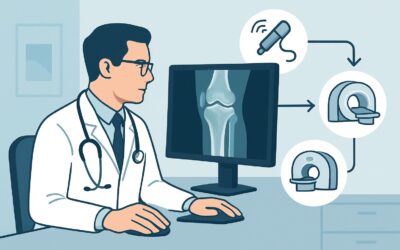
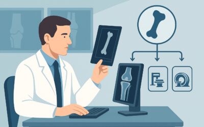
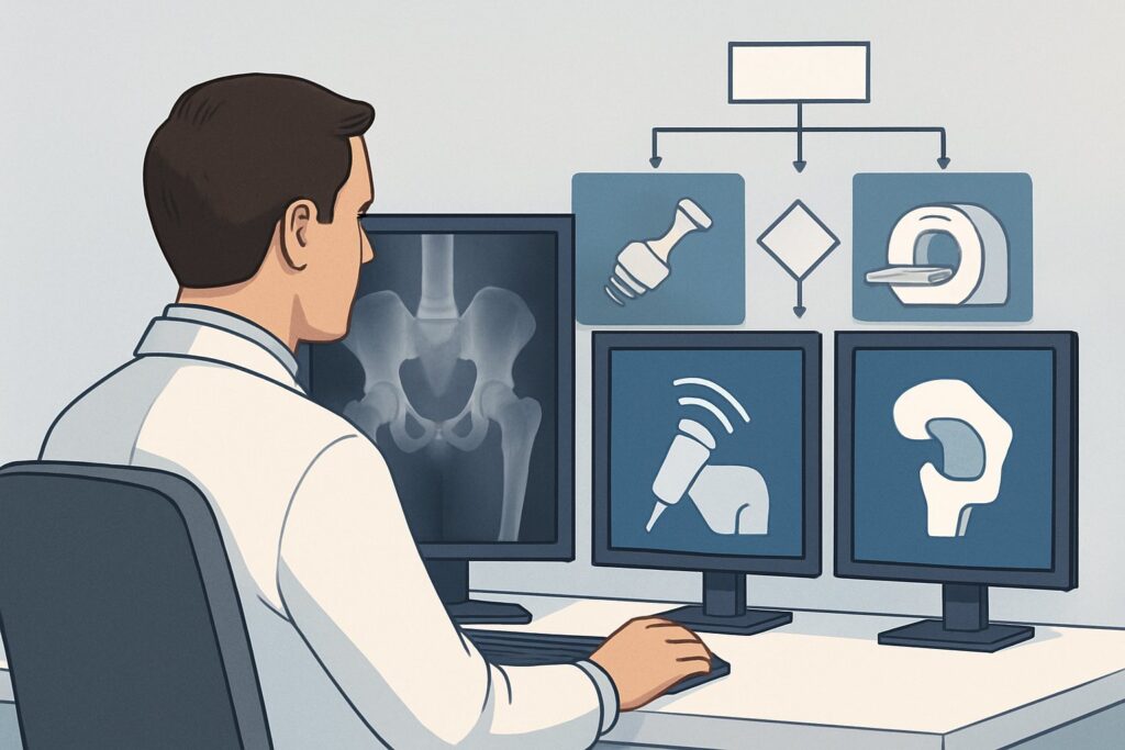
What’s Next After Radiographs? Downstream Workflow
The results of the initial radiographs will dictate the subsequent clinical pathway. The imaging findings create clear decision points for management or further investigation.
- Positive for Osteoarthritis: If radiographs show clear evidence of OA consistent with the patient’s symptoms, the diagnosis is established. The next steps are typically non-surgical management, including physical therapy, activity modification, weight management, and non-steroidal anti-inflammatory drugs (NSAIDs). Surgical consultation for arthroplasty is reserved for patients with severe symptoms refractory to conservative care.
- Negative or Nondiagnostic: If the radiographs are normal or show only equivocal findings, but the patient has persistent, disabling symptoms, further evaluation is warranted. The next step depends on the leading clinical suspicion.
- If you suspect a labral tear or FAI in a younger patient, the appropriate next study is often an MRI or MR arthrogram of the hip. This moves the patient into a different ACR scenario (“Suspect labral tear. Radiographs negative or nondiagnostic”).
- If you suspect an extra-articular cause like gluteal tendinopathy or trochanteric bursitis, a diagnostic ultrasound or MRI may be considered.
- If you suspect early-stage AVN, an MRI is the most sensitive test to order next.
- Indeterminate or Unexpected Findings: If radiographs reveal an unexpected finding, such as a potential stress fracture or a suspicious bone lesion, the next step is typically advanced imaging (CT or MRI) to better characterize the abnormality. Consultation with an orthopedic surgeon or musculoskeletal radiologist is often appropriate in these cases.
Pitfalls to Avoid (and When to Get Help)
When ordering and interpreting initial hip radiographs, several common pitfalls can lead to diagnostic delays or errors.
- Inadequate Views: Ordering only an AP pelvis view without a true lateral of the hip can cause you to miss FAI morphology (cam lesions) or subtle fractures of the femoral neck.
- Satisfaction of Search: Focusing on obvious OA in an older patient might lead to overlooking other findings, such as sacroiliitis or a subtle lytic lesion elsewhere in the pelvis.
- Misattributing Pain: Remember that radiographic findings do not always correlate with symptoms. Mild OA is a common incidental finding; ensure the clinical picture aligns before attributing all pain to it.
- Ignoring the Sacroiliac Joints: Pain in the buttock and posterior hip can originate from the sacroiliac (SI) joints. Always inspect the SI joints on a pelvic radiograph for signs of sacroiliitis.
If you encounter red flag symptoms (e.g., nocturnal pain, rapid progression, constitutional symptoms) or see a destructive or aggressive-appearing lesion on radiographs, escalate care promptly with an urgent referral to orthopedics or oncology.
Related ACR Topics and Tools
This article covers one specific scenario within the broader topic of Chronic Hip Pain. For a comprehensive overview of all related clinical variants and their recommended imaging pathways, please consult our parent guide. Additional tools can help you apply these guidelines in your practice.
- For breadth across all scenarios in Chronic Hip Pain, see our parent guide: Chronic Hip Pain: ACR Appropriateness Decoded.
- Explore other clinical presentations using the ACR Appropriateness Criteria Lookup.
- Review detailed imaging techniques in the Imaging Protocol Library.
- Discuss radiation exposure with patients using the Radiation Dose Calculator.
Frequently Asked Questions
Why order a pelvis radiograph instead of just a single hip radiograph?
An AP pelvis radiograph is often preferred because it allows for a direct side-by-side comparison with the asymptomatic hip, which is crucial for detecting subtle joint space narrowing or morphologic differences. It also provides a view of the sacroiliac joints and pelvic bones, which can be sources of referred pain or reveal other pathologies.
If I strongly suspect a labral tear, can I skip radiographs and go straight to an MRI?
No, the ACR recommends against this. Radiographs are the essential first step because they can identify underlying osseous abnormalities like femoroacetabular impingement (FAI) or dysplasia, which are often the root cause of labral tears. An MRI or MR arthrogram is considered the next appropriate step only after radiographs are found to be negative or non-diagnostic.
What specific radiographic views should I order for a chronic hip pain workup?
A standard initial series should include an anteroposterior (AP) view of the entire pelvis and a lateral view of the symptomatic hip. A frog-leg lateral or a cross-table lateral view is particularly important for assessing the femoral head-neck junction to evaluate for potential FAI.
Are radiographs useful if I suspect avascular necrosis (AVN)?
Yes, radiographs are the correct initial test. While they can be normal in the earliest stages of AVN, they are effective at diagnosing later-stage disease by showing findings like sclerosis, the ‘crescent sign,’ or femoral head collapse. If radiographs are negative but clinical suspicion for AVN remains high (e.g., in a patient on chronic steroids), an MRI is the appropriate next step as it is much more sensitive for early-stage disease.
Is a CT scan ever the right initial test for chronic hip pain?
No, for the initial, undifferentiated workup of chronic hip pain, a CT scan is rated as *Usually Not Appropriate* by the ACR. CT involves significantly more radiation than radiographs and is generally reserved for specific indications, such as characterizing a complex fracture seen on a radiograph or evaluating a bone lesion in greater detail.
Reviewed by Pouyan Golshani, MD, Interventional Radiologist — May 21, 2026