What Is the Right First Imaging Study for a Suspected Primary Bone Tumor?
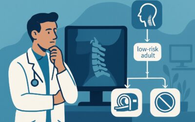
A 16-year-old high school athlete presents to your clinic with several weeks of persistent, localized pain in his distal femur that wakes him from sleep. It’s not improving with rest or NSAIDs, and on exam, you note subtle swelling and point tenderness. You are concerned about a primary bone tumor, but the differential is broad, ranging from benign cysts to aggressive malignancies like osteosarcoma. The immediate clinical question is not *if* you should order imaging, but *what* imaging to order first. This decision point is critical, as the initial study frames the entire subsequent diagnostic and therapeutic pathway. For this specific scenario—the initial imaging workup of a suspected primary bone tumor in an adult or child—the American College of Radiology (ACR) designates **Radiography of the area of interest** as **Usually Appropriate**.
Who Fits This Clinical Scenario?
This guidance applies to any patient, adult or child, with a new clinical suspicion of a primary bone tumor who has not yet had any imaging. The clinical presentation is often localized, persistent bone pain (especially pain at night), a new palpable mass, or unexplained swelling. It can also apply to the evaluation of a pathologic fracture, where a fracture occurs through weakened bone with minimal or no trauma.
It is crucial to distinguish this initial workup scenario from others that require a different imaging approach. This article does NOT apply if:
- The patient already has radiographs. If initial radiographs have been performed, the next step depends on their findings. The workflow then shifts to a different clinical scenario, such as evaluating a lesion with an indeterminate or aggressive appearance or one that appears clearly benign.
- A metastatic lesion is suspected. If the patient has a known history of a primary malignancy elsewhere (e.g., breast, lung, prostate cancer), the workup is for suspected metastatic disease, not a primary bone tumor, which follows a different diagnostic algorithm.
- Infection is the primary concern. If symptoms like fever, erythema, and elevated inflammatory markers strongly suggest osteomyelitis, the imaging pathway may differ, often involving Magnetic Resonance Imaging (MRI) earlier in the process.
What Diagnoses Are You Working Up in This Scenario?
The differential diagnosis for a suspected primary bone tumor is vast and varies significantly with patient age. The purpose of initial imaging is to narrow this differential and, most importantly, to triage lesions into three broad categories: clearly benign, indeterminate, or aggressive/likely malignant.
In children and adolescents, common benign lesions include non-ossifying fibroma, fibrous dysplasia, simple bone cysts, and osteochondromas. However, this is also the peak age for primary malignant bone tumors like osteosarcoma and Ewing sarcoma. Distinguishing between a benign process and an early malignancy is the central challenge.
In adults, the differential for benign lesions includes enchondromas and giant cell tumors of bone. Malignant possibilities shift toward chondrosarcoma and other sarcomas. It is also critical to consider multiple myeloma (a plasma cell malignancy) in adults over 40 with bone pain or a pathologic fracture, as it is the most common primary malignancy of bone.
Tumor-like conditions, such as aneurysmal bone cysts or Langerhans cell histiocytosis, can mimic true neoplasms and are also part of the differential. The initial radiograph provides the foundational anatomic and morphologic information needed to begin differentiating these possibilities.
Why Is Radiography the Recommended First Step for a Suspected Bone Tumor?
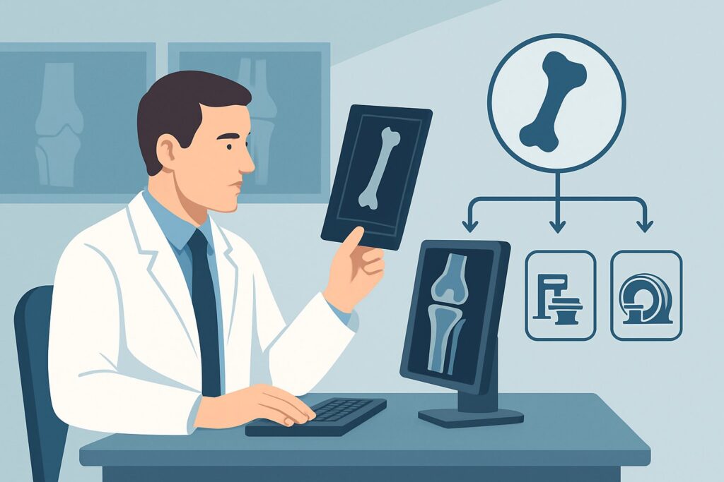
For the initial evaluation of a suspected primary bone tumor, radiography of the area of interest is rated as Usually Appropriate by the ACR. This recommendation is rooted in the exceptional value that plain films provide as a first-line diagnostic tool. Radiographs are inexpensive, rapidly accessible, and deliver a low radiation dose while offering a wealth of diagnostic information that is essential for lesion characterization.
A high-quality, two-view radiograph allows the clinician and radiologist to assess several critical features, known as the “radiographic predictors of aggressiveness”:
- Location: Where is the lesion within the bone (epiphysis, metaphysis, diaphysis) and which bone is involved?
- Margins: Is the border of the lesion sharp and well-defined (suggesting slow growth) or wide and permeative (suggesting an aggressive process)?
- Matrix: Does the lesion produce a mineralized substance, such as the cloud-like osteoid of osteosarcoma or the “rings and arcs” of a chondroid matrix in chondrosarcoma?
- Periosteal Reaction: How is the outer layer of the bone reacting? A smooth, solid reaction suggests a benign process, whereas a spiculated “sunburst” or layered “onion-skin” pattern indicates aggression.
Conversely, more advanced imaging modalities are rated as Usually Not Appropriate for the *initial* workup. Ordering MRI or Computed Tomography (CT) first is a common pitfall. Without the foundational context of a radiograph, findings on these advanced studies can be misleading. For example, the extensive edema seen on an MRI around a benign process could be misinterpreted as a sign of malignancy. CT (RRL: Varies) and MRI (RRL: O 0 mSv) are crucial for problem-solving and staging, but they are second-line studies, to be performed after a lesion has been identified and characterized on plain films.
Similarly, nuclear medicine studies like a whole-body bone scan (RRL: ☢☢☢ 1-10 mSv) or FDG-PET/CT (RRL: ☢☢☢☢ 10-30 mSv) are also Usually Not Appropriate as the first step. These are primarily used for staging—to look for multiple lesions or metastases—once a primary malignant tumor has been diagnosed or is highly suspected based on radiographs.
What’s Next After Radiography? Downstream Workflow
The results of the initial radiograph dictate the entire subsequent clinical pathway. The findings will route the patient to one of several distinct downstream workflows, often corresponding to other scenarios within the ACR Appropriateness Criteria.
- If the radiograph is negative: If the plain films are entirely normal but there is a high clinical suspicion for a tumor (e.g., persistent, severe night pain), the workup moves to the “Suspected primary bone tumor. No lesions on radiographs” scenario. In this case, MRI is often the appropriate next step to evaluate for a radiographically occult lesion or soft tissue pathology.
- If the radiograph shows a clearly benign lesion: If the findings are characteristic of a benign entity (like a non-ossifying fibroma), the workup proceeds down the “Lesion on radiographs. Benign appearance” pathway. This often involves clinical follow-up or no further imaging, providing reassurance to the patient and avoiding unnecessary procedures.
- If the radiograph shows an indeterminate or aggressive lesion: This is the most critical outcome. Any lesion with aggressive features (e.g., permeative margins, aggressive periosteal reaction) requires urgent further evaluation. The workflow shifts to the “Lesion on radiographs. Indeterminate or aggressive appearance” scenario. The next steps typically involve cross-sectional imaging (MRI for local staging, CT for matrix characterization) and prompt referral to an orthopedic oncologist for biopsy and definitive management.
This branching logic underscores why radiography is the indispensable first step. It is the primary triage tool that directs all future care, preventing both the under-investigation of a potential malignancy and the over-investigation of a benign finding.
Pitfalls to Avoid (and When to Get Help)
Navigating the initial workup of a suspected bone tumor requires careful attention to avoid common errors that can delay diagnosis or lead to inappropriate management.
A primary pitfall is ordering an MRI or CT scan as the initial study. This “skip” of radiography can lead to diagnostic confusion and may even compromise subsequent biopsy planning. Another common error is obtaining inadequate radiographs; always order at least two orthogonal views (e.g., anteroposterior and lateral) that include the entire lesion with a margin of normal bone. For pediatric patients, be wary of dismissing persistent, localized, or nocturnal bone pain as “growing pains,” as these can be red-flag symptoms for a malignancy.
If the initial radiograph reveals any features suggestive of an aggressive lesion—such as a permeative or moth-eaten appearance, a wide zone of transition, or an aggressive periosteal reaction—this is a critical finding that requires immediate escalation. The patient should be urgently referred to a musculoskeletal tumor specialist or a multidisciplinary tumor board before any further intervention, including biopsy, is considered.
Related ACR Topics and Tools
For a comprehensive overview of all clinical variants related to this topic, please consult our parent guide. Additional GigHz tools can help you apply these guidelines in your practice, from exploring adjacent scenarios to understanding imaging techniques and radiation dose.
- For breadth across all scenarios in Suspected Primary Bone Tumors, see our parent guide: Suspected Primary Bone Tumors: ACR Appropriateness Decoded.
- ACR Appropriateness Criteria Lookup — for adjacent scenarios
- Imaging Protocol Library — for technique on the recommended study
- Radiation Dose Calculator — for cumulative dose conversations
Frequently Asked Questions
Why not just order an MRI first, since it provides more detail than a radiograph?
While MRI provides excellent soft tissue contrast and detail, it is considered ‘Usually Not Appropriate’ as the first imaging study. Radiographs are superior for assessing key features like matrix mineralization and periosteal reaction, which are fundamental to the differential diagnosis. Interpreting an MRI without a corresponding radiograph can be misleading, as benign conditions can sometimes appear aggressive on MRI due to edema. The radiograph provides the essential framework for interpreting subsequent advanced imaging.
What if the patient has pain in multiple bones? Is a single radiograph still the first step?
If a patient presents with multifocal bone pain, the clinical suspicion may shift from a single primary bone tumor to a systemic process, such as metastatic disease, multiple myeloma, or a metabolic bone disorder. While radiographs of the most symptomatic area are still a reasonable starting point, a whole-body bone scan or PET/CT might be considered earlier in the workup depending on the full clinical picture. This presentation may fit a different clinical scenario than a single suspected primary tumor.
Does this guidance apply if I suspect a metastasis instead of a primary tumor?
No, this guidance is specifically for a suspected *primary* bone tumor. If a patient has a known history of a primary cancer (e.g., lung, breast, prostate) and develops new bone pain, the workup is for suspected metastatic disease. The imaging algorithm for that scenario is different and may involve modalities like bone scintigraphy or PET/CT earlier in the process to assess the full extent of disease.
Are two views, such as an AP and lateral, always necessary for the initial radiograph?
Yes, obtaining at least two orthogonal views is critical. A lesion’s appearance, or even its visibility, can change dramatically between different projections. A single view can miss subtle fractures, periosteal reactions, or cortical destruction. Two views provide a three-dimensional understanding of the lesion’s location and morphology, which is essential for accurate initial characterization.
The patient is a child, and I’m concerned about radiation. Is radiography still the best first choice?
Yes. Even with the heightened sensitivity to radiation in pediatric patients, a conventional radiograph remains the ‘Usually Appropriate’ first step. The radiation dose from a two-view radiograph of a limb is very low (RRL: Varies, but typically <1 mSv) and the diagnostic information it provides is indispensable. Starting with a higher-dose study like CT would be inappropriate, and MRI, while radiation-free, is not the correct initial test for the reasons mentioned above. The benefit of the radiograph far outweighs the minimal radiation risk.
Reviewed by Pouyan Golshani, MD, Interventional Radiologist — May 21, 2026