Should You Image a Low-Risk Adult After Acute Cervical Spine Trauma?
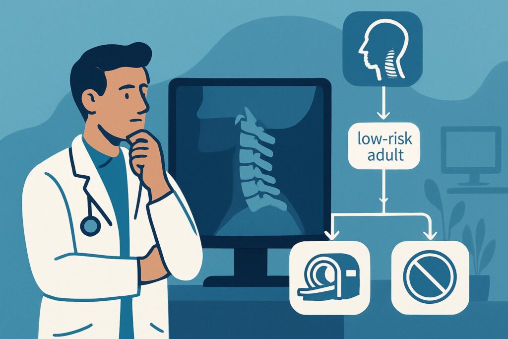
A 32-year-old driver is brought to the emergency department after a minor rear-end collision. He is alert, ambulatory, and complaining of mild neck stiffness but no significant pain. He denies any neurologic symptoms like numbness or weakness. On examination, he has no midline cervical tenderness, no focal neurologic deficits, a normal level of alertness, no evidence of intoxication, and no other painful, distracting injuries. The clinical question is immediate: does this patient need imaging to clear his cervical spine? This article provides a detailed workflow for this specific scenario—an adult patient between 16 and 65 years old with low-risk, blunt cervical spine trauma where clinical decision rules suggest imaging is not required. Based on the American College of Radiology (ACR) Appropriateness Criteria, for this presentation, all forms of imaging, including **Radiography cervical spine**, are rated **Usually not appropriate**.
Who Fits This Clinical Scenario?
This guidance applies to a very specific and common patient population: adults aged 16 to 65 who have experienced blunt trauma to the neck but are considered low-risk for a clinically significant cervical spine injury. The cornerstone of this determination is the validated application of clinical decision rules like the National Emergency X-Radiography Utilization Study (NEXUS) criteria or the Canadian C-Spine Rule (CCR).
To fit this scenario, the patient must meet **all** low-risk criteria from one of these rules. For NEXUS, this means the patient has:
* No posterior midline cervical spine tenderness
* No evidence of intoxication
* A normal level of alertness
* No focal neurologic deficit
* No painful distracting injury
If a patient meets these criteria, they are considered clinically cleared, and this workflow applies.
This guidance does **not** apply to patients who fail any of these criteria. For instance:
* **A patient with a focal neurologic deficit (e.g., new arm weakness):** This patient has a high-risk feature and immediately moves to a different imaging pathway, typically starting with CT.
* **An obtunded or intoxicated patient:** Their clinical examination is unreliable, and they cannot be cleared clinically. They require imaging.
* **A patient with a painful femur fracture:** This is a classic “distracting injury” that could mask neck pain from a fracture, making the clinical exam unreliable.
Properly identifying which patients belong in this low-risk category is the critical first step to avoiding unnecessary radiation and downstream testing.
What Diagnoses Are You Working Up in This Scenario?
In this clinical setting, the primary goal is not to work up a broad differential with imaging, but rather to use a validated clinical examination to confidently **exclude** life-threatening injuries. The power of the NEXUS and CCR criteria lies in their extremely high sensitivity and negative predictive value for unstable cervical spine fractures. When a patient meets the low-risk criteria, the pre-test probability of a significant injury is already exceedingly low.
**Cervical Spine Fracture:** This is the most feared diagnosis. An unstable fracture can lead to spinal cord injury, paralysis, or death. The clinical decision rules were specifically designed and validated to identify patients in whom the risk of such a fracture is so low that imaging provides no meaningful benefit. By confirming the absence of midline tenderness, neurologic deficits, and other risk factors, you are clinically ruling out this diagnosis.
**Significant Ligamentous Injury:** While plain radiographs are insensitive to ligamentous injury, and MRI is the definitive test, the clinical decision rules are also effective at screening for instability caused by these injuries. Patients with severe ligamentous disruption typically present with significant pain, tenderness, or neurologic findings, which would disqualify them from this low-risk pathway.
**Cervical Muscle Strain or Spasm:** This is the most common diagnosis in low-risk patients with neck pain or stiffness after trauma. It is a clinical diagnosis of exclusion. Once significant bony or ligamentous injury has been ruled out by the clinical exam, this benign, self-limiting condition can be diagnosed and managed with supportive care (e.g., NSAIDs, physical therapy) without the need for imaging.
Why Is Imaging Considered ‘Usually Not Appropriate’ for This Presentation?
For a patient who meets the low-risk criteria of NEXUS or the CCR, the ACR Appropriateness Criteria rate every single imaging modality—from plain X-rays to CT and MRI—as **Usually not appropriate**. This strong recommendation is based on high-quality evidence demonstrating that the risks and costs of imaging outweigh the negligible potential for benefit in this specific population.
The rationale is rooted in the proven performance of the clinical decision rules. Both NEXUS and the CCR have a negative predictive value approaching 100% for clinically significant cervical spine injuries. When a patient is successfully “cleared” by these rules, the probability of missing a serious injury is exceptionally small. Proceeding with imaging in this context offers little to no diagnostic value and introduces unnecessary downsides.
Let’s examine why specific modalities are not recommended:
* **Radiography cervical spine:** While it involves a relatively low radiation dose (ACR Relative Radiation Level ☢☢), it is still unnecessary exposure. Furthermore, radiographs have limitations, including poor visualization of the craniocervical junction (C1-C2) and the cervicothoracic junction (C7-T1). A technically inadequate or negative radiograph in a symptomatic patient can lead to uncertainty and pressure for further, higher-dose imaging like CT, creating an imaging cascade.
* **CT cervical spine without IV contrast:** This is the gold standard for detecting bony fractures, but it carries a significantly higher radiation dose (ACR RRL ☢☢☢) than radiography. Ordering a CT on a clinically-cleared patient exposes them to this radiation without a clear indication, increasing lifetime cancer risk for no diagnostic gain.
* **MRI cervical spine without IV contrast:** MRI is excellent for evaluating soft tissues, ligaments, and the spinal cord, and it involves no ionizing radiation (ACR RRL O). However, it is a costly, time-consuming study with a high rate of incidental findings (e.g., disc bulges, degenerative changes) that are often clinically irrelevant in the acute trauma setting but may trigger anxiety and further unnecessary interventions.
In summary, for the low-risk, clinically-cleared patient, the diagnostic yield of any imaging is too low to justify the associated radiation, cost, time, and risk of incidentalomas.
What’s Next After Deciding Not to Image? Downstream Workflow
The workflow for a patient who is clinically cleared of cervical spine injury is focused on patient education, appropriate discharge, and clear return precautions. The decision not to image is an active, evidence-based clinical choice, not a passive omission.
* **If the decision is made not to image:** The patient can have their cervical collar removed. The primary diagnosis is typically cervical strain. Management includes reassurance, symptomatic treatment with analgesics (e.g., NSAIDs), and advice on gentle range-of-motion exercises as tolerated. Crucially, the discharge instructions must be explicit about reasons to return to the emergency department. These include the development of new or worsening neck pain, any new neurologic symptoms (weakness, numbness, tingling in the extremities), or difficulty with balance or walking.
* **If the clinical exam changes:** A patient’s status can evolve. If a patient who was initially non-tender develops clear, focal midline tenderness hours later, they no longer fit this low-risk scenario. At that point, the patient should be re-evaluated, potentially placed back in a cervical collar, and considered for imaging according to the appropriate ACR variant for a patient with high-risk features.
* **If imaging is performed despite meeting clearance criteria:** If for some reason (e.g., institutional policy, extreme patient anxiety) imaging is still obtained and is negative, it serves to confirm the clinical assessment. The patient can be managed as above. If the imaging reveals an incidental finding, such as mild degenerative disc disease, it is vital to explain to the patient that this is a chronic finding and not the cause of their acute symptoms, preventing further unnecessary workup.
Pitfalls to Avoid (and When to Get Help)
Successfully applying this “no imaging” pathway requires careful and rigorous clinical assessment. Here are common pitfalls to avoid:
* **Incomplete Application of Decision Rules:** Do not “cherry-pick” criteria. If a patient has a distracting injury or is even mildly intoxicated, their exam is unreliable, and they cannot be cleared clinically. All criteria must be met.
* **Ignoring a Change in Exam:** A patient’s exam is not static. Re-assess patients who are being observed, especially if their pain or symptoms change over time.
* **Failure to Document:** Meticulously document every component of the NEXUS or CCR assessment in the medical record. This documentation justifies the decision not to image and is critical for medical-legal purposes.
* **Underestimating “Distracting Injury”:** A truly distracting injury is one severe enough to potentially mask a cervical spine injury. This is a clinical judgment, but err on the side of caution with severe injuries like long-bone fractures, visceral injuries, or extensive burns.
If there is any doubt about the reliability of the clinical exam or the proper application of the decision rules, escalate by obtaining imaging (typically a non-contrast CT of the cervical spine) or consulting a trauma or spine specialist.
Related ACR Topics and Tools
This article covers one specific, low-risk scenario. For a comprehensive overview of imaging recommendations across all presentations of spinal trauma, or for tools to help with study selection and patient communication, please refer to the following resources.
* For breadth across all scenarios in Acute Spinal Trauma, see our parent guide: Acute Spinal Trauma: ACR Appropriateness Decoded.
* ACR Appropriateness Criteria Lookup — for adjacent scenarios
* Imaging Protocol Library — for technique on the recommended study
* Radiation Dose Calculator — for cumulative dose conversations
Frequently Asked Questions
What exactly qualifies as a ‘painful distracting injury’ in the NEXUS criteria?
A painful distracting injury is any other injury severe enough to potentially distract the patient from a second injury in their neck. There is no absolute list, but common examples include long bone fractures, visceral organ injury, extensive burns, or large lacerations. A minor ankle sprain or a small cut on the hand would typically not be considered distracting. The determination requires clinical judgment regarding the severity of the second injury and its potential to mask cervical spine pain.
Do the NEXUS or Canadian C-Spine Rules apply to patients with a history of prior cervical spine surgery?
The original validation studies for these clinical decision rules largely excluded patients with known spinal disease, including prior surgery. While some subsequent research suggests they may still be safe, caution is warranted. Many clinicians choose to have a lower threshold for imaging in patients with prior fusions or instrumentation, as their biomechanics are altered. This decision should be made on a case-by-case basis.
If a patient meets low-risk criteria but has persistent neck stiffness for several days, is imaging then indicated?
If the patient continues to meet all low-risk criteria (no midline tenderness, no neurologic deficits), imaging is still generally not indicated for persistent stiffness, which is likely muscular. However, if the clinical picture changes—for example, if they develop new midline tenderness or radicular symptoms—they should be re-evaluated, and imaging may become appropriate. For persistent mechanical pain without red flags, physical therapy is often the best next step.
Is there any role for flexion-extension radiographs in this acute, low-risk setting?
No. Flexion-extension views are used to assess for ligamentous instability, but they are not recommended in the acute trauma setting. Performing these maneuvers on a potentially unstable spine can be dangerous. Furthermore, muscle spasm in the acute phase can limit motion, leading to a non-diagnostic study. These views are sometimes considered in the subacute or chronic setting for patients with persistent symptoms after an initial negative advanced imaging study, but they have no role in the initial evaluation of a low-risk patient.
If a patient was wearing a helmet during the trauma (e.g., a bicycle accident), does that change the application of these rules?
The presence of a helmet does not change the application of the NEXUS or CCR criteria. The clinical assessment is performed after the helmet has been safely removed. The decision to image or not is based entirely on the patient’s clinical findings (tenderness, neurologic status, etc.), regardless of what protective gear they were wearing.
Reviewed by Pouyan Golshani, MD, Interventional Radiologist — May 21, 2026