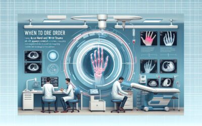
Should You Order Radiographs for a Minor Knee Injury if the Patient Can Walk?
A 34-year-old patient presents to your clinic on a Tuesday afternoon after twisting their knee during a weekend soccer game. They report a mild, diffuse ache but were able to walk off the field and have been ambulating since, albeit with some caution. Your physical exam is reassuring: there is no visible effusion, no focal bony tenderness over the patella, fibular head, or condyles, and the patient can flex the knee to 90 degrees and bear their full weight for several steps. The clinical question is whether initial imaging is warranted. For this specific presentation—an acute knee trauma with a low-suspicion clinical exam—the American College of Radiology (ACR) rates knee radiography as *May be appropriate*, reflecting the nuanced decision-making required when the pre-test probability of a significant injury is low.
Who Fits This Clinical Scenario?
This guidance applies to a specific and common patient presentation: an adult or a child aged 5 years or older who has experienced a fall or an acute twisting injury to the knee. The defining clinical features that place a patient in this category are the absence of significant red flags on physical examination.
**Inclusion criteria for this workflow:**
* **Mechanism:** An acute traumatic event, such as a fall or a twisting motion.
* **Age:** 5 years or older.
* **Physical Exam:** All of the following must be true:
* No focal (point) tenderness over bony structures.
* No knee effusion (swelling).
* The patient is able to walk and bear weight.
It is critical to distinguish this low-suspicion scenario from similar presentations that require a different imaging approach. This workflow does **not** apply if the patient has one or more of the following findings, which would place them in a separate ACR variant:
* **Focal tenderness or effusion:** If the patient has point tenderness over the patella or fibular head, or if a joint effusion is present, the clinical suspicion for a fracture rises, and imaging is more strongly indicated.
* **Inability to bear weight:** A patient who cannot take four consecutive steps is considered at higher risk for a clinically significant fracture.
* **High-energy mechanism:** Significant trauma, such as from a motor vehicle accident or a knee dislocation, warrants a more aggressive imaging workup, often starting with radiographs and potentially escalating to advanced imaging.
What Diagnoses Are You Working Up in This Scenario?
In a patient with a minor knee injury who is ambulatory and lacks focal tenderness or effusion, the differential diagnosis is narrow, and the primary goal of considering imaging is to rule out an unlikely but clinically important bony injury. The pre-test probability of a significant finding on an x-ray is low.
The most common diagnosis in this setting is a **knee sprain or strain**. This involves stretching or minor tearing of the ligaments or musculotendinous units around the knee. These are soft-tissue injuries that will not be visualized on a radiograph, and management is conservative with rest, ice, and activity modification.
A **bone contusion (bruise)** is also possible. While a severe bone bruise can be painful, it does not typically represent a structural failure of the bone and is also managed conservatively. Radiographs are usually normal in the setting of a simple contusion.
The key diagnosis that imaging aims to exclude is an **occult or non-displaced fracture**. While the absence of focal tenderness, effusion, and the ability to bear weight makes a fracture much less likely (forming the basis of clinical decision tools like the Ottawa Knee Rules), it does not reduce the probability to zero. A radiograph serves as a definitive, low-cost screen to rule out this possibility before committing to a diagnosis of a simple sprain.
Why Is Knee Radiography Only ‘May Be Appropriate’ for This Presentation?
The ACR rating of *May be appropriate* for knee radiography in this low-suspicion scenario is a deliberate and nuanced recommendation. It signifies that while radiographs are a valid option, the decision to image should be based on clinical judgment, as forgoing imaging altogether is also a reasonable pathway.
The rationale is rooted in balancing the low probability of a positive finding against the costs and radiation exposure of the test. In this patient population, the vast majority of radiographs will be negative for fracture. The primary value of the radiograph is its high negative predictive value—a normal x-ray provides strong reassurance that no significant bony injury is present.
The ACR rates more advanced imaging modalities as *Usually not appropriate* for this initial workup:
* **MRI knee without IV contrast:** While MRI is the gold standard for evaluating soft tissues like ligaments (ACL, PCL), menisci, and cartilage, it is not the appropriate first-line test for an acute injury when the primary question is the presence of a fracture. It is a high-cost, resource-intensive study that is unnecessary for this initial screen. If symptoms of a ligamentous or meniscal injury persist despite conservative management after a negative radiograph, an MRI may become appropriate later in the clinical course.
* **CT knee without IV contrast:** CT provides excellent bony detail but imparts a higher radiation dose than radiography without offering a significant diagnostic advantage for the initial evaluation in this low-risk scenario. Its use is typically reserved for characterizing complex fractures that have already been identified on a radiograph.
The radiation dose from knee radiography is minimal. For an adult, the relative radiation level (RRL) is ☢ (<0.1 mSv), and for a child, it is even lower at ☢ (<0.03 mSv). This low dose makes it a safe screening tool when deemed clinically necessary.
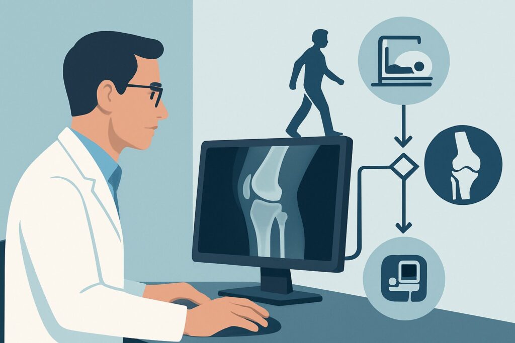
What’s Next After Knee Radiography? Downstream Workflow
The downstream management path diverges based on the imaging results, though in this scenario, the most common outcome is a negative study.
* **If the radiograph is negative:** This is the expected result. The patient can be confidently diagnosed with a knee sprain or contusion. Management consists of conservative measures: rest, ice, compression, elevation (RICE), and analgesics as needed. Crucially, the patient should be given clear instructions on when to follow up—typically if pain does not improve within a week or two, or if new symptoms like instability, locking, or swelling develop. Persistent symptoms may warrant a clinical re-evaluation and potential referral for an MRI to assess for soft-tissue injury.
* **If the radiograph is positive for a fracture:** This finding, while less likely, immediately changes the management plan. The patient should be made non-weight-bearing, the knee should be immobilized in a splint or immobilizer, and an urgent referral to orthopedic surgery is required for definitive fracture management.
* **If the radiograph is indeterminate or suspicious for an occult fracture:** On rare occasions, the initial radiograph may be inconclusive, or the clinical suspicion for a fracture may remain high despite a negative x-ray (e.g., the patient develops focal tenderness after the initial exam). In this case, the patient’s clinical scenario has changed. They now fit the ACR variant for a “suspected occult fracture after negative radiographs,” which appropriately triggers consideration for advanced imaging, such as an MRI, to identify a radiographically occult fracture or bone marrow edema.
Pitfalls to Avoid in Low-Suspicion Knee Injuries
Navigating this common clinical scenario involves avoiding several potential missteps that can lead to either over-testing or missed diagnoses.
* **Pitfall 1: Routine imaging of all minor knee trauma.** The *May be appropriate* rating explicitly discourages a reflexive order for an x-ray on every patient with a twisted knee. Applying clinical judgment and validated decision rules can help safely reduce unnecessary imaging, saving cost and radiation exposure.
* **Pitfall 2: Equating a negative radiograph with a “normal” knee.** A negative x-ray only rules out a fracture. It provides no information about the ligaments, menisci, or cartilage. Avoid telling the patient their knee is “fine”; instead, explain that there is no broken bone, but a soft-tissue sprain may take time to heal.
* **Pitfall 3: Inadequate follow-up instructions.** The most important part of managing a negative radiograph is providing a clear safety net. The patient must understand what symptoms warrant a return visit (e.g., persistent pain, new mechanical symptoms, or worsening instability), as this may be the first sign of a significant soft-tissue injury that requires further workup.
If a patient’s symptoms evolve—for instance, if they develop a significant effusion or focal tenderness days after the injury—it is essential to escalate care and re-evaluate, as their clinical scenario has changed.
Related ACR Topics and Tools
For a comprehensive overview of imaging recommendations across all related knee trauma scenarios, and for tools to help with study selection and patient communication, the following GigHz resources are available:
- For breadth across all scenarios in Acute Trauma to the Knee, see our parent guide: Acute Trauma to the Knee: ACR Appropriateness Decoded.
- To explore other clinical presentations, use the ACR Appropriateness Criteria Lookup.
- For details on imaging techniques, consult the Imaging Protocol Library.
- To discuss radiation exposure with patients, the Radiation Dose Calculator can help contextualize the dose from medical imaging.
Frequently Asked Questions
Why isn’t knee radiography ‘Usually Appropriate’ for this scenario?
The rating is ‘May be appropriate’ because the clinical suspicion for a fracture is very low in a patient who can walk and has no focal tenderness or effusion. Clinical decision rules, like the Ottawa Knee Rules, often suggest that no imaging is necessary for these patients. The ACR rating reflects that while an x-ray is a reasonable option to definitively exclude a fracture, forgoing imaging in favor of clinical follow-up is also a valid and often preferred approach.
If I skip the x-ray, could I miss a significant injury?
While possible, it is unlikely to miss a clinically significant fracture in a patient who meets all the low-risk criteria (ambulatory, no effusion, no focal bony tenderness). The more common injuries in this group are soft-tissue sprains. The key is to provide excellent follow-up instructions, ensuring the patient returns if their symptoms worsen or fail to improve as expected.
Should I order an MRI instead to check the ligaments and meniscus?
No, an MRI is rated ‘Usually not appropriate’ as the initial imaging test for this specific presentation. The first step is to rule out a bony injury. An MRI is a high-cost study that should be reserved for cases where there is persistent pain, instability, or mechanical symptoms after an initial period of conservative management, and after a fracture has been excluded.
Does this guidance apply to a 4-year-old child?
No, this specific ACR variant applies to adults and children 5 years of age or older. Younger children have different injury patterns and considerations, such as physeal (growth plate) injuries, which may require a different diagnostic approach.
What specific views should I order for a knee radiograph?
A standard trauma knee series typically includes an anteroposterior (AP) view, a lateral view, and often sunrise (or merchant) views for the patella. Some protocols may also include oblique views. It is best to order a ‘trauma knee series’ and allow the radiology department to perform their standard protocol.
Reviewed by Pouyan Golshani, MD, Interventional Radiologist — May 21, 2026