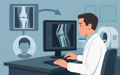
What Is the Right Initial Imaging for an Acute Knee Injury With Swelling or Tenderness?

A 45-year-old lands awkwardly after a rebound in a pickup basketball game, feeling a pop in their right knee. In the urgent care clinic, they have a moderate effusion, focal tenderness over the lateral joint line, and cannot bear weight. You suspect a fracture or a significant internal derangement, but what is the appropriate first imaging study to order? This common clinical scenario requires a clear, evidence-based approach to avoid unnecessary radiation and cost while ensuring a timely diagnosis. For an adult or child over five with acute knee trauma presenting with these specific findings, the American College of Radiology (ACR) designates Radiography knee as Usually Appropriate for initial evaluation.
Who Fits This Clinical Scenario?
This guidance applies to a specific and common patient presentation: an adult or a child aged five or older who has experienced an acute fall or twisting injury to the knee. The key clinical indicators that place a patient in this workflow are the presence of one or more of the following signs:
- Focal tenderness: Tenderness to palpation over a specific bony structure (e.g., the patella, fibular head, or tibial plateau).
- Effusion: Clinically detectable swelling within the knee joint.
- Inability to bear weight: The patient cannot take four steps, both immediately after the injury and at the time of evaluation.
This workflow is designed for the initial imaging decision. It is crucial to distinguish this scenario from others that may appear similar but require a different diagnostic pathway.
This guidance does NOT apply if:
- The patient has no concerning signs: If an individual has a similar mechanism of injury but lacks focal tenderness, effusion, and is able to bear weight, imaging is often not indicated.
- Radiographs have already been performed and are negative: If initial radiographs show no fracture but there is high clinical suspicion for an occult fracture or significant soft tissue injury, the workup proceeds to a different ACR variant, often involving MRI.
- The injury involves very high energy: Patients with knee trauma from a major event like a motor vehicle accident or a suspected knee dislocation fall under a separate, more aggressive imaging protocol.
What Diagnoses Are You Working Up in This Scenario?
When a patient presents with these signs after acute knee trauma, the primary goal of initial imaging is to identify or exclude urgent orthopedic conditions. The differential diagnosis is focused on injuries that would alter immediate management.
Fracture is the most critical diagnosis to rule out. Radiographs are the primary tool for identifying fractures of the distal femur, proximal tibia (including the tibial plateau), proximal fibula, or patella. A finding of a fracture almost always changes management from conservative care to orthopedic consultation for possible reduction or surgical fixation.
Significant Ligamentous or Meniscal Injury is also a key consideration. While plain radiographs cannot directly visualize soft tissues like the Anterior Cruciate Ligament (ACL), menisci, or collateral ligaments, they can reveal secondary signs of a severe underlying injury. A joint effusion, particularly a lipohemarthrosis (fat and blood in the joint), is highly suggestive of an intra-articular fracture and associated soft tissue damage.
Osteochondral Injury is a less common but important possibility, where a fragment of articular cartilage and underlying bone is sheared off during the traumatic event. These fragments can sometimes be visualized on radiographs and may cause mechanical symptoms like locking or catching, often requiring surgical intervention.
Spontaneously Reduced Patellar or Knee Dislocation can also present this way. Even after the joint has returned to its normal position, radiographs can show subtle avulsion fractures (like a Segond fracture, associated with ACL tears) or other signs that point to a significant, unstable underlying injury that requires further evaluation.
Why Is Knee Radiography the Recommended First Step for This Presentation?
The ACR designates Radiography knee as Usually Appropriate because it is the most effective initial test for answering the most urgent clinical question: is there a fracture? Radiographs are fast, widely available, inexpensive, and highly sensitive for detecting most acute fractures and joint dislocations. Clinical decision aids like the Ottawa Knee Rules and the Pittsburgh Decision Rules are built around this principle, using clinical findings to identify patients who are most likely to have a fracture and would benefit from radiography.
The radiation dose is minimal. For an adult, a standard knee series delivers a radiation dose of ☢ <0.1 mSv, and for a child, the dose is even lower at ☢ <0.03 mSv [ped]. This is a very small exposure, equivalent to a few days of natural background radiation.
Why Other Studies Are Not Appropriate for Initial Imaging
Advanced imaging modalities are powerful but are not the correct first step in this specific scenario, as they introduce unnecessary complexity, cost, and potential delays without changing immediate management.
- MRI knee without IV contrast is rated Usually not appropriate as an initial study. While MRI is the gold standard for evaluating ligaments, menisci, and cartilage, it is not needed to rule out the primary concern of a fracture. It is often the correct next step if radiographs are negative but clinical suspicion for a significant soft tissue injury remains high. Ordering it first is an inefficient use of resources.
- CT knee without IV contrast is also Usually not appropriate for initial evaluation. CT provides excellent bone detail but delivers a higher radiation dose than radiography (☢ <0.1 mSv in adults, but a more significant ☢☢ 0.03-0.3 mSv [ped] in children) and is generally reserved for characterizing complex fractures (like a tibial plateau fracture) that have already been identified on a plain film to aid in surgical planning.
What’s Next After Knee Radiography? Downstream Workflow
The results of the initial knee radiograph will guide your next steps and determine whether the patient needs immediate orthopedic referral or can proceed down a different diagnostic pathway.
- If the radiograph is POSITIVE for a fracture: The immediate next step is consultation with an orthopedic surgeon. The type, location, and displacement of the fracture will dictate management, which may range from immobilization to surgical fixation. For complex articular fractures, such as a tibial plateau fracture, the surgeon will likely order a CT scan for preoperative planning, which aligns with a separate ACR Appropriateness Criteria variant.
- If the radiograph is NEGATIVE for a fracture: The decision-making process hinges on the severity of the clinical signs. If the patient has persistent mechanical symptoms (locking, instability), a large effusion, or continued inability to bear weight despite a negative radiograph, the concern shifts toward a significant soft tissue injury (e.g., ACL tear, meniscal tear) or an occult (hidden) fracture. This moves the patient into a different clinical scenario: “No fracture seen on radiographs. Suspect occult fracture or internal derangement.” In that case, MRI without IV contrast often becomes the next appropriate study.
- If the radiograph shows INDIRECT signs of injury: Findings like a large joint effusion or lipohemarthrosis in the absence of a clear fracture line are red flags. A lipohemarthrosis, for instance, is virtually diagnostic of an intra-articular fracture, even if one is not visible. Similarly, certain small avulsion fractures (e.g., a Segond fracture) are pathognomonic for major ligamentous tears. These findings warrant a low threshold for ordering a follow-up MRI to fully characterize the extent of the injury.
Pitfalls to Avoid (and When to Get Help)
Navigating this common scenario effectively requires avoiding a few key pitfalls that can delay diagnosis or lead to unnecessary testing.
- Pitfall: Ordering an incomplete radiographic series. A standard knee series should include an anteroposterior (AP), a lateral, and often oblique or patellar (sunrise) views. Missing a view can lead to a missed fracture.
- Pitfall: Dismissing a “negative” radiograph with positive clinical signs. A radiograph that is negative for fracture does not mean the knee is uninjured. Overlooking soft tissue signs like an effusion or lipohemarthrosis can delay the diagnosis of a major ligamentous injury requiring surgical repair.
- Pitfall: Forgetting pediatric considerations. In skeletally immature patients, be mindful of growth plates (physes), which can be mistaken for fractures. Comparison views of the uninjured knee may sometimes be helpful, and any fracture involving a growth plate requires prompt orthopedic evaluation.
If you identify a displaced or open fracture, or if there is any concern for neurovascular compromise (e.g., diminished distal pulses, foot drop), escalate immediately for an emergent orthopedic consultation.
Related ACR Topics and Tools
For a comprehensive overview of all clinical scenarios related to acute knee trauma and for tools to help with imaging decisions, the following resources are available.
- For breadth across all scenarios in Acute Trauma to the Knee, see our parent guide: Acute Trauma to the Knee: ACR Appropriateness Decoded.
- ACR Appropriateness Criteria Lookup — for adjacent scenarios
- Imaging Protocol Library — for technique on the recommended study
- Radiation Dose Calculator — for cumulative dose conversations
Frequently Asked Questions
Do I have to apply the Ottawa Knee Rules before ordering a radiograph in this scenario?
While not mandatory, using a validated clinical decision rule like the Ottawa Knee Rules or Pittsburgh Decision Rules is highly recommended. These rules help identify patients at very low risk of a clinically significant fracture, for whom radiographs can be safely avoided. This scenario’s inclusion criteria (focal tenderness, effusion, inability to bear weight) overlap significantly with these rules, meaning most patients who fit this scenario will also meet the criteria for imaging.
Why not just order an MRI first to see everything, including ligaments and cartilage?
Ordering an MRI as the initial test is rated ‘Usually Not Appropriate’ by the ACR for this presentation. The most urgent clinical question is whether a fracture is present, which would change immediate management (e.g., making the patient non-weight-bearing, splinting, orthopedic referral). Radiographs answer this question quickly, cheaply, and with very low radiation. An MRI is more expensive, less readily available, and not necessary if a simple fracture is found. It becomes the right tool later in the workflow if radiographs are negative but a significant soft tissue injury is still suspected.
What if the patient is a child under 5 years old?
This specific ACR guidance applies to children aged 5 and older. Younger children, particularly toddlers, have different injury patterns (e.g., toddler’s fractures) and may not be able to communicate symptoms or cooperate with a physical exam in the same way. While radiography is still the primary modality for suspected fractures, the clinical threshold for ordering it may be different and requires careful clinical judgment.
If the radiograph shows a large joint effusion but no fracture, what should I do?
A large effusion, especially in the setting of acute trauma, is a significant finding that indicates substantial intra-articular injury. It strongly suggests a ligament tear, meniscal tear, or an occult fracture not visible on plain films. The patient should be managed with conservative measures initially (RICE, crutches) and referred for orthopedic follow-up. This presentation now fits the ‘suspect internal derangement’ scenario, and an MRI is often the appropriate next step to make a definitive diagnosis.
Is a CT scan ever the right first choice for an acute knee injury?
For this specific scenario of a fall or twisting injury, CT is ‘Usually Not Appropriate’ as the initial imaging test. However, in cases of very high-energy trauma, such as a motor vehicle accident or a fall from a significant height where complex fractures are highly suspected, CT may be considered as part of a broader trauma evaluation. More commonly, CT is used to better characterize a complex fracture (like a tibial plateau fracture) that has already been identified on a radiograph to guide surgical planning.
Reviewed by Pouyan Golshani, MD, Interventional Radiologist — May 21, 2026