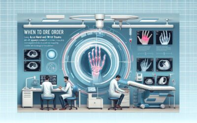
When to Order Imaging for Acute Spinal Trauma: ACR Appropriateness Decoded

When to Order Imaging for Acute Spinal Trauma: ACR Appropriateness Decoded
It’s a busy shift in the emergency department. A patient arrives on a backboard after a motor vehicle collision. They are hemodynamically stable but have midline cervical tenderness. You need to clear their cervical spine, but which imaging study is the right first step? Do they meet criteria for imaging at all? Choosing between radiography and computed tomography (CT), or deciding when to escalate to magnetic resonance imaging (MRI), requires balancing diagnostic yield with radiation exposure and resource utilization. This guide decodes the American College of Radiology (ACR) Appropriateness Criteria for acute spinal trauma to help you make a confident, evidence-based decision.
What Does ACR Acute Spinal Trauma Cover?
The ACR Appropriateness Criteria for Acute Spinal Trauma focus on imaging for patients aged 16 and older who have sustained blunt traumatic injury to the cervical, thoracic, or lumbar spine. The guidelines are structured around common clinical scenarios, from the initial evaluation of a low-risk patient to follow-up imaging for persistent pain or suspected neurologic or vascular injury. These criteria are designed to guide the initial and subsequent imaging workup in the acute setting.
This topic specifically addresses blunt trauma. It does not cover imaging for penetrating trauma, chronic spinal pain, degenerative disease, suspected infection, or oncologic processes. The recommendations are predicated on the use of validated clinical decision rules, such as the National Emergency X-Radiography Utilization Study (NEXUS) criteria and the Canadian C-Spine Rule (CCR), to first determine if any imaging is warranted for cervical spine injuries.
What Imaging Should I Order for Acute Spinal Trauma? Recommendations by Clinical Scenario
The appropriate imaging for acute spinal trauma depends entirely on the clinical context, including the patient’s age, mechanism of injury, clinical stability, and neurologic status. The ACR provides clear guidance for these distinct scenarios.
For an adult patient (age 16-65) with acute cervical spine blunt trauma who is considered low-risk—meaning imaging is not indicated by CCR or NEXUS clinical criteria—the ACR rates all initial imaging modalities, including radiography and CT, as Usually Not Appropriate. In these cases, clinical clearance alone is sufficient, and the risks of radiation exposure from imaging outweigh the potential benefits.
Conversely, for an adult patient with acute cervical spine blunt trauma where imaging is indicated by CCR or NEXUS criteria, the recommendation is clear. CT cervical spine without IV contrast is rated Usually Appropriate. This is the preferred initial study due to its high sensitivity for detecting clinically significant fractures. Plain radiography, in this context, is considered Usually Not Appropriate.
In the thoracolumbar spine, for a high-risk or unexaminable patient with acute blunt trauma, CT of the area of interest without IV contrast is also rated Usually Appropriate as the initial imaging study. Radiography is deemed Usually Not Appropriate for this high-risk population.
If there is a clinical suspicion for arterial injury (e.g., from specific fracture patterns or neurologic deficits), CTA head and neck with IV contrast is the Usually Appropriate next step to evaluate for blunt cerebrovascular injury (BCVI).
For suspected or confirmed ligamentous injury, spinal cord injury, or nerve root impingement, MRI is the modality of choice. In these cases, MRI of the spine area of interest without IV contrast is rated Usually Appropriate. This is often performed after an initial negative CT when clinical suspicion for a soft tissue injury remains high.
A challenging scenario involves an obtunded patient with acute cervical spine trauma whose initial non-contrast CT is negative. Here, the ACR notes disagreement among the panel. MRI cervical spine without IV contrast is rated May be appropriate (Disagreement) as the next imaging study to definitively exclude unstable ligamentous injury before collar removal.
ACR Imaging Recommendations Table
| Clinical Scenario | Top Procedure | ACR Rating | Adult RRL | Pediatric RRL |
|---|---|---|---|---|
| Age ≥16 and <65 years. Acute cervical spine blunt trauma; imaging not indicated by CCR or NEXUS clinical criteria. Low-risk criteria. Initial imaging. | Radiography cervical spine | Usually not appropriate | ☢ ☢ 0.1-1mSv | ☢ ☢ 0.03-0.3 mSv [ped] |
| Age ≥16 years. Acute cervical spine blunt trauma. Imaging indicated by CCR or NEXUS clinical criteria. Initial imaging. | CT cervical spine without IV contrast | Usually appropriate | ☢ ☢ ☢ 1-10 mSv | ☢ ☢ ☢ ☢ 3-10 mSv [ped] |
| Age ≥16 years. Acute cervical spine blunt trauma. No unstable injury demonstrated initially, but kept in collar for neck pain. No new neurologic symptoms. Follow-up imaging. | CT cervical spine without IV contrast | Usually appropriate | ☢ ☢ ☢ 1-10 mSv | ☢ ☢ ☢ ☢ 3-10 mSv [ped] |
| Age ≥16 years. Acute cervical spine blunt trauma. Suspected arterial injury with or without positive cervical spine CT. Next imaging study. | CTA head and neck with IV contrast | Usually appropriate | ☢ ☢ ☢ 1-10 mSv | ☢ ☢ ☢ ☢ 3-10 mSv [ped] |
| Age ≥16 years. Acute cervical, thoracic or lumbar spine blunt trauma. Suspected or confirmed ligamentous, spinal cord or nerve root injury, with or without trauma identified on CT. Next imaging study. | MRI spine area of interest without IV contrast | Usually appropriate | O 0 mSv | O 0 mSv [ped] |
| Age ≥16 years. Acute thoracic or lumbar spine blunt trauma in a high-risk or unexaminable patient. Initial imaging. | CT spine area of interest without IV contrast | Usually appropriate | Varies | Varies |
| Age ≥16 years. Acute cervical spine blunt trauma. Obtunded. No trauma identified on cervical spine CT without IV contrast. Next imaging study. | MRI cervical spine without IV contrast | May be appropriate (Disagreement) | O 0 mSv | O 0 mSv [ped] |
Adult vs. Pediatric Acute Spinal Trauma Imaging: Radiation Dose Tradeoffs
While these ACR guidelines are specified for patients aged 16 and older, the principles of radiation safety are paramount, especially when considering imaging in younger adults and adolescents. The concept of As Low As Reasonably Achievable (ALARA) is a cornerstone of medical imaging. Children and young adults have a longer life expectancy, providing a larger window for the potential stochastic effects of ionizing radiation to manifest. Their developing tissues are also inherently more radiosensitive than those of older adults.
The provided Relative Radiation Level (RRL) data highlights this. For a CT of the cervical spine, the adult dose is in the 1-10 mSv range (☢ ☢ ☢), while the pediatric equivalent is in the 3-10 mSv range (☢ ☢ ☢ ☢), reflecting a higher relative level for the same absolute dose due to increased risk. This underscores the critical importance of strictly adhering to clinical decision rules like NEXUS and CCR to avoid unnecessary CT scans in low-risk patients. When CT is necessary, protocols should be optimized to use the lowest possible radiation dose that still achieves diagnostic image quality.
Imaging Protocol Details for Acute Spinal Trauma
Once you’ve decided on the right study, the specific imaging protocol is critical for diagnostic accuracy. Key considerations include the scan range, slice thickness, use of intravenous contrast, and reconstruction algorithms. Our protocol guides provide detailed, practical information for the key studies recommended in these guidelines.
- CT Cervical Spine
- CTA Head and Neck (Carotid + COW)
- MRI Cervical Spine Without Contrast
- MRI Lumbar Spine Without Contrast
Tools to Help You Order the Right Study
Navigating imaging guidelines can be complex, but several tools can streamline the process. These resources are designed to provide quick access to evidence-based information at the point of care, helping you select the most appropriate study and communicate effectively with patients and colleagues.
The ACR Appropriateness Criteria Lookup provides a searchable interface to the full library of ACR guidelines, covering hundreds of clinical scenarios beyond acute spinal trauma. It’s a valuable resource for ensuring your imaging orders align with national standards.
Our Imaging Protocol Library offers detailed, step-by-step guides for performing various imaging studies. These are useful for trainees learning the technical aspects of scans and for any clinician wanting to understand what a specific order entails.
The Radiation Dose Calculator is a practical tool for estimating cumulative radiation exposure for patients undergoing multiple imaging studies. It can be instrumental in patient counseling, especially when discussing the risks and benefits of follow-up CT scans.
What is the difference between the NEXUS criteria and the Canadian C-Spine Rule (CCR)?
Both are validated clinical decision rules used to determine which patients with potential cervical spine injury require imaging. NEXUS is based on five low-risk criteria: no posterior midline cervical tenderness, no evidence of intoxication, normal level of alertness, no focal neurologic deficit, and no painful distracting injuries. If a patient meets all five, their spine is considered clinically cleared. CCR is an algorithm that considers high-risk factors (age ≥65, dangerous mechanism, paresthesias), low-risk factors allowing for safe range of motion assessment, and the patient’s ability to actively rotate their neck. CCR has been shown to have slightly higher sensitivity and specificity than NEXUS.
Why is CT preferred over radiography for initial imaging in high-risk cervical spine trauma?
CT is significantly more sensitive than plain radiography for detecting clinically significant cervical spine fractures. Studies have shown that radiography can miss a substantial number of fractures, particularly at the craniocervical junction and cervicothoracic junction, which are difficult to visualize on X-ray. While CT involves more radiation, its superior diagnostic accuracy in high-risk patients is considered essential to avoid missing an unstable injury that could lead to catastrophic neurologic damage.
When should I order an MRI as the first-line study in acute spinal trauma?
MRI is rarely the first-line study in the initial evaluation of acute spinal trauma. Its primary role is to evaluate for soft tissue injuries like ligamentous disruption, spinal cord contusion, epidural hematoma, or traumatic disc herniation. Therefore, MRI is typically ordered as a second-line study in patients with a neurologic deficit, or after a CT scan that is negative for fracture but where there is high clinical suspicion for an unstable ligamentous injury (e.g., in an obtunded patient or a patient with persistent, severe neck pain).
Is intravenous contrast necessary for the initial CT scan in acute spinal trauma?
No, for the initial evaluation of bony injury, a non-contrast CT is the standard of care and is rated “Usually Appropriate” by the ACR. Intravenous contrast does not improve the visualization of bone and adds potential risks (e.g., contrast-induced nephropathy, allergic reaction). Contrast is only used when there is a specific indication to evaluate for vascular injury, in which case a CT Angiography (CTA) protocol is performed.
What is a “distracting injury” in the context of NEXUS criteria?
A distracting injury is any injury thought to be painful enough to distract the patient from a second, less painful injury, such as a cervical spine fracture. Examples include a long bone fracture (e.g., femur), a visceral injury requiring surgery, a large laceration, or extensive burns. The presence of such an injury makes the clinical examination of the cervical spine unreliable, and therefore, imaging is required for clearance regardless of other findings.
Reviewed by Pouyan Golshani, MD, Interventional Radiologist — May 12, 2026