What Imaging Is Best for Chronic Ankle Instability After Normal X-Rays?
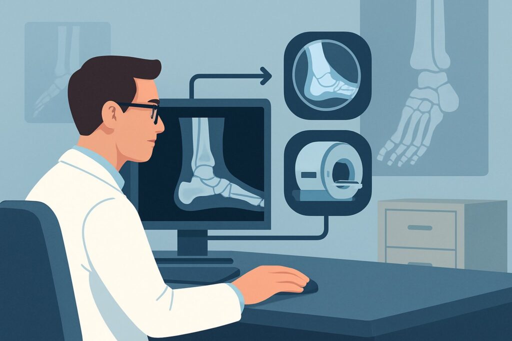
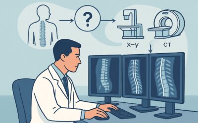
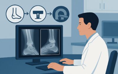
A 34-year-old patient presents to your clinic with six months of persistent right ankle pain and a sensation of “giving way,” especially on uneven ground. They recall multiple ankle sprains playing soccer in college, but the current symptoms are more chronic and bothersome. You’ve already obtained standard ankle radiographs, which came back normal, showing no fracture, dislocation, or significant degenerative change. Now you face the key clinical question: what is the next best imaging study to evaluate for suspected chronic ankle instability? This article provides a detailed workflow for this specific scenario, grounded in the American College of Radiology (ACR) Appropriateness Criteria. For this presentation, MR arthrography of the ankle is rated Usually appropriate.
Who Fits This Clinical Scenario?
This guidance is for a specific patient population: adults or adolescents with chronic ankle pain (typically lasting three months or longer) where the clinical history is suspicious for mechanical instability. Key features include a history of recurrent ankle sprains, a subjective feeling of the ankle “giving out” or being unreliable, and pain localized to the lateral ankle, though it can be more diffuse. Crucially, this workflow applies only after initial ankle radiographs have been performed and are either entirely normal or show only nonspecific findings like minor soft tissue swelling.
This pathway is not intended for:
- Patients with acute trauma: This scenario is for chronic, ongoing symptoms, not a new injury.
- Patients with clear radiographic evidence of advanced arthritis: If radiographs show significant degenerative joint disease in the hindfoot, a different diagnostic algorithm applies. This is a distinct ACR scenario focused on characterizing the extent of arthritis.
- Patients where the primary suspicion is a tendon abnormality: If the pain and physical exam point specifically to the peroneal or Achilles tendons (e.g., pain with resisted eversion, palpable tendon thickening), the imaging workup is tailored differently. This also represents a separate ACR scenario.
Correctly identifying your patient’s presentation ensures you order the most diagnostically useful and appropriate study from the start.
What Diagnoses Are You Working Up in This Scenario?
When you suspect chronic ankle instability despite normal radiographs, your differential diagnosis centers on soft-tissue and cartilaginous injuries that standard X-rays cannot visualize. The primary goal of advanced imaging is to identify the specific anatomical cause of the instability.
Chronic Lateral Ligament Complex Injury: This is the most common cause of chronic ankle instability. It typically involves attenuation, scarring, or complete tearing of the anterior talofibular ligament (ATFL) and, less commonly, the calcaneofibular ligament (CFL). A prior severe sprain may have healed inadequately, leading to persistent laxity.
Syndesmotic Injury: A “high ankle sprain” involves the ligaments connecting the tibia and fibula. If not properly healed, it can lead to chronic pain and instability, particularly with activities involving rotation. These injuries can be subtle and are often missed in the acute phase.
Osteochondral Lesion of the Talus (OLT): An injury to the cartilage and underlying bone of the talar dome, often a result of the impaction forces during an ankle sprain. Small or nondisplaced OLTs are frequently invisible on radiographs but can be a significant source of chronic, deep ankle pain and mechanical symptoms.
Sinus Tarsi Syndrome: This is a clinical syndrome characterized by pain in the sinus tarsi, the space between the talus and calcaneus. It is often associated with subtalar joint instability and inflammation of the synovial tissues within the sinus, frequently stemming from injury to the interosseous and cervical ligaments.
Why Is MR Arthrography the Recommended Study for This Presentation?
For a patient with suspected chronic ankle instability and normal radiographs, both MR arthrography ankle and MRI ankle without IV contrast are rated Usually appropriate by the ACR. However, MR arthrography often provides the most definitive evaluation in this specific context.
The primary advantage of MR arthrography is the injection of dilute gadolinium-based contrast directly into the tibiotalar joint. This distends the joint capsule, allowing for exquisite visualization of the ligaments, capsule, and cartilage surfaces. The contrast will leak out of the joint through any full-thickness ligamentous or capsular tear, providing a direct sign of injury that can be missed on a non-contrast study. This is particularly valuable for assessing the integrity of the ATFL and CFL, the key stabilizers of the lateral ankle.
While a high-quality non-contrast MRI is also very effective, it relies on secondary signs of ligament injury, such as thickening, thinning, or abnormal signal. MR arthrography turns a potential inference into a direct observation, increasing diagnostic confidence for subtle or partial-thickness tears and for evaluating the integrity of surgically repaired ligaments.
Let’s consider the alternatives and why they are rated lower for this scenario:
- Radiography ankle stress views are rated May be appropriate. These can demonstrate functional instability by showing talar tilt or anterior drawer under applied stress. However, they are operator-dependent, can be painful for the patient, and do not visualize the specific soft-tissue structures that are injured. They show that instability exists but not why.
- US ankle (ultrasound) is also rated May be appropriate. In experienced hands, dynamic ultrasound can be a powerful tool for assessing ligament integrity. However, it is highly operator-dependent, has a limited field of view compared to MRI, and can be less effective for evaluating intra-articular pathology like osteochondral lesions.
From a safety perspective, both recommended MRI options are excellent choices as they involve no ionizing radiation (0 mSv). CT arthrography, rated May be appropriate, is an alternative if MRI is contraindicated but involves a small amount of radiation (☢ <0.1 mSv).
Once you’ve decided on an MRI-based study, our protocol guide covers the technical details, contrast considerations, and key reading principles. For a deep dive into the procedure itself, see our guide: MRI Ankle/Foot.
What’s Next After MR Arthrography? Downstream Workflow
The results of the MR arthrogram will guide your next steps, typically leading to either conservative management or surgical consultation. The goal is to move from a diagnosis to a definitive treatment plan.
If the study is positive for a significant ligamentous tear (e.g., ATFL, CFL): This finding confirms the anatomic basis for the patient’s instability. The next step is typically a referral to an orthopedic surgeon or a sports medicine specialist. Treatment options range from intensive physical therapy focused on proprioception and peroneal strengthening to surgical intervention, such as a ligament repair or reconstruction (e.g., Broström-Gould procedure).
If the study is negative: A high-quality negative MR arthrogram makes a significant structural cause of instability much less likely. The focus should shift to non-anatomic causes. This may involve a referral to physical therapy to focus on neuromuscular control, balance, and proprioceptive training. If pain persists without a clear cause, you may be transitioning into the “Chronic ankle pain, pain of uncertain etiology” clinical scenario, which has its own workup.
If the study shows an alternative diagnosis (e.g., an osteochondral lesion): This changes the management pathway. The patient should be referred to an orthopedic surgeon for discussion of treatments specific to OLTs, which can include microfracture, cartilage transfer procedures, or fragment fixation, depending on the size and stability of the lesion.
Pitfalls to Avoid (and When to Get Help)
Navigating the workup for chronic ankle instability requires careful clinical correlation. Here are a few common pitfalls to avoid:
- Underestimating the clinical exam: Don’t rely on imaging alone. A positive anterior drawer or talar tilt test on physical exam strongly suggests instability, even if imaging findings are subtle.
- Not specifying the clinical question: When ordering the MRI, clearly state “evaluate for chronic instability” and mention the specific ligaments of concern (ATFL, CFL). This helps the radiologist tailor their protocol and focus their interpretation.
- Stopping at a non-contrast MRI if suspicion remains high: If a non-contrast MRI is equivocal but your clinical suspicion for a ligament tear is very strong, an MR arthrogram may still be warranted to provide a definitive answer.
- Ignoring the syndesmosis: Remember to consider a high ankle sprain in the differential, especially if the patient has a history of an external rotation injury and pain superior to the ankle joint.
If the diagnosis remains elusive after appropriate imaging or if the patient’s symptoms are worsening, escalation to an orthopedic or musculoskeletal subspecialist is always the correct next step.
Related ACR Topics and Tools
This article is a deep dive into one specific clinical scenario. For a broader view of imaging for all types of chronic ankle pain, from initial workup to post-operative evaluation, please consult our comprehensive parent guide. It provides a hub-and-spoke model to help you navigate all related ACR variants.
- For breadth across all scenarios in Chronic Ankle Pain, see our parent guide: Chronic Ankle Pain: ACR Appropriateness Decoded.
- To explore other ACR scenarios or different clinical topics, use the Imaging Appropriateness Selector.
- For detailed procedural techniques on recommended studies, visit the Imaging Protocol Library.
- To discuss radiation exposure with patients, especially when considering CT, use the Radiation Dose Calculator.
Frequently Asked Questions
Why not just order a standard MRI without contrast first?
A standard MRI of the ankle without contrast is also rated ‘Usually appropriate’ by the ACR for this scenario and is an excellent study. However, MR arthrography, which involves injecting contrast into the joint, is often superior for detecting subtle or partial ligament tears and capsular defects. It distends the joint, making it easier to see contrast leak through a tear, which provides a more definitive diagnosis of the cause of instability.
What are stress radiographs, and why are they only ‘May be appropriate’?
Stress radiographs involve taking X-rays of the ankle while a manual force is applied (simulating an anterior drawer or talar tilt test). They can demonstrate functional instability by showing excessive joint space opening. However, they are rated ‘May be appropriate’ because they are operator-dependent, can be painful, and do not visualize the soft tissues. They show that the ankle is unstable but not the specific ligament that is torn, which is what MRI shows.
Is an MR arthrogram painful for the patient?
The procedure involves a small needle injection of contrast material into the ankle joint, typically performed by a radiologist using fluoroscopy or ultrasound guidance. The area is numbed with a local anesthetic first. Patients may feel some pressure or discomfort during the injection and some joint fullness for a day or two afterward, but it is generally well-tolerated.
What if my patient has a contraindication to MRI or a gadolinium allergy?
If a patient cannot undergo an MRI (e.g., due to an incompatible implanted device), CT arthrography is a reasonable alternative and is rated ‘May be appropriate’. It also uses an intra-articular injection but involves ionizing radiation. For a known severe allergy to gadolinium-based contrast agents, a non-contrast MRI would be the preferred next step, or CT arthrography using iodinated contrast.
Does this guidance apply to an acute ankle sprain?
No. This workflow is specifically for chronic ankle pain (symptoms >3 months) where instability is suspected. Acute ankle sprains are typically managed clinically, and imaging is usually reserved for cases where a fracture is suspected (following the Ottawa Ankle Rules) or if there is a failure to improve with conservative management.
Reviewed by Pouyan Golshani, MD, Interventional Radiologist — May 26, 2026