How Should You Follow Up Low Bone Density in Younger Adults? An ACR-Guided Workflow
A 42-year-old woman with a history of celiac disease and long-term glucocorticoid use for a separate autoimmune condition returns to your clinic. Two years ago, an initial bone density scan revealed osteopenia, and you initiated calcium and vitamin D supplementation. Now, you need to determine if her bone mineral density (BMD) has remained stable, improved, or worsened. This is not a screening scenario; it is a targeted follow-up for a known condition in a premenopausal female with significant risk factors. The American College of Radiology (ACR) provides clear guidance for this exact situation, rating Dual-energy X-ray Absorptiometry (DXA) of the lumbar spine and hip(s) as Usually appropriate. This article details the clinical workflow for this specific follow-up scenario.
Who Fits This Clinical Scenario?
This guidance applies specifically to the follow-up imaging of premenopausal females and males under 50 years of age who have established risk factors for bone loss and have already been diagnosed with low bone mineral density on a prior study. The key elements are the patient’s age (younger than the typical screening population), the presence of secondary risk factors, and the clinical context of surveillance rather than initial diagnosis.
Inclusion criteria include:
- Premenopausal female or male less than 50 years of age.
- Known risk factors for secondary osteoporosis (e.g., chronic glucocorticoid use, hypogonadism, malabsorption syndromes, hyperparathyroidism, chronic inflammatory disease).
- A previously documented low bone mineral density (osteopenia or osteoporosis).
It is crucial to distinguish this scenario from others. This workflow does not apply to:
- Initial screening: The first-time evaluation of a patient in this demographic is a separate clinical question. This article is strictly for follow-up.
- Postmenopausal females or males over 50: These patients fall under standard osteoporosis screening and follow-up guidelines, which have different considerations.
- Patients without risk factors: The presence of a clear secondary cause for bone loss is a prerequisite for this specific scenario.
What Diagnoses Are You Working Up in This Scenario?
When ordering a follow-up bone density scan in a younger adult with known low BMD, you are primarily assessing the trajectory of their bone health to guide management. The differential is less about a new diagnosis and more about quantifying change over time.
Progressive Secondary Osteoporosis: This is the primary concern. The follow-up scan aims to detect a statistically significant decrease in bone mineral density, which would indicate that the underlying condition or its treatment is inadequate to prevent further bone loss. This finding would prompt a re-evaluation of the treatment plan, potentially escalating therapy or more aggressively managing the root cause.
Stable or Improving Bone Mineral Density: An equally important finding is stability. If the BMD has not worsened or has shown improvement, it suggests that the current management strategy (e.g., supplementation, treatment of the underlying disease, antiresorptive therapy) is effective. This result supports continuing the current plan and establishes a new baseline for future comparisons.
Treatment-Induced Changes: For patients who have been started on specific osteoporosis medications (e.g., bisphosphonates, denosumab), the follow-up DXA serves as a direct measure of therapeutic response. The goal is to confirm that the medication is having the desired effect of increasing or stabilizing bone mass.
Underlying Osteomalacia: While DXA measures mineral density, it cannot distinguish between low bone mass (osteoporosis) and defective bone mineralization (osteomalacia). If a patient’s BMD continues to decline despite appropriate osteoporosis management, and they have risk factors like vitamin D deficiency or malabsorption, the possibility of coexisting osteomalacia should be considered. This would require a different diagnostic workup, including specific laboratory tests.
Why Is DXA of the Lumbar Spine and Hip(s) the Recommended Study?
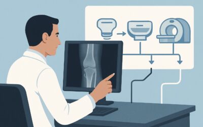
The ACR designates DXA of the lumbar spine and hip(s) as Usually appropriate for this follow-up scenario because it is the gold standard for monitoring changes in bone mineral density with high precision and very low radiation exposure.
The rationale for its top rating is multifactorial. DXA provides a quantitative measurement of areal BMD (g/cm²), which is highly reproducible. For follow-up studies, this precision is critical to confidently detect small but clinically significant changes over time. The standard measurement sites—the lumbar spine and proximal femur—are chosen because they are common sites of osteoporotic fractures and reflect both trabecular and cortical bone health.
The radiation dose from a DXA scan is extremely low (ACR Relative Radiation Level ☢ <0.1 mSv), which is comparable to daily background radiation. This makes it exceptionally safe for serial monitoring, a key consideration in this younger patient population who may require multiple scans over their lifetime.
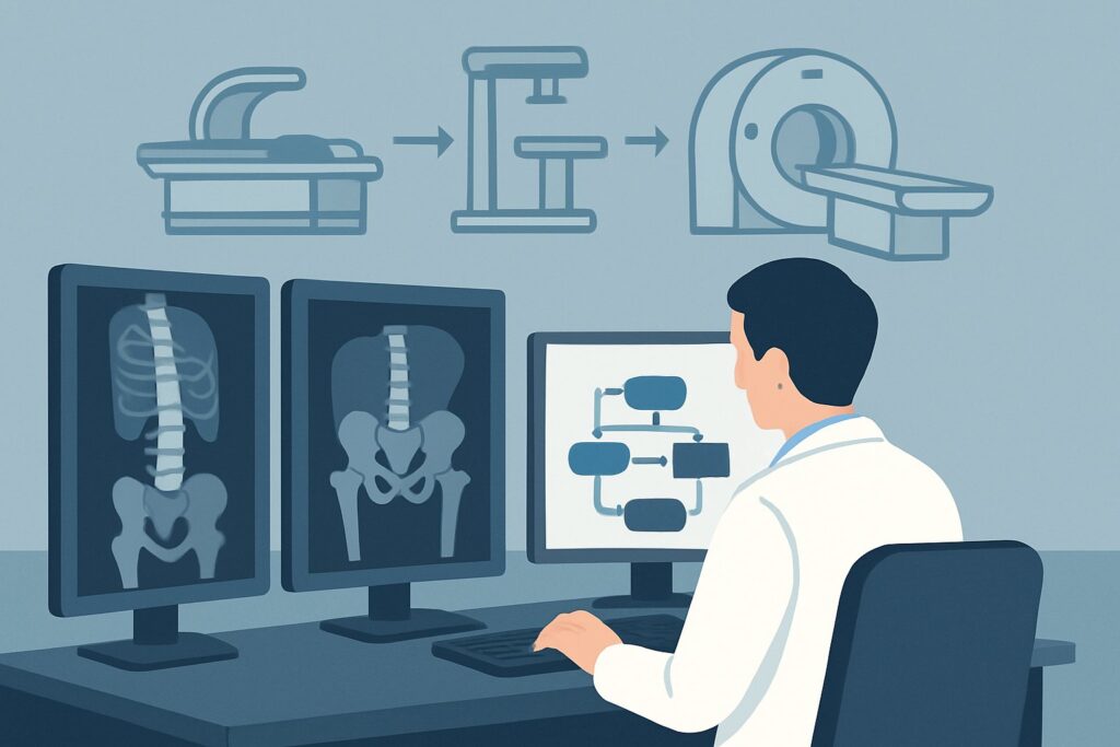
Alternative studies are rated lower for specific reasons in this context:
- Quantitative Computed Tomography (QCT) of the lumbar spine and hip is rated May be appropriate. QCT provides true volumetric BMD and can isolate trabecular bone, which is more metabolically active. However, it imparts a substantially higher radiation dose (ACR RRL ☢☢☢ 1-10 mSv) and is generally less available and more costly. Its use is typically reserved for complex cases where DXA results are confounded by severe degenerative changes or aortic calcification.
- Quantitative Ultrasound (QUS) of the calcaneus is rated Usually not appropriate for follow-up. While QUS is a radiation-free screening tool for fracture risk, it lacks the precision and standardization required to monitor changes in BMD or response to therapy. It cannot be used to diagnose osteoporosis based on World Health Organization T-score criteria or to track a patient’s progress.
What’s Next After DXA? Downstream Workflow
The results of the follow-up DXA scan directly inform the next steps in patient management. The key is to compare the new results to the prior study, noting the least significant change (LSC) calculated by the imaging facility to determine if any change is real versus measurement variability.
- If BMD is stable or has increased: This is a favorable outcome. It suggests the current management plan is effective. The recommendation is typically to continue the current regimen (e.g., supplementation, lifestyle modifications, treatment of the secondary cause) and schedule the next follow-up DXA in 2-3 years, depending on the clinical severity and stability.
- If BMD has significantly decreased: This is an adverse finding that requires action. The first step is to re-evaluate the patient for adherence to therapy and to investigate for new or worsening secondary causes of bone loss. This may involve a laboratory workup for vitamin D deficiency, hyperparathyroidism, or other endocrine abnormalities. A referral to an endocrinologist or rheumatologist is often warranted to consider initiating or escalating pharmacologic therapy for osteoporosis.
- If the patient develops new, severe, localized back pain: A significant drop in lumbar spine BMD accompanied by new, acute back pain should raise suspicion for a vertebral compression fracture. While initial evaluation may start with radiographs, an MRI is highly sensitive for detecting acute or subacute fractures and associated bone marrow edema, which can alter management. For a detailed review of the imaging technique, see our protocol guide: MRI Lumbar Spine Without Contrast.
Pitfalls to Avoid (and When to Get Help)
Navigating follow-up for low BMD in younger adults has several potential pitfalls. A primary error is inconsistent follow-up; ensure scans are performed on the same manufacturer’s machine whenever possible to improve precision. Another pitfall is failing to calculate or consider the least significant change (LSC); small fluctuations in BMD may be within the margin of error and not represent true biological change. Also, avoid attributing all bone loss to the known risk factor without periodically reassessing for other contributing causes. If a patient experiences continued bone loss despite appropriate management or develops an unexpected fracture, it is critical to escalate care and seek consultation from a bone metabolism specialist or endocrinologist.
Related ACR Topics and Tools
This article focuses on a single clinical scenario. For a comprehensive overview of all related presentations and imaging modalities, please consult our parent guide. For further exploration of imaging criteria, protocols, and radiation safety, the following GigHz resources are available.
- For breadth across all scenarios in Osteoporosis and Bone Mineral Density, see our parent guide: Osteoporosis and Bone Mineral Density: ACR Appropriateness Decoded.
- Imaging Appropriateness Selector — for adjacent scenarios
- Imaging Protocol Library — for technique on the recommended study
- Radiation Dose Calculator — for cumulative dose conversations
Frequently Asked Questions
How often should a follow-up DXA scan be performed in a premenopausal woman with risk factors?
The optimal interval varies, but a common approach is every 2-3 years after the initial diagnosis. The frequency may be increased to 1 year after initiating new pharmacologic therapy to assess response, or decreased if bone density is stable over several cycles. The decision should be individualized based on the severity of bone loss and the underlying risk factors.
Why is the forearm sometimes scanned with DXA?
DXA of the distal forearm is rated as ‘May be appropriate’. It is typically performed when the hip or spine cannot be accurately measured or interpreted. This can occur in patients with severe degenerative arthritis of the spine, bilateral hip replacements, or those who exceed the weight limit of the DXA table.
Can I use a Z-score instead of a T-score for this patient population?
Yes, for premenopausal women and men under 50, the Z-score is the clinically preferred metric. The Z-score compares the patient’s BMD to that of an age- and sex-matched reference population. A Z-score of -2.0 or lower is defined as ‘low bone mass for age.’ T-scores, which compare to a young adult reference, are used for diagnosing osteoporosis in postmenopausal women and men over 50.
What if the patient’s risk factor, like glucocorticoid use, has been discontinued?
If the secondary cause of bone loss has been resolved, follow-up DXA is still important to assess for recovery of bone mass. Some bone density may be regained after stopping glucocorticoids, for example. The follow-up scan helps determine if the patient’s BMD has returned to a normal trajectory or if residual low bone mass persists, requiring continued monitoring.
Does Trabecular Bone Score (TBS) have a role in this scenario?
For this specific ACR scenario, Trabecular Bone Score (TBS) is rated ‘Usually not appropriate’ for follow-up. TBS is a software analysis of DXA images that provides an indirect measure of bone microarchitecture. While it can enhance fracture risk prediction, its role in monitoring treatment response or changes over time is less established than standard BMD measurement from DXA.
Reviewed by Pouyan Golshani, MD, Interventional Radiologist — May 26, 2026