Which Imaging Is Best for Suspected Hip Bursitis or Tendonitis After Negative X-rays?
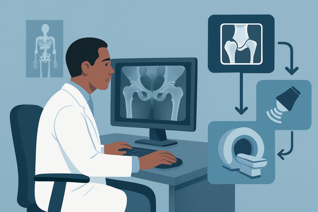
A 48-year-old patient presents with five months of persistent, aching pain over their right lateral hip, which worsens when they lie on that side at night or rise from a chair. The physical exam reveals exquisite point tenderness over the greater trochanter. You’ve already obtained radiographs of the hip, which are unremarkable, showing no fracture, osteoarthritis, or other osseous abnormality. Your leading diagnosis is an extra-articular issue like gluteal tendinopathy or trochanteric bursitis. This article details the American College of Radiology (ACR) recommended imaging workflow for this specific clinical scenario. For a patient with suspected noninfectious extra-articular hip pain and negative radiographs, `US hip` is rated Usually appropriate.
Who Fits This Clinical Scenario?
This guidance is for patients with chronic hip pain (typically lasting three months or longer) where the clinical suspicion points away from the hip joint itself and toward the surrounding soft tissues. The key inclusion criteria are:
- A clinical history and physical exam suggesting an extra-articular source of pain, such as tendonitis, tendinosis, or bursitis. Common locations include the greater trochanter (gluteal tendons, trochanteric bursa), anterior hip (iliopsoas tendon/bursa), or posterior hip (hamstring origin).
- Initial radiographs have been performed and are either negative or nondiagnostic for the cause of the patient’s symptoms.
- There are no “red flag” symptoms suggesting infection (e.g., fever, erythema) or malignancy.
This workflow is distinct from other chronic hip pain scenarios. This guidance does not apply if you primarily suspect an intra-articular problem. For instance, if the patient reports mechanical symptoms like clicking, locking, or instability, the workup for a suspected labral tear or femoroacetabular impingement (FAI) follows a different pathway, often involving MR arthrography. Similarly, if radiographs show evidence of osteoarthritis, the imaging goal shifts to evaluating articular cartilage, which is another distinct ACR variant.
What Diagnoses Are You Working Up in This Scenario?
When initial radiographs are negative, the diagnostic focus shifts to the complex network of muscles, tendons, and bursae surrounding the hip. The goal of subsequent imaging is to pinpoint the specific soft-tissue generator of the patient’s pain.
Greater Trochanteric Pain Syndrome (GTPS): This is the most common diagnosis in this scenario, particularly for lateral hip pain. Once broadly termed “trochanteric bursitis,” it is now understood to be a spectrum of pathology most often driven by tendinopathy, tearing, or calcification of the gluteus medius and minimus tendons. The trochanteric bursa may be secondarily inflamed, but tendon pathology is frequently the primary issue.
Iliopsoas Tendinopathy or Bursitis: For patients presenting with chronic anterior hip or groin pain, inflammation or degeneration of the iliopsoas tendon and its associated bursa is a primary consideration. This can sometimes cause a “snapping hip” sensation as the tendon moves over the pelvic brim.
Proximal Hamstring Tendinopathy: Pain localized to the buttock or ischial tuberosity, often worsened by prolonged sitting, points toward pathology at the origin of the hamstring muscles. This is a common overuse injury in runners and other athletes.
Ischiofemoral Impingement: A less common cause of posterior hip and buttock pain, this condition results from the narrowing of the space between the ischial tuberosity and the lesser trochanter of the femur, leading to compression of the quadratus femoris muscle.
Why Is US hip the Recommended Next Study for This Presentation?
For a patient with suspected extra-articular hip pain and negative radiographs, both `US hip` and `MRI hip without IV contrast` are rated Usually appropriate by the ACR. However, ultrasound often serves as the ideal first choice for several reasons.
Ultrasound provides excellent high-resolution imaging of superficial soft tissues, making it highly effective for evaluating tendons, muscles, and bursae. Its key advantage is the ability to perform a dynamic assessment. The sonographer can have the patient move their hip, directly visualizing tendon movement and identifying areas of impingement or subluxation. Furthermore, the examiner can use the transducer to apply pressure to the area of reported pain (sonographic palpation), correlating the patient’s symptoms directly with the anatomic structures on the screen. This real-time feedback is invaluable for confirming a diagnosis like tendinopathy or bursitis.
From a safety and practical standpoint, ultrasound involves no ionizing radiation (0 mSv) and is widely available, generally less expensive, and quicker to perform than an MRI. It also allows for a straightforward transition to a therapeutic procedure; if bursitis or tendinopathy is identified, an ultrasound-guided corticosteroid injection can often be performed in the same session.
While also Usually appropriate, `MRI hip without IV contrast` provides a more global, panoramic view of both the intra-articular and extra-articular structures. It is an excellent problem-solving tool if the ultrasound is negative or equivocal, or if there is suspicion of a deeper pathology (like a stress fracture or ischiofemoral impingement) that ultrasound cannot visualize well.
In contrast, other studies are rated lower for this specific scenario. `MR arthrography hip` is Usually not appropriate because it is an invasive procedure designed to distend the joint capsule to evaluate for intra-articular pathology like a labral tear, which is not the primary clinical suspicion here. Similarly, all `CT` variants are Usually not appropriate due to their poor soft-tissue contrast and unnecessary radiation exposure (☢☢☢ 1-10 mSv) for a workup focused on tendons and bursae.
What’s Next After US hip? Downstream Workflow
The results of the hip ultrasound will guide your next clinical steps in a branching pathway.
If the study is positive: A definitive finding of gluteal tendinopathy, trochanteric bursitis, or another specific extra-articular abnormality confirms the clinical diagnosis. This allows for targeted treatment, which may include focused physical therapy, activity modification, or NSAIDs. If these measures fail, the positive imaging provides a clear target for a therapeutic injection. The ACR rates `Image-guided anesthetic +/- corticosteroid injection` as May be appropriate, and this is often performed under ultrasound guidance to ensure accurate needle placement and improve efficacy.
If the study is negative: A negative or nondiagnostic ultrasound in a patient with persistent, debilitating pain should prompt consideration of alternative diagnoses. The most logical next step is to obtain the other Usually appropriate study: `MRI hip without IV contrast`. An MRI can identify pathology that may be too deep for ultrasound to visualize, such as a femoral neck stress fracture, early avascular necrosis, or ischiofemoral impingement. It also provides a comprehensive assessment of the intra-articular structures, in case the pain is referred from an occult joint problem.
If the study is indeterminate: Occasionally, ultrasound findings may be ambiguous. In these cases, similar to a negative study, proceeding to an `MRI hip without IV contrast` is the recommended next step to clarify the anatomy and reach a definitive diagnosis.
Pitfalls to Avoid (and When to Get Help)
Navigating the workup for extra-articular hip pain requires careful clinical correlation. Here are a few common pitfalls to avoid:
- Anchoring on “Bursitis”: Be aware that Greater Trochanteric Pain Syndrome is more often caused by underlying gluteus medius or minimus tendinopathy than by primary bursal inflammation. The imaging report should be read carefully to guide the most appropriate physical therapy.
- Forgetting Dynamic Assessment: When ordering an ultrasound, ensure the performing site is comfortable with dynamic musculoskeletal imaging. Simply taking static pictures may miss key findings that are only evident with patient movement.
- Ordering the Wrong MRI: For suspected extra-articular pain, a non-contrast MRI is sufficient. Ordering an MR arthrogram is an unnecessary invasive procedure for this indication, while ordering an MRI with IV contrast is not needed unless there is a specific concern for infection, tumor, or inflammatory arthritis.
If a patient develops systemic symptoms such as fever, chills, or unexplained weight loss, or if imaging suggests an aggressive process, escalate care promptly for a workup of potential infection or malignancy.
Related ACR Topics and Tools
This article covers one specific scenario in depth. For a comprehensive overview of all clinical variants and imaging modalities for chronic hip pain, please consult our parent guide. For additional tools to help with ordering and patient communication, see the resources below.
- For breadth across all scenarios in Chronic Hip Pain, see our parent guide: Chronic Hip Pain: ACR Appropriateness Decoded.
- To look up other clinical scenarios, use the Imaging Appropriateness Selector.
- To review technical details for the recommended studies, see the Imaging Protocol Library.
- To discuss radiation exposure with patients, use the Radiation Dose Calculator.
Frequently Asked Questions
Why is ultrasound often preferred over MRI if both are ‘Usually Appropriate’ for suspected bursitis?
Ultrasound is often preferred as the initial study due to its lower cost, wider availability, and lack of ionizing radiation. Its key advantage is the ability to perform a dynamic, real-time assessment, allowing the clinician to correlate the patient’s exact point of pain with the underlying anatomy and observe tendon movement. It also facilitates a seamless transition to an image-guided injection if needed.
What if my patient has a high Body Mass Index (BMI) that might limit ultrasound quality?
This is an important consideration. In patients with a high BMI, subcutaneous tissue can limit the depth and clarity of ultrasound imaging. In such cases, MRI hip without IV contrast becomes a stronger first-choice option, as its image quality is not typically degraded by body habitus and it can provide a clear view of both deep and superficial structures.
When should I consider an image-guided injection for this condition?
An image-guided injection is rated ‘May be appropriate’ and is typically considered after a diagnosis of tendinopathy or bursitis is confirmed on imaging and the patient has failed a course of conservative management (e.g., physical therapy, activity modification, NSAIDs). Using ultrasound or fluoroscopic guidance ensures the medication is delivered precisely to the site of inflammation, which can increase efficacy and diagnostic confidence.
My patient’s pain is more in the groin area. Does this workflow still apply?
Yes, this workflow applies to suspected extra-articular pain around the hip, including the anterior (groin) region. In this case, the clinical suspicion would be for diagnoses like iliopsoas tendinopathy or bursitis. Both ultrasound and MRI are excellent at evaluating these anterior structures.
If the ultrasound is negative, is it necessary to get an MRI?
If a high-quality ultrasound is definitively negative but the patient remains significantly symptomatic, an MRI is the logical and recommended next step. The MRI can evaluate for causes of pain that are beyond the reach or capability of ultrasound, such as a femoral neck stress fracture, avascular necrosis, ischiofemoral impingement, or occult intra-articular pathology.
Reviewed by Pouyan Golshani, MD, Interventional Radiologist — May 26, 2026