What’s the Next Imaging Step for Persistent Ankle Pain After Negative Radiographs?
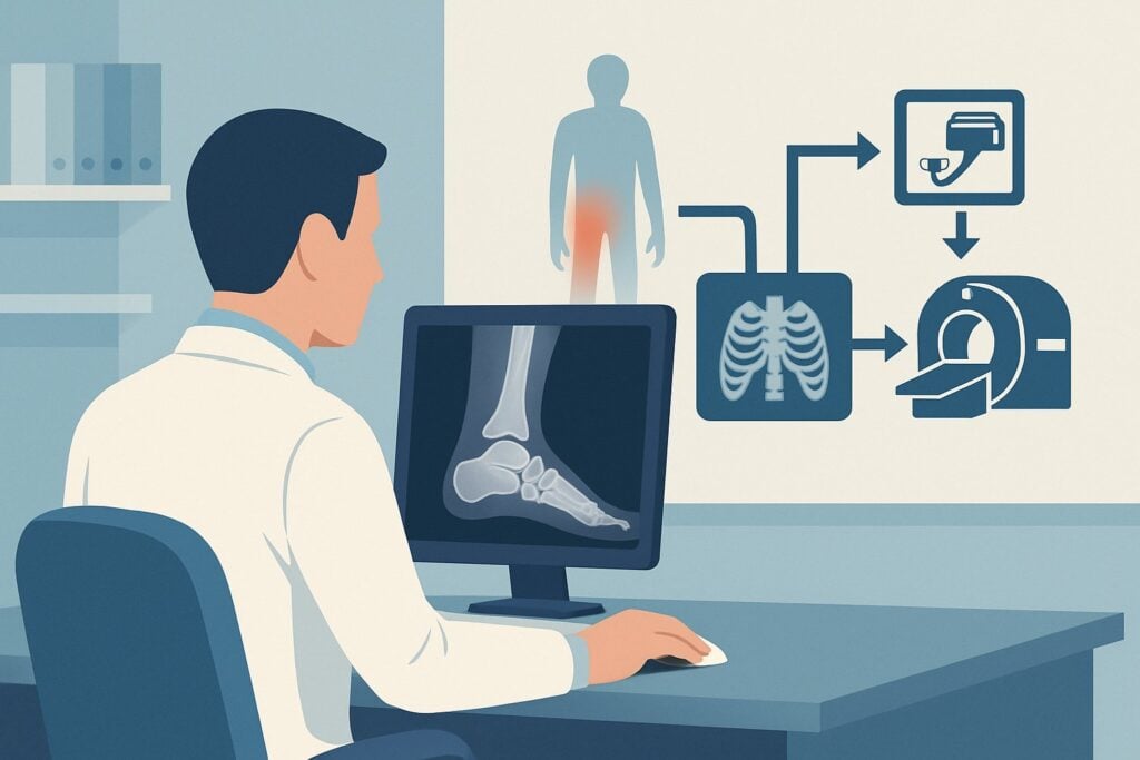
A 28-year-old patient returns to your clinic two weeks after twisting their ankle during a basketball game. The initial radiographs from an urgent care center were negative for fracture. Despite following RICE (rest, ice, compression, elevation) protocols, they report persistent lateral ankle pain, swelling, and difficulty with full weight-bearing. The initial diagnosis of a simple sprain seems insufficient given the lack of improvement. You are now faced with a decision: continue conservative management or order advanced imaging to investigate a potential occult injury. This article details the American College of Radiology (ACR) Appropriateness Criteria for this exact scenario, explaining why a specific study is recommended to clarify the diagnosis and guide treatment. For this presentation, MRI ankle without IV contrast is rated Usually Appropriate.
Who Fits This Clinical Scenario for Persistent Ankle Pain?
This guidance applies to a specific and common clinical situation: an adult or child aged 5 years or older who has experienced acute ankle trauma and continues to have pain for more than one week but less than three weeks. A critical component of this scenario is that initial ankle radiographs were performed and interpreted as negative for an acute fracture. The patient has no exclusionary criteria, such as a known neurologic disorder, open fracture, or signs of vascular compromise.
It is crucial to distinguish this subacute presentation from other related scenarios that follow different diagnostic pathways:
- Initial Acute Trauma (< 1 week): This workflow is not for the immediate evaluation of an ankle injury where the Ottawa Ankle Rules would first determine the need for initial radiography.
- Positive Initial Radiographs: If the first set of X-rays demonstrated a fracture or dislocation, the imaging workup would shift to characterizing that known injury, often with CT for surgical planning.
- High Suspicion of Syndesmotic Injury: If physical exam findings (e.g., a positive squeeze test) strongly suggest a high ankle sprain despite negative radiographs, the ACR has a separate variant that may prioritize stress radiographs or MRI sooner.
- Chronic Pain (> 3 weeks): Pain persisting beyond this subacute window may involve a different differential diagnosis, including chronic instability or inflammatory conditions, which can alter imaging priorities.
This article is exclusively for the patient with persistent symptoms in the 1- to 3-week post-injury window after negative initial X-rays.
What Diagnoses Are You Working Up with Persistent Post-Traumatic Ankle Pain?
When initial radiographs are negative but pain persists, the clinical question shifts from identifying an obvious fracture to detecting the occult injuries that X-rays cannot visualize. The differential diagnosis is focused on the soft tissues and subtle osseous injuries that are common causes of prolonged disability after an ankle sprain.
A primary concern is a significant ligamentous injury. While a simple “sprain” implies ligamentous stretch or partial tear, persistent symptoms may indicate a high-grade or complete tear of the lateral ligament complex—most commonly the anterior talofibular ligament (ATFL)—or the medial deltoid ligament. A syndesmotic injury, or “high ankle sprain,” involving the ligaments connecting the tibia and fibula, is a consequential diagnosis that can be missed initially and requires a longer recovery.
Another key consideration is an occult osseous or osteochondral injury. Radiographs can miss non-displaced fractures, particularly of the lateral process of the talus, the anterior process of the calcaneus, or the base of the fifth metatarsal. Furthermore, impaction or shear forces during the injury can cause an osteochondral lesion (OCL) of the talar dome, where a piece of cartilage and underlying bone is damaged. These injuries are a well-known cause of persistent pain and mechanical symptoms.
Less commonly, the differential includes tendon pathology, such as a tear or subluxation of the peroneal tendons, which run behind the lateral malleolus. Finally, extensive bone marrow edema (bone bruise) without a discrete fracture line can be a significant pain generator and is only visible on advanced imaging.
Why Is MRI Ankle Without IV Contrast Usually Appropriate for This Presentation?
For a patient with persistent ankle pain one to three weeks after trauma and negative radiographs, the ACR designates MRI ankle without IV contrast as Usually Appropriate. This recommendation is based on MRI’s unparalleled ability to evaluate the full spectrum of potential injuries in this scenario.
The primary advantage of MRI is its superior soft-tissue contrast. It provides detailed, multiplanar visualization of all the structures on the differential diagnosis list:
- Ligaments: MRI can accurately grade tears of the lateral, medial, and syndesmotic ligaments, which is critical for prognosis and treatment planning.
- Tendons: It can clearly depict tendinosis, tenosynovitis, and full or partial-thickness tears.
- Cartilage and Bone: MRI is highly sensitive for detecting osteochondral lesions of the talar dome and can identify occult fractures and bone marrow edema that are invisible on radiographs.
Intravenous contrast is rated Usually not appropriate for this indication. In the subacute trauma setting, the inherent fluid and inflammation from the injury create high signal on fluid-sensitive MRI sequences (like T2-weighted or proton density-weighted images with fat suppression), providing excellent intrinsic contrast. Adding gadolinium-based contrast agents does not typically improve diagnostic yield for these injuries and introduces unnecessary cost and potential risks.
In contrast, other modalities are rated lower for this comprehensive evaluation:
- CT ankle without IV contrast is also rated Usually Appropriate but serves a different purpose. CT is superior to MRI for delineating the complex anatomy of a subtle fracture but provides very limited information about ligaments, tendons, or cartilage. It is an excellent choice if the pre-test suspicion is almost exclusively for an occult fracture. However, it involves ionizing radiation (adult RRL=☢ <0.1 mSv; pediatric RRL=☢☢ 0.03-0.3 mSv).
- Ultrasound (US) ankle is rated Usually not appropriate. While ultrasound can be used to evaluate specific superficial tendons or ligaments, it is highly operator-dependent, has a limited field of view, and cannot assess for bone marrow edema or intra-articular osteochondral lesions. It is not suited for a global assessment of undifferentiated, persistent post-traumatic pain.
Given its ability to comprehensively assess bone, cartilage, ligaments, and tendons without using ionizing radiation (RRL=O 0 mSv), MRI without contrast is the most effective and efficient next step to reach a definitive diagnosis.
What’s the Next Step After the Ankle MRI Results?
The results of the ankle MRI will directly guide the subsequent clinical workflow, moving the patient from a state of diagnostic uncertainty to a clear treatment plan.
- If the MRI is positive for a significant injury:
- High-grade ligament tear (e.g., complete ATFL/CFL tear) or syndesmotic injury: This finding typically warrants a referral to an orthopedic surgeon or a sports medicine specialist. Management may involve a period of non-weight-bearing in a boot or cast, followed by structured physical therapy. In cases of significant instability or syndesmotic disruption, surgical intervention may be considered.
- Occult fracture or osteochondral lesion (OCL): These findings almost always require orthopedic consultation. Treatment for an OCL can range from conservative management with restricted weight-bearing to surgical procedures like microfracture or cartilage transplantation, depending on the size, location, and stability of the lesion.
- If the MRI is negative or shows only minor findings:
- A negative MRI is a powerful finding, effectively ruling out significant structural injury. This provides reassurance to both the clinician and the patient. The diagnosis is likely a low-grade sprain with associated synovitis or a bone bruise, which can take several weeks to resolve. The next step is to focus on conservative management, including a progressive physical therapy program to restore range of motion, strength, and proprioception.
- If the MRI is indeterminate:
- This is uncommon with modern MRI. However, if a finding is equivocal (e.g., questioning the stability of an OCL), the next step is typically a discussion with the interpreting radiologist and consultation with an orthopedic specialist, who may proceed with diagnostic arthroscopy.
Pitfalls to Avoid (and When to Get Help)
Navigating this clinical scenario requires avoiding a few common pitfalls to ensure timely and accurate diagnosis.
1. Prolonged “Wait and See” Approach: While many ankle sprains improve within one to two weeks, persistent pain and functional limitation beyond this point should trigger consideration for advanced imaging. Delaying an MRI can postpone the diagnosis of an injury (like an OCL or syndesmotic sprain) that benefits from earlier, specific treatment.
2. Ordering the Wrong MRI Protocol: Be specific on the imaging requisition. “MRI ankle without IV contrast” is sufficient. Ordering it “with and without contrast” is unnecessary for this indication and adds cost and time.
3. Over-relying on Ultrasound: Do not order an ultrasound as a screening tool for undifferentiated, persistent post-traumatic ankle pain. Its inability to assess bone and deep intra-articular structures makes it an incomplete examination for this scenario.
4. Ignoring Pediatric Considerations: In skeletally immature patients, be mindful of physeal (growth plate) injuries, which can be radiographically occult and are well-visualized on MRI.
If a patient develops mechanical symptoms like locking or catching, or if there is persistent, focal bony tenderness despite a negative MRI, an orthopedic consultation is warranted to evaluate for subtle instability or other pathology.
Related ACR Topics and Tools
For a comprehensive overview of imaging for all ankle trauma scenarios, from initial injury to post-operative follow-up, please consult our parent guide. For tools to help with ordering and interpreting these studies, see the resources below.
- For breadth across all scenarios in Acute Trauma to the Ankle, see our parent guide: Acute Trauma to the Ankle: ACR Appropriateness Decoded.
- Imaging Appropriateness Selector — for adjacent scenarios
- Imaging Protocol Library — for technique on the recommended study
- Radiation Dose Calculator — for cumulative dose conversations
Frequently Asked Questions
Why not just repeat the ankle radiographs in another week?
Repeating radiographs is rated as ‘May be appropriate’ by the ACR. While it can sometimes reveal a previously occult fracture as bone resorption occurs at the fracture line, it provides no information about the ligaments, tendons, or cartilage. If pain persists beyond a week, an MRI offers a much more comprehensive and definitive evaluation, preventing further diagnostic delay.
Is CT a reasonable alternative to MRI if my patient is claustrophobic?
Yes, CT ankle without IV contrast is also rated ‘Usually appropriate’ and can be a good alternative for patients who cannot undergo MRI. It is excellent for detecting subtle fractures. However, it will not visualize soft tissue injuries like ligament or tendon tears. The decision should be based on the highest clinical suspicion—if it’s an occult fracture, CT is sufficient; if it’s a ligament or cartilage injury, every effort should be made to obtain an MRI, perhaps with sedation or at an open MRI facility.
Does this guidance apply to a child under 5 years old?
No, this specific ACR variant is for children 5 years of age or older and adults. Younger children have different injury patterns, such as toddler’s fractures (non-displaced spiral fractures of the tibia), and the clinical evaluation can be more challenging. Imaging decisions in very young children often require consultation with a pediatric orthopedic specialist or radiologist.
What if the pain has been present for over three weeks?
Pain lasting longer than three weeks is generally considered chronic rather than subacute. While MRI is still often the best imaging modality, the differential diagnosis expands to include conditions like sinus tarsi syndrome, stress fractures, avascular necrosis, or inflammatory arthropathies. The clinical history and exam become even more important in guiding the imaging protocol and interpretation in the chronic setting.
Should I order ankle stress views before an MRI?
Ankle stress views are rated ‘May be appropriate’ and are used specifically to assess for ligamentous instability, particularly of the syndesmosis. They are most useful when there is high clinical suspicion for instability despite normal static radiographs. However, they can be painful for the patient and provide no information on other potential causes of pain like osteochondral lesions or tendon tears. MRI provides a more complete, non-provocative assessment of all relevant structures.
Reviewed by Pouyan Golshani, MD, Interventional Radiologist — June 27, 2026