What Is the Next Imaging Study for Chronic Elbow Pain with Normal Radiographs?
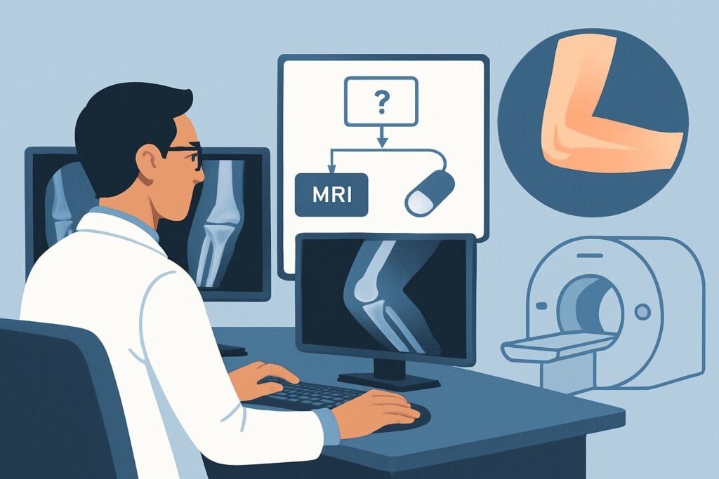
A 17-year-old baseball pitcher presents with three months of persistent, deep posteromedial elbow pain that worsens with throwing. He has focal tenderness over the olecranon, but his range of motion is full. You ordered radiographs two weeks ago, which were read as normal. Despite rest, the pain continues, and you suspect an occult stress fracture or other underlying bone injury that isn’t visible on the initial films. What is the most appropriate next step to confirm the diagnosis and guide management? This article provides a clinical workflow for this specific scenario, grounded in the American College of Radiology (ACR) Appropriateness Criteria. For this presentation, an MRI elbow without IV contrast is rated Usually Appropriate.
## Who Fits This Clinical Scenario?
This guidance applies to a specific patient population: individuals with chronic elbow pain (typically lasting more than three months) where the clinical suspicion points toward an underlying bone abnormality that is not apparent on initial radiographs.
Inclusion criteria for this workflow:
- Chronic, localized pain: The pain is persistent and often associated with focal bony tenderness (e.g., over the olecranon, capitellum, or medial epicondyle).
- History of repetitive stress: The patient is often an athlete involved in overhead or weight-bearing activities (e.g., throwing, gymnastics, weightlifting) or a manual laborer.
- Normal or nonspecific radiographs: Initial X-rays have been performed and show no clear fracture, dislocation, or advanced arthritic changes.
It is critical to distinguish this scenario from similar presentations that follow different diagnostic pathways. This workflow is not for patients whose primary symptoms are:
- Mechanical locking or clicking: These symptoms suggest an intra-articular loose body or cartilage defect, which falls under a different ACR variant.
- Pain localized to the epicondyles with classic signs of tendinopathy: If the presentation is clearly lateral or medial epicondylalgia (tennis or golfer’s elbow), the workup follows the pathway for suspected tendon tears.
- Neurologic symptoms: Pain accompanied by numbness, tingling, or weakness in the hand or forearm suggests a nerve abnormality (e.g., cubital tunnel syndrome) and requires a different imaging approach.
## What Diagnoses Are You Working Up in This Scenario?
When radiographs are negative in a patient with chronic, focal elbow pain, the differential diagnosis shifts to conditions that affect subchondral bone and marrow. The goal of advanced imaging is to identify these occult pathologies.
Occult Stress Fracture
This is the most common and critical diagnosis to consider, particularly in athletes. Repetitive microtrauma from activities like throwing can overwhelm the bone’s capacity for repair, leading to a stress reaction (bone marrow edema) that can progress to a cortical fracture. The olecranon is a classic location in throwing athletes.
Osteochondritis Dissecans (OCD)
Prevalent in adolescent athletes, OCD is a condition affecting the subchondral bone and the overlying articular cartilage. Repetitive compressive forces can disrupt the blood supply to a segment of bone, causing it to soften and potentially separate. The capitellum is the most common site in the elbow.
Bone Contusion or Marrow Edema
This represents a less severe injury on the stress-injury spectrum. While not a fracture, significant bone marrow edema from a direct blow or repetitive microtrauma can be a source of significant pain and a precursor to a more severe stress injury if the offending activity is not modified.
Avascular Necrosis (AVN)
Though less common in the elbow than in the hip or shoulder, AVN can occur, leading to bone death from a compromised blood supply. It may be associated with a history of trauma, high-dose corticosteroid use, or other systemic conditions.
## Why Is MRI Elbow Without IV Contrast the Recommended Study for This Presentation?
The ACR rates MRI elbow without IV contrast as Usually Appropriate for this scenario because of its superior ability to visualize bone marrow and soft tissues without using ionizing radiation.
The primary rationale for this recommendation includes:
- High Sensitivity for Bone Marrow Edema: MRI is the most sensitive imaging modality for detecting bone marrow edema, which is the earliest sign of a stress reaction, bone contusion, or OCD. It can identify these injuries long before cortical changes become visible on radiographs or even CT. This early detection is crucial for initiating proper treatment and preventing progression to a complete fracture or cartilage collapse.
- Comprehensive Anatomic Detail: Beyond bone marrow, MRI provides excellent visualization of articular cartilage, ligaments, and tendons. This allows for the simultaneous assessment of associated injuries, such as cartilage delamination in OCD or adjacent ligamentous strain.
- No Ionizing Radiation: The recommended study, MRI, has a relative radiation level of zero (0 mSv). This is a significant advantage, especially since the patient population often includes children and young adults who are more sensitive to the cumulative effects of radiation.
Why are other studies rated lower for this specific scenario?
- CT elbow without IV contrast is also rated Usually Appropriate and is an excellent alternative if MRI is contraindicated or unavailable. CT provides exquisite detail of the cortical bone and is superior for defining the exact location and extent of a fracture line. However, it is less sensitive than MRI for detecting early stress reactions that only manifest as marrow edema. It also involves a low but non-zero dose of ionizing radiation (ACR RRL=☢☢, 0.1-1 mSv).
- A 3-phase bone scan is rated May be appropriate (Disagreement). While highly sensitive for detecting areas of increased bone metabolism (a “hot spot”), it is not specific. A positive finding could represent a fracture, infection, inflammation, or tumor, often requiring follow-up with MRI or CT for definitive characterization. It also carries a higher radiation dose (ACR RRL=☢☢☢, 1-10 mSv).
- Ultrasound is rated Usually not appropriate for this indication. Ultrasound is superb for evaluating tendons, ligaments, and fluid collections but cannot penetrate bone to visualize marrow edema, making it unsuitable for diagnosing a stress fracture or OCD.
Intravenous contrast is not needed for the primary differential diagnoses in this scenario. Therefore, MRI elbow without and with IV contrast is rated Usually not appropriate, as the addition of contrast provides no significant diagnostic benefit for stress injuries or OCD and adds unnecessary cost and potential risk.
## What’s Next After MRI Elbow Without IV Contrast? Downstream Workflow
The MRI results will directly guide the subsequent clinical management. The workflow typically branches based on whether the findings are positive, negative, or indeterminate.
- If the MRI is positive for a stress fracture or OCD: The next step is typically a referral to an orthopedic or sports medicine specialist. Management will focus on activity modification, which may include a complete cessation of the offending sport or activity, immobilization (casting or bracing), and a structured physical therapy program. For high-grade OCD lesions or displaced fractures, surgical intervention may be necessary.
- If the MRI is negative for bone abnormality: This finding makes a significant osseous injury unlikely. The clinical focus should pivot to other potential causes of chronic elbow pain. Re-evaluate the patient for soft tissue pathologies that may have been overlooked, such as refractory epicondylalgia, collateral ligament sprain, or nerve entrapment. This may lead you to a different ACR workflow, such as the one for a suspected collateral ligament tear or nerve abnormality.
- If the MRI is indeterminate or shows an unexpected finding: In rare cases, the MRI might reveal an unexpected lesion, such as a bone tumor or signs of infection (osteomyelitis). In these situations, the next step depends on the specific finding. This may involve obtaining an MRI with IV contrast for better tissue characterization, a CT scan for bony detail, or a direct referral to an orthopedic oncologist or infectious disease specialist for biopsy and further management.
## Pitfalls to Avoid (and When to Get Help)
Navigating this clinical scenario requires avoiding several common pitfalls to ensure timely and accurate diagnosis.
- Pitfall 1: Over-reliance on negative radiographs. A normal X-ray does not rule out a significant bone injury, especially an early stress fracture. Delaying advanced imaging in a patient with persistent, focal pain and a high-risk history can lead to progression of the injury.
- Pitfall 2: Ordering the wrong advanced study. Choosing ultrasound to look for a stress fracture or ordering an MR arthrogram when an intra-articular loose body is not suspected can lead to non-diagnostic results and unnecessary costs.
- Pitfall 3: Forgetting the pediatric-specific differential. In skeletally immature patients, be highly suspicious of pathology at the growth plates (physes), such as apophysitis or Salter-Harris stress injuries, which have different management implications.
- Pitfall 4: Misinterpreting bone marrow edema. While often indicative of a stress injury, marrow edema can also be seen with infection, tumors, or inflammatory arthritis. The pattern of edema and the clinical context are key to an accurate diagnosis.
If the clinical picture and imaging findings are discordant, or if an aggressive lesion like a tumor or infection is suspected, it is crucial to escalate care. This typically involves a discussion with the interpreting radiologist and a prompt referral to an appropriate orthopedic subspecialist.
## Related ACR Topics and Tools
For a comprehensive overview of all clinical variants related to chronic elbow pain, from initial imaging to specific soft-tissue concerns, please see our parent guide. It provides a breadth-first look at the entire ACR topic.
- Parent Topic Hub: For breadth across all scenarios in Chronic Elbow Pain, see our parent guide: Chronic Elbow Pain: ACR Appropriateness Decoded.
To explore adjacent scenarios, compare imaging techniques, or discuss radiation dose with your patients, the following GigHz tools may be helpful:
- Imaging Appropriateness Selector — for adjacent scenarios
- Imaging Protocol Library — for technique on the recommended study
- Radiation Dose Calculator — for cumulative dose conversations
Frequently Asked Questions
Why is MRI preferred over CT if both are rated ‘Usually Appropriate’?
While both are appropriate, MRI is often preferred because it is more sensitive for detecting the earliest signs of a bone stress injury, specifically bone marrow edema. It also provides superior evaluation of cartilage and surrounding soft tissues without using ionizing radiation, which is a key consideration in the young athletes who frequently present with this condition. CT is an excellent alternative if MRI is contraindicated or if precise visualization of a cortical fracture line is needed.
Is an MR arthrogram ever needed for this scenario?
For this specific scenario—suspected occult fracture or bone abnormality—an MR arthrogram is rated ‘Usually not appropriate.’ An arthrogram involves injecting contrast directly into the joint and is primarily used to evaluate for intra-articular loose bodies, cartilage defects, or ligament tears, particularly of the ulnar collateral ligament. If the clinical suspicion shifts to those diagnoses (e.g., the patient develops mechanical locking), then an arthrogram might be considered under a different clinical variant.
What if my patient has a pacemaker or other contraindication to MRI?
If a patient has an absolute contraindication to MRI, such as a non-compatible pacemaker or certain metallic implants, CT elbow without IV contrast is the best alternative. It is also rated ‘Usually Appropriate’ by the ACR for this scenario and provides excellent detail of the bone cortex to identify a subtle fracture line.
How long should a patient rest before getting an MRI for a suspected stress fracture?
There is no mandatory rest period required before obtaining an MRI. In fact, imaging is often performed to confirm the diagnosis and guide the duration of required rest. If a stress fracture is suspected based on persistent, activity-related pain and focal tenderness despite negative radiographs, proceeding with the MRI is the appropriate next step to avoid diagnostic delay.
Does a finding of ‘bone marrow edema’ on MRI automatically mean a stress fracture?
Not necessarily. Bone marrow edema is a nonspecific finding that indicates fluid accumulation within the bone. While it is the hallmark of a stress reaction or fracture, it can also be caused by a bone contusion (bruise), infection (osteomyelitis), inflammatory arthritis, or a tumor. The radiologist will interpret the pattern and location of the edema in the context of your clinical information to arrive at the most likely diagnosis.
Reviewed by Pouyan Golshani, MD, Interventional Radiologist — May 26, 2026