What Imaging Is Best for Acute Foot Trauma When Ottawa Rules Don’t Apply?
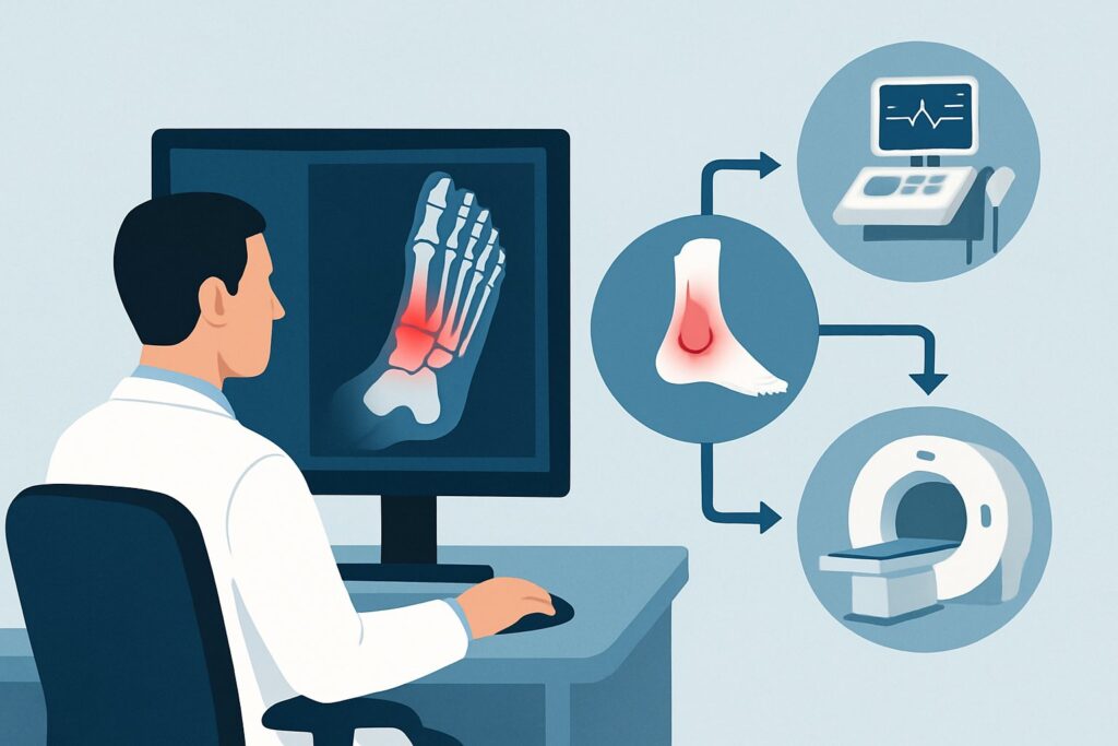
A 10-year-old boy is brought to the urgent care clinic after falling off his scooter. He has significant pain and swelling over the dorsum of his right foot and is unwilling to bear weight. Due to his distress and inability to fully cooperate with the exam, you cannot reliably apply the Ottawa foot rules. You need to decide on the appropriate initial imaging to evaluate for a fracture, balancing diagnostic yield with radiation exposure in a pediatric patient. This article outlines the American College of Radiology (ACR) recommended workflow for this specific clinical scenario, where a reliable physical exam is not possible. For this presentation, the ACR designates Radiography foot as Usually Appropriate.
Who Fits This Clinical Scenario for Acute Foot Trauma?
This guidance applies to a specific subset of patients: adults or children older than 5 years who have sustained acute trauma to the foot, but for whom the Ottawa Ankle Rules (which include a foot examination component) cannot be reliably evaluated. The inability to apply these rules is the key differentiator for this workflow.
Common reasons the Ottawa rules are excluded include:
- Altered Mental Status: Intoxication, head injury, or metabolic derangement.
- Inability to Cooperate: Young age, developmental delay, or severe pain preventing a reliable examination.
- Diminished Sensation: Pre-existing peripheral neuropathy (e.g., from diabetes) that confounds the assessment of point tenderness.
- Distracting Injuries: A significant, painful injury elsewhere (e.g., a femur fracture) that makes the foot examination unreliable.
- Gross Swelling: Severe edema that physically prevents palpation of the key bony landmarks.
This workflow is distinct from scenarios where the Ottawa rules can be applied and are either positive or negative. It also differs from cases with a high pre-test suspicion for a specific complex injury, such as a Lisfranc dislocation or penetrating trauma with a retained foreign body, which may require a different initial imaging approach.
What Diagnoses Are You Working Up When Ottawa Rules Are Excluded?
When a reliable physical exam is off the table, imaging serves as the primary tool to exclude clinically significant injuries that require specific management. The differential diagnosis is broad, but the initial imaging is focused on identifying osseous or articular pathology.
Metatarsal Fractures: These are among the most common foot fractures. The focus is often on the fifth metatarsal, which can sustain several fracture types, including avulsion fractures at the base (pseudo-Jones) and transverse fractures of the proximal diaphysis (Jones fracture), the latter of which can have a higher rate of nonunion. Fractures of the other metatarsals are also common, particularly from direct blows or crush injuries.
Tarsal Bone Fractures: Fractures of the midfoot and hindfoot bones, such as the navicular, cuboid, cuneiforms, talus, and calcaneus, are less common but can carry significant morbidity. Calcaneal and talar fractures, often resulting from high-energy mechanisms like falls from a height, can be complex and frequently require advanced imaging and surgical intervention.
Phalangeal Fractures: Fractures of the toes are very common and often result from direct impact (“stubbed toe”). While many are managed non-operatively with buddy taping, identifying the fracture, its location, and any associated dislocation is crucial for proper treatment and patient counseling.
Dislocations and Subluxations: Traumatic joint dislocations can occur at any level of the foot, from the tarsometatarsal joints (a Lisfranc injury) to the metatarsophalangeal or interphalangeal joints. Radiographs are the essential first step in identifying these injuries, which often represent orthopedic emergencies.
Why Is Foot Radiography the Recommended First Step in This Scenario?
In the setting of an unreliable clinical examination, imaging becomes the primary diagnostic tool. The ACR designates Radiography foot as Usually appropriate because it provides an excellent balance of diagnostic capability, accessibility, speed, and safety for the initial evaluation of acute foot trauma.
Standard foot radiographs, typically including anteroposterior (AP), oblique, and lateral views, are highly effective at identifying the vast majority of fractures and dislocations. The study is widely available in nearly all acute care settings, can be performed quickly, and delivers a very low radiation dose. For adults, the relative radiation level (RRL) is minimal (☢ <0.1 mSv), and for children, it is even lower (☢ <0.03 mSv [ped]). This makes it an ideal screening tool, especially in the pediatric population. Alternative imaging modalities are rated lower for this initial workup for several key reasons:
- CT foot without IV contrast is rated May be appropriate. While CT offers superior detail for complex fractures and can detect occult fractures missed on radiographs, it is not the recommended first-line study. Its use is reserved for situations where radiographs are negative or inconclusive but clinical suspicion for a significant injury (like a subtle tarsal fracture or Lisfranc injury) remains high. The radiation dose, while still low for adults (☢ <0.1 mSv), is notably higher for children (☢☢ 0.03-0.3 mSv [ped]) compared to radiography.
- MRI foot without IV contrast is rated Usually not appropriate for the initial evaluation. MRI provides excellent detail of soft tissues (ligaments, tendons) and bone marrow edema but is more expensive, less available in emergent settings, and takes significantly longer to perform. It is a powerful problem-solving tool used downstream to evaluate for suspected ligamentous tears, stress fractures, or osteochondral injuries after initial radiographs have been obtained.
- US foot is rated Usually not appropriate for a general fracture workup. Ultrasound is highly operator-dependent and cannot adequately visualize the complex osseous anatomy of the entire foot. Its role is limited to specific indications, such as evaluating for tendon rupture, abscess, or a superficial foreign body.
What Is the Downstream Workflow After Initial Foot Radiographs?
The results of the initial foot radiographs will guide the subsequent clinical pathway. The goal is to ensure appropriate treatment and follow-up while avoiding unnecessary additional imaging.
- If the radiograph is POSITIVE for a simple, non-displaced fracture (e.g., a single metatarsal shaft or toe fracture): The typical next step is non-operative management. This usually involves immobilization in a walking boot or post-operative shoe, weight-bearing restrictions, and scheduled follow-up with primary care or orthopedics to monitor healing.
- If the radiograph is POSITIVE for a complex injury (e.g., a displaced or intra-articular fracture, multiple fractures, dislocation, or suspected Lisfranc injury): This requires urgent orthopedic consultation. Advanced imaging, most commonly a CT foot without IV contrast, is often the next step to fully characterize the fracture pattern, assess for articular involvement, and guide surgical planning.
- If the radiograph is NEGATIVE but clinical suspicion remains high: This is a critical decision point. If the patient continues to have severe pain, focal tenderness (if it can be elicited later), or inability to bear weight, an occult fracture is possible. The next step often involves a period of conservative management (immobilization and restricted weight-bearing) with plans for repeat clinical evaluation. If symptoms do not improve, or if a high-risk injury like a navicular stress fracture or subtle Lisfranc injury is suspected, further imaging with CT or MRI may be warranted. This moves the patient into a different clinical scenario, often “Suspect Lisfranc injury, tendon injury, or occult fracture.”
Pitfalls to Avoid (and When to Get Help)
Navigating this scenario requires careful consideration to avoid common diagnostic and management errors.
- Pitfall 1: Underestimating a “negative” radiograph. A normal-appearing initial radiograph does not definitively rule out a significant injury, especially in the midfoot. Maintain a high index of suspicion if the clinical picture (e.g., persistent inability to bear weight) does not match the imaging findings.
- Pitfall 2: Missing a Lisfranc injury. These tarsometatarsal joint injuries can be subtle on non-weight-bearing radiographs. Look for any widening between the first and second metatarsal bases or malalignment of the second metatarsal with the middle cuneiform. If suspected, obtain weight-bearing views if possible or proceed to CT.
- Pitfall 3: Not obtaining adequate views. A standard three-view series is essential. Incomplete imaging can easily miss fractures. Ensure the technologist provides high-quality AP, lateral, and oblique images.
- Pitfall 4: Forgetting pediatric considerations. Be mindful of growth plates (physes) in children, which can mimic fractures. Comparison views of the contralateral foot may be helpful in ambiguous cases.
If you identify a complex fracture pattern, dislocation, or open fracture, or if you have a strong suspicion for a Lisfranc injury despite initial radiographs, immediate orthopedic consultation is the appropriate escalation.
Related ACR Topics and Tools
For a comprehensive overview of all clinical scenarios related to acute foot trauma and for tools to assist in your clinical decision-making, please refer to the following resources.
- For breadth across all scenarios in Acute Trauma to the Foot, see our parent guide: Acute Trauma to the Foot: ACR Appropriateness Decoded.
- To explore other clinical situations, use the ACR Appropriateness Criteria Lookup.
- For details on imaging techniques, consult the Imaging Protocol Library.
- To discuss radiation exposure with patients, the Radiation Dose Calculator can be a helpful aid.
Frequently Asked Questions
Why can’t I just order a CT scan first to be safe, since the exam is unreliable?
While CT is more sensitive for subtle fractures, it is rated ‘May be appropriate’ rather than ‘Usually appropriate’ as the initial study. This is because standard radiography effectively diagnoses the vast majority of clinically significant fractures with a much lower radiation dose, especially important in children. The ACR recommends a stepwise approach, reserving CT for cases where radiographs are negative or inconclusive but a high clinical suspicion for fracture remains.
What if the patient is a child under 5 years old?
This specific ACR variant applies to children older than 5 years. Younger children have different injury patterns and a higher prevalence of cartilaginous structures that are not well-visualized on radiographs. While radiography is still often the first step, the threshold for consultation with a pediatric orthopedic specialist or for alternative imaging may be lower. Always consult pediatric-specific guidelines for this age group.
Does this guidance apply if I suspect an Achilles tendon rupture?
No. This workflow is for a general evaluation of acute foot trauma with a focus on bony injury. If you specifically suspect a major tendon injury like an Achilles rupture, the diagnostic pathway is different. Clinical exam (e.g., the Thompson test) is paramount, and ultrasound or MRI would be the imaging modalities of choice, making that a separate clinical scenario.
If the initial radiograph is negative, when should I consider getting weight-bearing views?
Weight-bearing views are extremely valuable for assessing ligamentous stability, particularly for a suspected Lisfranc injury. If the patient can tolerate it, and initial non-weight-bearing films are equivocal for midfoot alignment, obtaining weight-bearing AP and lateral views can unmask instability that is not apparent on resting images. This should only be attempted if a displaced fracture has been ruled out.
Is an MRI ever the right first choice in acute foot trauma?
For the vast majority of acute traumatic injuries, MRI is not the appropriate initial imaging study and is rated ‘Usually not appropriate’ by the ACR. Its primary role is as a secondary, problem-solving tool for assessing soft tissue structures (ligaments, tendons), cartilage, or bone marrow edema (stress fractures/bone bruises) after initial radiographs have been performed.
Reviewed by Pouyan Golshani, MD, Interventional Radiologist — May 29, 2026