What Is the Next Imaging Study for Suspected Wrist Trauma with Negative X-Rays?
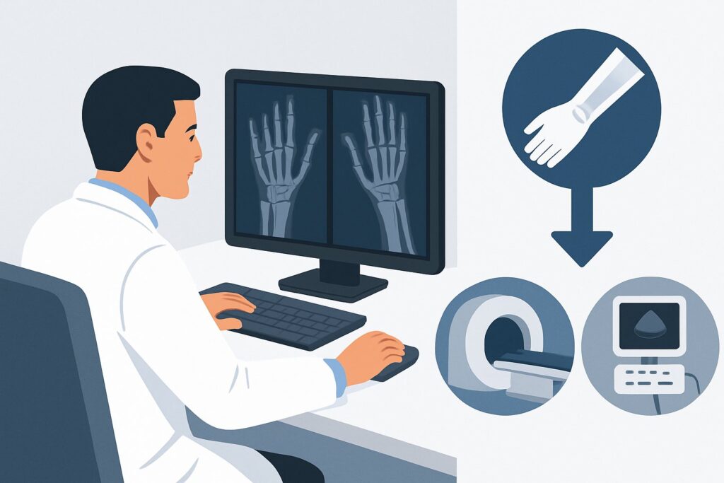
A 24-year-old falls onto an outstretched hand while skateboarding and presents to the emergency department with significant wrist pain. On examination, there is marked tenderness in the anatomic snuffbox. You order a standard three-view series of the wrist, but the initial radiographs are read as negative for acute fracture or dislocation. Despite the normal imaging, your clinical suspicion for an occult scaphoid fracture remains high. What is the most appropriate next step in the imaging workup to avoid missing a consequential injury?
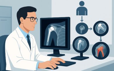
This scenario—persistent suspicion of acute hand or wrist trauma despite negative or equivocal initial radiographs—is a common clinical challenge. The decision between immediate advanced imaging and a period of conservative management followed by repeat imaging has significant implications for cost, radiation exposure, and time to diagnosis. According to the American College of Radiology (ACR) Appropriateness Criteria, the primary recommendation of Radiography area of interest repeat in 10-14 days is rated as Usually Appropriate.
Who Fits This Clinical Scenario for Negative Initial Radiographs?
This clinical workflow is designed for a specific patient population: those with a clear history of acute trauma to the hand or wrist where the initial radiographic studies are unrevealing. The key inclusion criterion is a high index of clinical suspicion for an occult fracture that outweighs the negative initial imaging report. This suspicion is typically based on physical exam findings like focal tenderness over a specific bony structure, such as the scaphoid (“snuffbox” tenderness), triquetrum (dorsal ulnar-sided pain), or hook of the hamate (pain at the hypothenar eminence).
It is critical to distinguish this scenario from several similar but distinct clinical presentations that follow different diagnostic pathways:
- Patients with a confirmed fracture on initial radiographs: If a fracture is already identified, the next imaging question often involves assessing for associated soft tissue injuries. This falls under a different ACR variant, such as Acute wrist fracture on radiographs. Suspect wrist tendon or ligament trauma.
- Patients with obvious malalignment: If the initial exam or radiographs show carpal malalignment or joint dislocation, even without a clear fracture line, the primary concern shifts to ligamentous instability. This is addressed in the variant Initial radiographs showing distal radioulnar joint or carpal malalignment in the absence of fracture.
- Patients with suspected penetrating trauma: When the mechanism involves penetration and there is concern for a retained foreign body, the imaging goals are different. This is covered by the scenario Suspect penetrating trauma with a foreign body in the soft tissues in the hand or wrist.
This guide is exclusively for the patient with a traumatic injury, a concerning physical exam, and unhelpful initial X-rays.
What Diagnoses Are You Working Up with Negative Initial Radiographs?
When initial radiographs are negative, the imaging strategy is aimed at identifying injuries that are commonly radiographically occult in the acute setting. The differential diagnosis guides the choice and timing of the next study.
The most common and consequential concern is an occult scaphoid fracture. The scaphoid’s complex three-dimensional anatomy and orientation mean that non-displaced fracture lines can be easily missed on standard radiographic views. Failure to diagnose and properly immobilize a scaphoid fracture can lead to serious complications, including nonunion, avascular necrosis (AVN) of the proximal pole due to its tenuous retrograde blood supply, and subsequent degenerative arthritis (scaphoid nonunion advanced collapse, or SNAC wrist).
While the scaphoid is the classic culprit, other occult carpal fractures can present similarly. These include fractures of the triquetrum (often dorsal chip fractures from impaction), the hook of the hamate (common in sports involving bats, clubs, or racquets), or the capitate. Like scaphoid fractures, these may not be visible until bone resorption occurs at the fracture site or until cross-sectional imaging is performed.
A significant bone contusion, or bone bruise, is another key consideration. This represents a trabecular microfracture with associated hemorrhage and edema within the bone marrow, but without a discrete cortical fracture line. While it does not carry the same risk of nonunion as a true fracture, a bone contusion can be a source of significant, prolonged pain and disability. It is only visible on MRI.
Finally, a purely ligamentous injury, such as a tear of the scapholunate or lunotriquetral ligaments, can mimic the presentation of an occult fracture. While this scenario prioritizes the search for a fracture, persistent pain despite negative repeat radiographs would shift the diagnostic focus toward these soft-tissue injuries.
Why Are Repeat Radiographs in 10-14 Days Usually Appropriate?
The ACR panel rates three different imaging studies as Usually Appropriate in this scenario, but the recommendation to repeat radiographs serves as a practical and effective default pathway for many patients. The rationale is based on a balance of diagnostic yield, resource utilization, and radiation safety.
The primary reason Radiography area of interest repeat in 10-14 days is recommended is the biological process of fracture healing. In the 10 to 14 days following an injury, osteoclasts begin to resorb bone at the fracture margins. This process slightly widens the fracture line, making a previously invisible fracture become apparent on new radiographs. This approach is highly specific if positive, widely available, low-cost, and involves a minimal amount of ionizing radiation (ACR Relative Radiation Level: Varies, but low). It is a well-established and validated strategy, particularly for suspected scaphoid fractures.
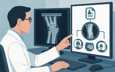
However, two advanced imaging modalities are also rated Usually Appropriate and are often the better choice in specific contexts:
- MRI area of interest without IV contrast: MRI is extremely sensitive for detecting occult fractures and bone marrow edema within hours of an injury. It is the most accurate study for ruling out a fracture definitively. If an MRI is negative for fracture or bone edema, an occult fracture is virtually excluded. This is the preferred next step for high-performance athletes or individuals whose occupation requires a rapid and definitive diagnosis to guide treatment and return-to-activity timelines. It has the significant advantage of using no ionizing radiation (RRL: O 0 mSv).
- CT area of interest without IV contrast: CT provides exquisite detail of cortical bone and is excellent for delineating fracture lines that are equivocal on radiographs. It is faster and often more accessible than MRI. CT is a strong alternative when MRI is contraindicated (e.g., incompatible hardware) or when the primary question is solely the presence of a fracture line, not bone edema or ligamentous injury. It does involve ionizing radiation (RRL: Varies).
In contrast, Bone scan area of interest is rated Usually Not Appropriate. While highly sensitive for bone turnover, it is notoriously non-specific; arthritis, infection, and other conditions can cause a positive result. Furthermore, it has been largely supplanted by the superior anatomic detail of MRI and CT and carries a higher radiation dose (RRL: ☢☢☢ 1-10 mSv).
What Is the Downstream Workflow After Repeat Radiographs?
The results of the follow-up study dictate the next steps in management. The clinical decision tree branches based on whether the imaging confirms, excludes, or remains uncertain about a fracture.
- If repeat radiographs are positive for a fracture: The diagnosis is confirmed. The patient should be referred for orthopedic management. Treatment will depend on the specific bone involved and the fracture pattern (e.g., thumb spica cast for a non-displaced scaphoid waist fracture vs. potential surgical fixation for a displaced or proximal pole fracture). No further diagnostic imaging is typically needed at this stage.
- If repeat radiographs are negative but symptoms persist: A negative follow-up radiograph significantly lowers the probability of a fracture, but does not entirely exclude it, nor does it address potential soft-tissue injury. At this point, the patient’s ongoing symptoms warrant further investigation. The next logical step is to proceed to one of the other Usually Appropriate studies: MRI without IV contrast. MRI can definitively rule out an occult fracture and simultaneously evaluate for bone contusion, ligament tears, or tendinopathy that could be causing the persistent pain.
- If repeat radiographs are equivocal: If the new films show a questionable lucency or sclerosis, the next step is typically CT without IV contrast. CT’s superior spatial resolution is ideal for clarifying ambiguous radiographic findings and confirming or refuting the presence of a cortical breach.
This staged approach, starting with repeat radiographs, effectively triages patients, reserving more expensive and resource-intensive advanced imaging for those who truly need it.
Pitfalls to Avoid (and When to Get Help)
Navigating this scenario requires careful clinical judgment to avoid common diagnostic errors.
- Pitfall 1: Disregarding clinical suspicion. The most significant error is dismissing a patient’s symptoms after a single negative radiograph, especially with classic findings like snuffbox tenderness. This can lead to a missed scaphoid fracture and its associated long-term complications.
- Pitfall 2: Failure to immobilize. While awaiting follow-up imaging, a patient with a high clinical suspicion for a scaphoid fracture should be placed in a thumb spica splint or cast. Treating the patient as if they have a fracture until proven otherwise is the standard of care.
- Pitfall 3: Inadequate follow-up instructions. Patients must understand the importance of returning for their follow-up appointment or repeat imaging. Clearly explain the risk of a missed injury and the rationale for the 10-14 day waiting period.
If a patient presents with signs of neurovascular compromise, such as severe swelling, paresthesias, or diminished pulses, this constitutes a clinical emergency. Escalate immediately for an urgent orthopedic or hand surgery consultation, as this may indicate a compartment syndrome or an unstable injury not apparent on initial films.
Related ACR Topics and Tools
For a comprehensive overview of all clinical variants related to this topic, please see our parent guide. Additional GigHz tools can help you apply these criteria in your daily practice.
- Parent Topic Hub: For breadth across all scenarios in Acute Hand and Wrist Trauma, see our parent guide: Acute Hand and Wrist Trauma: ACR Appropriateness Decoded.
- ACR Appropriateness Criteria Lookup: https://gighz.com/acr-appropriateness-criteria/
- Imaging Protocol Library: https://gighz.com/imaging-protocol-library/
- Radiation Dose Calculator: https://gighz.com/radiation-dose-calculator/
Frequently Asked Questions
Why wait 10-14 days for repeat X-rays instead of getting an MRI right away?
Waiting 10-14 days allows for bone resorption at the fracture site, which can make a previously hidden fracture visible on a standard radiograph. This is a cost-effective and low-radiation strategy. However, obtaining an MRI immediately is also rated ‘Usually Appropriate’ by the ACR and is the preferred approach for elite athletes or when a rapid, definitive diagnosis is required to minimize time in a cast or guide immediate treatment.
If I suspect a scaphoid fracture, should I order a dedicated ‘scaphoid view’ radiograph initially?
Yes. A standard initial radiographic series for suspected scaphoid fracture should include posteroanterior (PA), lateral, oblique, and a PA view with ulnar deviation. The ulnar deviation view elongates the scaphoid and can help reveal subtle waist fractures. This article’s workflow applies after this complete initial series is negative or equivocal.
Is there a role for ultrasound in this scenario?
According to the ACR Appropriateness Criteria, ultrasound of the area of interest is rated ‘Usually Not Appropriate’ for this specific scenario. While ultrasound can be useful for evaluating soft tissues like tendons and ligaments, its ability to detect occult carpal fractures is limited and highly operator-dependent. Radiographs, CT, and MRI are the standard modalities for assessing for occult fractures.
What if the patient cannot get an MRI due to a pacemaker?
If MRI is contraindicated, CT without IV contrast is an excellent alternative and is also rated ‘Usually Appropriate.’ CT provides superb bony detail and is highly effective at identifying occult fracture lines. It is the modality of choice when MRI cannot be performed.
Does this guidance apply to children?
Yes, the principles are similar for pediatric patients, and the ACR ratings for MRI and repeat radiography apply. However, clinical evaluation in children can be more challenging, and the differential diagnosis includes physeal (growth plate) injuries. Consultation with a pediatric radiologist or orthopedic surgeon is often beneficial, and care should be taken to minimize radiation dose (ALARA principle).
Reviewed by Pouyan Golshani, MD, Interventional Radiologist — May 29, 2026