What Is the Next Imaging Study for a Wrist Fracture with Suspected Ligament Injury?
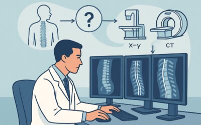
A 45-year-old patient presents to the emergency department after a fall onto an outstretched hand. Radiographs confirm a distal radius fracture, but your clinical exam raises red flags. There is marked point tenderness over the scapholunate interval and a positive Watson test, suggesting significant carpal instability beyond the fracture itself. You know the fracture management is straightforward, but an unstable wrist from a missed ligament tear could lead to long-term disability. The immediate question is which advanced imaging study will best evaluate the ligaments and tendons without delaying care. This article provides a clinical workflow for this specific scenario, where the American College of Radiology (ACR) Appropriateness Criteria rate US wrist as Usually Appropriate for the initial workup of suspected soft tissue injury in the setting of a known wrist fracture.
Who Fits This Clinical Scenario?
This guidance is for a specific patient population: adults or children who have already had initial radiographs confirming an acute wrist fracture, but for whom there is a concurrent, strong clinical suspicion of an associated tendon or ligament injury. This suspicion may arise from the mechanism of injury (e.g., high-energy trauma), specific physical exam findings (e.g., localized tenderness over the scapholunate or lunotriquetral intervals, palpable clunks with provocative maneuvers), or signs of neurologic compromise (e.g., acute carpal tunnel syndrome symptoms).
This workflow does not apply to several similar-but-distinct clinical situations:
- Initial radiographs are negative or equivocal: If x-rays show no fracture but you still suspect an occult fracture or pure ligamentous injury, the imaging pathway is different. This patient falls under the “Suspect acute hand or wrist trauma. Initial radiographs negative or equivocal” variant.
- The fracture is in the hand, not the wrist: If radiographs show a metacarpal or phalangeal fracture with suspected tendon injury (e.g., a boxer’s fracture with concern for extensor tendon disruption), that scenario has its own dedicated workflow.
- Malalignment without fracture: If initial x-rays show carpal malalignment or a widened distal radioulnar joint (DRUJ) but no clear fracture line, the primary concern is pure ligamentous disruption, which follows a separate ACR variant.
The key distinction for this article’s scenario is the coexistence of a confirmed fracture and a suspected soft tissue injury in the wrist.
What Diagnoses Are You Working Up in This Scenario?
When a wrist fracture is accompanied by signs of further instability or dysfunction, the differential diagnosis expands beyond simple bone healing. The goal of advanced imaging is to identify these consequential soft tissue injuries that can alter surgical planning and long-term prognosis.
Scapholunate (SL) Ligament Tear: This is the most common and clinically significant carpal ligamentous injury. A complete tear leads to dorsal intercalated segment instability (DISI), a predictable pattern of carpal collapse that results in scapholunate advanced collapse (SLAC) wrist arthritis if left untreated. It is a critical diagnosis to make, as the management of a distal radius fracture may change significantly if concomitant SL instability is present.
Triangular Fibrocartilage Complex (TFCC) Tear: The TFCC is the primary stabilizer of the distal radioulnar joint (DRUJ). Tears are very common in association with distal radius fractures, particularly those involving the ulnar styloid. Identifying a significant, unstable TFCC tear is crucial for addressing ulnar-sided wrist pain and DRUJ instability after the fracture has healed.
Lunotriquetral (LT) Ligament Tear: While less common than SL tears, LT ligament disruption can cause ulnar-sided wrist pain and a volar intercalated segment instability (VISI) deformity. It is an important consideration in the workup of post-traumatic carpal instability.
Tendon Injury or Entrapment: Tendons can be directly injured by fracture fragments or become entrapped within the fracture site. A classic, though less common, complication is the delayed rupture of the Extensor Pollicis Longus (EPL) tendon after a non-displaced distal radius fracture due to attrition over a sharp bone edge. Imaging can identify tendon sheath fluid, displacement, or frank discontinuity.
Acute Median Nerve Compromise: Swelling and hematoma from the fracture can cause an acute carpal tunnel syndrome. While primarily a clinical diagnosis, imaging can help identify a structural cause, such as a fracture fragment impinging on the carpal tunnel, which may necessitate more urgent surgical intervention.
Why Is Ultrasound the Recommended First Step for Suspected Soft Tissue Injury?
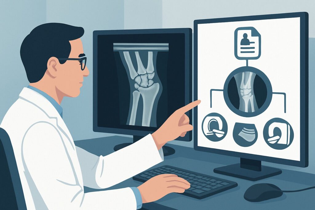
For a patient with a confirmed wrist fracture and suspected soft tissue injury, the ACR designates several modalities as Usually Appropriate, including Ultrasound (US), Magnetic Resonance Imaging (MRI) without contrast, and MR Arthrography. However, ultrasound often serves as the best initial choice due to its unique advantages in this specific context.
Ultrasound of the wrist is a powerful, dynamic imaging tool that provides high-resolution visualization of superficial soft tissues like tendons, extrinsic ligaments, and the median nerve. Its key benefit is the ability to perform a real-time dynamic assessment, observing tendon gliding and ligamentous stability with stress maneuvers. This can be invaluable for confirming suspected instability. Furthermore, US involves no ionizing radiation (0 mSv) and is often more readily accessible and cost-effective than MRI. It is particularly adept at evaluating for EPL tendon integrity and assessing median nerve swelling or compression.
While US is excellent, other modalities are also rated Usually Appropriate and serve important roles:
- MRI wrist without IV contrast (0 mSv): MRI provides a comprehensive, global assessment of the wrist’s anatomy. It is superior to ultrasound for evaluating the intrinsic carpal ligaments (like the scapholunate and lunotriquetral ligaments) and detecting bone marrow edema or occult fractures. If the primary concern is intrinsic carpal instability, MRI is often the definitive non-invasive study.
- MR Arthrography (0 mSv) and CT Arthrography (<0.1 mSv): These are considered the gold standard for diagnosing ligamentous tears, especially partial tears or perforations of the TFCC. By injecting contrast directly into the joint, the study can visualize extravasation through a ligamentous defect. However, they are invasive procedures and are typically reserved for cases where non-invasive imaging is equivocal or for pre-operative planning.
Conversely, several studies are rated Usually Not Appropriate. A CT wrist without IV contrast is excellent for characterizing complex fracture patterns but provides very poor visualization of tendons and ligaments. Adding intravenous contrast (CT with IV contrast) does not improve the evaluation of these non-vascular structures. Similarly, a bone scan is non-specific and would not be helpful in differentiating fracture healing from a ligamentous injury.
What’s Next After Wrist Ultrasound? Downstream Workflow
The results of the initial wrist ultrasound will guide the subsequent clinical and imaging pathway. The goal is to confirm or exclude a significant soft tissue injury that requires a change in management from standard fracture care.
If the ultrasound is positive for a significant injury:
A definitive finding, such as a full-thickness EPL tendon tear or clear evidence of dynamic scapholunate instability, often provides sufficient information for surgical planning. The patient should be referred urgently to an orthopedic or hand surgeon. The surgeon may or may not require additional pre-operative imaging like an MRI or CT arthrogram depending on the specific injury and their preference.
If the ultrasound is negative or inconclusive:
A negative or equivocal ultrasound does not fully exclude an intrinsic ligament injury, particularly a tear of the scapholunate or lunotriquetral ligaments, which can be difficult to visualize completely with US. If high clinical suspicion for carpal instability persists despite a non-diagnostic ultrasound, the next appropriate step is to proceed to MRI wrist without IV contrast. MRI offers a more panoramic view and superior intrinsic ligament detail, making it the ideal problem-solving tool in this situation.
If there is a contraindication to MRI:
For patients who cannot undergo an MRI (e.g., due to an incompatible implanted device), CT arthrography becomes the next best option. While it involves a small amount of ionizing radiation (adult RRL ☢ <0.1 mSv), it provides excellent detail of ligamentous structures when contrast is injected into the joint and is a well-established alternative for diagnosing tears.
Pitfalls to Avoid (and When to Get Help)
Navigating this clinical scenario requires careful integration of exam findings and imaging results. Here are a few common pitfalls to avoid:
- Stopping the workup at the fracture: The most significant error is failing to recognize the clinical signs of ligamentous instability and treating only the bone injury. Always perform a thorough soft tissue exam.
- Relying solely on a negative ultrasound: Remember that ultrasound is operator-dependent and has limitations for deep intrinsic ligaments. Maintain a high index of suspicion for carpal instability if the clinical story fits, and be prepared to order an MRI if the US is unrevealing.
- Ordering the wrong advanced study: A non-contrast CT is the wrong test for ligaments and tendons. Ordering it will expose the patient to radiation without answering the clinical question, delaying the correct diagnosis.
- Ignoring signs of acute nerve compression: Paresthesias or motor weakness in the median nerve distribution after a wrist fracture may represent an orthopedic emergency requiring immediate decompression.
If you identify a complete ligament tear, significant instability on dynamic imaging, or signs of acute nerve compromise, an urgent consultation with a hand or orthopedic surgeon is warranted.
Related ACR Topics and Tools
The ACR Appropriateness Criteria are a living resource, and understanding adjacent scenarios can help refine your clinical decision-making. For breadth across all scenarios in Acute Hand and Wrist Trauma, see our parent guide: Acute Hand and Wrist Trauma: ACR Appropriateness Decoded.
For additional decision support and technical details, the following GigHz tools are available:
- ACR Appropriateness Criteria Lookup — for quickly checking ratings on adjacent or alternative clinical scenarios.
- Imaging Protocol Library — for detailed technical specifications on how studies like wrist MRI or US are performed.
- Radiation Dose Calculator — for discussing cumulative radiation exposure with patients when considering CT-based studies.
Frequently Asked Questions
Why not just get an MRI on everyone with a wrist fracture?
While MRI is excellent for soft tissues, it is not necessary for the vast majority of wrist fractures, which can be managed based on radiographs alone. Advanced imaging is reserved for cases with specific clinical suspicion of a concomitant, treatment-altering injury like a complete ligament tear. Ultrasound is often a faster, more accessible, and equally effective first step for many of these concerns, such as tendon integrity.
What specific physical exam findings should make me suspect a ligament injury in the setting of a fracture?
Key findings include point tenderness directly over the scapholunate (SL) interval (the ‘soft spot’ just distal to Lister’s tubercle) or lunotriquetral (LT) interval. Provocative tests like the Watson scaphoid shift test, where a ‘clunk’ may be felt, are highly suggestive of SL instability. Significant swelling, weakness, or a sense of instability reported by the patient should also raise suspicion.
Is MR Arthrography better than a non-contrast MRI for this scenario?
MR Arthrography is generally considered more sensitive and specific for detecting ligamentous tears, especially partial-thickness tears or TFCC perforations. However, it is an invasive procedure involving a needle injection into the joint. For this reason, a high-quality non-contrast MRI is often preferred as the initial advanced study after an inconclusive ultrasound, as it can provide an excellent diagnosis for most complete tears without the associated risks and discomfort of arthrography.
Can I use a CT scan to look for ligament injuries if I’m already getting one for fracture planning?
A standard CT scan, even with intravenous contrast, is very poor for directly visualizing ligaments and tendons. While it can show secondary signs of instability like carpal malalignment or widened joint spaces, it cannot confirm or rule out a tear. If ligamentous injury is the primary question, MRI or an arthrographic study (CT or MR) is required.
How does the patient’s age affect the choice of imaging?
The principles are largely the same for both pediatric and adult patients. However, there is an even stronger preference for non-ionizing radiation modalities like ultrasound and MRI in children. It’s also important to consider physeal (growth plate) injuries in skeletally immature patients, which are well-visualized on MRI.
Reviewed by Pouyan Golshani, MD, Interventional Radiologist — May 29, 2026