What Imaging Is Best for Routine Asymptomatic Shoulder Arthroplasty Follow-Up?
A 74-year-old patient arrives for his two-year follow-up after a left total shoulder arthroplasty. He is an avid golfer and reports he is thrilled with the outcome, demonstrating excellent range of motion and no pain. As the managing clinician, you consider whether surveillance imaging is necessary to assess the implant’s integrity, even in the absence of symptoms. This common clinical decision point—evaluating a well-functioning joint replacement—requires a clear, evidence-based approach to avoid unnecessary radiation and cost. This article details the American College of Radiology (ACR) workflow for this specific scenario. For routine follow-up of an asymptomatic primary shoulder arthroplasty, the ACR designates Radiography shoulder as Usually Appropriate.
Who Fits This Clinical Scenario for Asymptomatic Arthroplasty Follow-Up?
This guidance applies specifically to patients who have undergone a primary shoulder arthroplasty—including total shoulder arthroplasty (TSA), reverse total shoulder arthroplasty (rTSA), or hemiarthroplasty—and are now presenting for routine, scheduled follow-up. The defining characteristic of this scenario is the complete absence of new or worsening symptoms. The patient should report no pain, weakness, instability, clicking, grinding, or loss of function related to the operative shoulder.
This workflow is intended for surveillance, not diagnosis. It is crucial to distinguish this patient from those with similar histories but different presentations. This guidance does not apply if:
- The patient is symptomatic. Any new onset of pain, weakness, or mechanical symptoms places the patient in a different diagnostic category, typically requiring initial radiographs followed by potential advanced imaging.
- Infection is suspected. Clinical signs such as fever, erythema, warmth, or drainage around the shoulder joint necessitate a distinct workup focused on periprosthetic joint infection.
- There has been recent trauma. A fall or direct blow to the shoulder requires an evaluation for periprosthetic fracture, which follows a separate imaging pathway.
Applying this asymptomatic surveillance protocol to a symptomatic patient can delay the diagnosis of clinically significant complications.
What Diagnoses Are You Monitoring For in Routine Asymptomatic Follow-Up?
Even when a patient feels well, surveillance imaging aims to detect subclinical signs of potential long-term problems before they become symptomatic and more difficult to manage. The differential for findings on routine follow-up radiographs is focused on the biomechanical stability and longevity of the implant.
Component Loosening or Migration
This is a primary concern in long-term arthroplasty follow-up. Aseptic (non-infectious) loosening is the most common reason for late-term revision surgery. Radiographs are highly effective at detecting subtle changes in component position, such as subsidence of the humeral stem or tilting of the glenoid component, which may precede the onset of pain.
Progressive Periprosthetic Lucency
Radiographs allow for the assessment of the bone-implant or bone-cement interface. The development or progression of radiolucent lines greater than 2 mm in width, particularly in certain zones around the glenoid or humeral components, is a key indicator of potential loosening that requires closer monitoring.
Polyethylene Wear
While the polyethylene liner itself is radiolucent, its wear can be inferred indirectly on radiographs. Asymmetric positioning of the humeral head relative to the glenoid component (eccentricity) suggests uneven wear of the polyethylene, a process that can lead to instability and osteolysis over time.
Hardware Failure
Though uncommon, catastrophic failure of the implant itself, such as a fracture of the humeral stem or dissociation of a modular component, can occur. Routine radiographs can identify signs of implant fatigue or failure before a catastrophic event.
Heterotopic Ossification
The formation of bone in the soft tissues surrounding the joint can occur after arthroplasty. While often asymptomatic, extensive heterotopic ossification can eventually lead to stiffness and loss of motion, and its progression is easily monitored with radiographs.
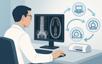
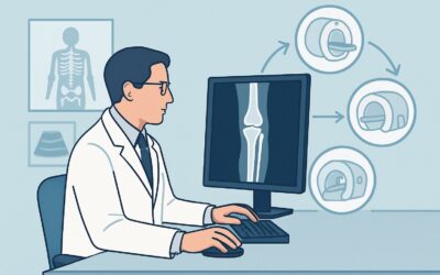
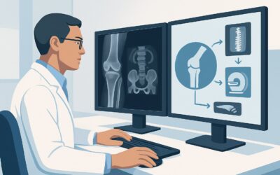
Why Are Shoulder Radiographs the Recommended Study for Routine Follow-Up?
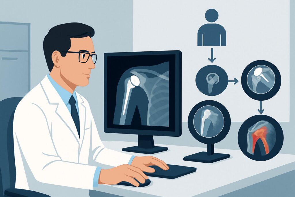
The American College of Radiology (ACR) has designated Radiography shoulder as Usually Appropriate for the routine follow-up of an asymptomatic patient with a primary shoulder arthroplasty. This recommendation is based on the modality’s high utility for assessing the most relevant potential complications in this specific clinical context, balanced against its low risk and cost.
A standard shoulder arthroplasty series, typically including a true anteroposterior (AP) view in the scapular plane (Grashey view) and an axillary lateral view, provides a comprehensive assessment. These views are critical for evaluating component alignment, detecting periprosthetic lucency, assessing for wear, and ensuring hardware integrity. The comparison with prior examinations is paramount for detecting subtle, progressive changes that signal early implant failure.
The radiation dose associated with shoulder radiography is minimal. With a relative radiation level (RRL) of ☢ <0.1 mSv, the exposure is significantly lower than the average annual background radiation in the United States. This makes it a safe and repeatable examination for long-term surveillance.
Why Other Modalities Are Not Recommended
In contrast to radiography, more advanced imaging modalities are rated as Usually Not Appropriate for this asymptomatic screening scenario.
- CT shoulder (without or with contrast): While excellent for detailed bone assessment, CT delivers a substantially higher radiation dose (☢☢☢ 1-10 mSv). Its use is not justified in an asymptomatic patient when radiographs can effectively screen for the primary concerns of loosening and wear. CT is reserved for cases where radiographs are equivocal or when a specific problem, like subtle osteolysis, is suspected based on radiographic findings.
- MRI and US shoulder: These modalities are primarily used to evaluate soft tissues, such as the rotator cuff tendons and muscles. In a patient with no symptoms, there is no clinical indication to assess for soft tissue pathology. Furthermore, metal artifact from the arthroplasty components significantly degrades image quality on MRI, limiting its diagnostic value even when indicated. Both MRI and US are rated Usually Not Appropriate for this routine surveillance task.
What’s the Next Step After Routine Shoulder Radiographs?
The results of surveillance radiographs guide the subsequent management plan and follow-up interval. The workflow branches based on whether the findings are stable, show concerning changes, or are indeterminate.
If Radiographs Are Stable and Unchanged
When the radiographs show no change compared to previous studies—meaning the components are well-fixed, there are no new or progressive radiolucent lines, and no signs of eccentric wear—the finding is reassuring. The appropriate next step is to continue routine clinical follow-up as scheduled by the orthopedic surgeon. No further imaging is warranted until the next planned surveillance interval (e.g., in 2-5 years, per institutional protocol).
If Radiographs Show Concerning Changes
If the images reveal new or progressive findings, such as widening radiolucent lines, a shift in component position (subsidence or tilt), or significant eccentric wear, action is required even if the patient remains asymptomatic. These findings may represent early, subclinical implant failure. The next step is typically a consultation with the operating surgeon. Depending on the specific finding, the surgeon may recommend shortening the follow-up interval for closer monitoring or proceeding to advanced imaging, such as a CT scan, to better characterize the extent of bone loss or loosening. The patient’s clinical scenario effectively shifts from “routine follow-up” to “suspected loosening.”
If Radiographs Are Indeterminate
Occasionally, technical factors like poor patient positioning or improper radiographic technique can render an exam difficult to interpret. In this case, the first and most logical step is to repeat the radiographs with careful attention to technique to obtain diagnostic-quality images. Persistently indeterminate findings are rare but would warrant discussion with the radiologist and orthopedic surgeon to decide on the best course of action.
Pitfalls to Avoid (and When to Get Help)
Navigating asymptomatic surveillance requires discipline to avoid common missteps that can lead to either missed diagnoses or unnecessary testing.
- Pitfall 1: Inconsistent Views. Failing to obtain a standardized series, especially a true AP Grashey and an axillary lateral view, severely limits the ability to compare with prior studies and detect subtle component migration.
- Pitfall 2: Over-reliance on Patient Symptoms. Do not dismiss subtle but progressive radiographic changes (e.g., a new radiolucent line) simply because the patient feels well. The goal of surveillance is to detect these issues before they cause symptoms.
- Pitfall 3: Premature Advanced Imaging. Ordering a CT or MRI for an asymptomatic patient “just in case” is contrary to ACR guidelines, exposing the patient to needless radiation or cost and often yielding incidental findings that complicate management.
If radiographs show clear evidence of component shift, progressive and significant radiolucency, or hardware fracture, escalate immediately to the orthopedic surgeon for management, as these findings may require intervention even in an asymptomatic patient.
Related ACR Topics and Tools
For a comprehensive overview of all clinical scenarios related to shoulder arthroplasty imaging, further reading and specialized tools can provide additional context. The resources below are designed to assist in evidence-based ordering and dose management.
- For breadth across all scenarios in Imaging After Shoulder Arthroplasty, see our parent guide: Imaging After Shoulder Arthroplasty: ACR Appropriateness Decoded.
- To explore other clinical presentations, use the Imaging Appropriateness Selector.
- For details on imaging techniques, consult the Imaging Protocol Library.
- To discuss radiation exposure with patients, use the Radiation Dose Calculator.
Frequently Asked Questions
How often should routine radiographs be performed after shoulder arthroplasty in an asymptomatic patient?
There is no universal consensus, and protocols vary by surgeon and institution. A common schedule includes imaging immediately post-op, at 6 weeks, 1 year, 2 years, 5 years, and then every 3-5 years thereafter, provided the patient remains asymptomatic and prior radiographs are stable.
Does this guidance apply to both total shoulder and reverse total shoulder arthroplasty?
Yes. The ACR Appropriateness Criteria for this scenario apply to all common forms of primary shoulder arthroplasty, including anatomic total shoulder, reverse total shoulder, and hemiarthroplasty. The principle of using radiographs for routine asymptomatic surveillance is the same.
What specific radiographic views are most important for arthroplasty follow-up?
A standard arthroplasty series should include at a minimum: a true anteroposterior (AP) view in the scapular plane (Grashey view) to assess the glenohumeral articulation without overlap, and an axillary lateral view. The axillary view is crucial for evaluating glenoid component loosening, version, and posterior humeral head subluxation.
If a patient becomes symptomatic between scheduled follow-ups, should I wait for the routine imaging appointment?
No. The onset of new symptoms such as pain, instability, weakness, or clicking fundamentally changes the clinical scenario from ‘routine follow-up’ to a diagnostic workup. The patient should be evaluated promptly, and the imaging pathway will follow the ACR guidelines for a symptomatic patient, not this surveillance protocol.
Is there any role for ultrasound in asymptomatic follow-up of a shoulder replacement?
According to the ACR, ultrasound is rated ‘Usually Not Appropriate’ for this specific scenario. Ultrasound’s primary utility is in evaluating soft tissues like the rotator cuff. In an asymptomatic patient with a well-functioning arthroplasty, there is no clinical indication to assess the soft tissues, and the implant can cause acoustic shadowing that limits the exam.
Reviewed by Pouyan Golshani, MD, Interventional Radiologist — May 26, 2026