What Imaging Is Best for a Limping Child with Nonlocalized Symptoms and Suspected Infection?
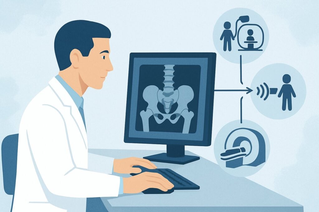
A 4-year-old is brought to the emergency department with a two-day history of refusing to bear weight on his right leg. He has a low-grade fever, is irritable, and cannot point to a specific area of pain, gesturing vaguely at his entire leg and hip. Laboratory studies reveal an elevated C-reactive protein (CRP) and erythrocyte sedimentation rate (ESR), heightening concern for an infectious process like osteomyelitis or septic arthritis. You need to choose the initial imaging study that can accurately identify the source of infection without localizing signs to guide you. This article provides a detailed clinical workflow for this exact scenario, based on the American College of Radiology (ACR) Appropriateness Criteria.
For a young child with an acute limp, nonlocalized symptoms, and concern for infection, the ACR rates MRI lower extremity without and with IV contrast as Usually Appropriate.
Who Fits This Clinical Scenario?
This guidance is specifically for children up to age 5 presenting with an acute limp where the source of pain is unclear. The key inclusion criteria are:
- Age: Up to 5 years old.
- Presentation: Acute limp or refusal to bear weight.
- Symptoms: Nonlocalized. The child cannot or will not point to a specific site of pain. The examination does not reveal a clear focal point of tenderness.
- Clinical Suspicion: Concern for an infectious etiology, supported by systemic signs like fever or laboratory evidence such as elevated inflammatory markers (e.g., CRP, ESR, or white blood cell count).
This workflow is distinct from other similar presentations. This guidance does not apply if:
- Symptoms are localized: If the child has focal tenderness, swelling, or limited range of motion at a specific joint like the hip or knee, you should consult the ACR variant for symptoms localized to the hip or lower extremity.
- There is no concern for infection: If the child is afebrile and inflammatory markers are normal, the differential diagnosis shifts away from infection, and a different imaging pathway is indicated.
- There is a clear history of significant trauma: In cases of a known fall or injury, radiographs are typically the first-line imaging modality to evaluate for fracture.
What Diagnoses Are You Working Up in This Scenario?
When a young child presents with a limp and signs of infection but no localizing symptoms, the differential diagnosis is broad. The goal of imaging is to pinpoint the location and nature of the pathology, which most commonly includes musculoskeletal infections.
Osteomyelitis: This infection of the bone is a primary concern. In young children, it most often affects the metaphyses of long bones like the femur or tibia. Hematogenous spread is the typical mechanism. Early diagnosis is critical to prevent bone destruction and chronic infection. MRI is highly sensitive for detecting the early bone marrow edema that precedes changes visible on radiographs.
Septic Arthritis: An infection within a joint space, most commonly the hip or knee in this age group. This is a true orthopedic emergency, as the inflammatory process can rapidly destroy articular cartilage. A child may not localize pain to the hip, instead presenting with a limp or generalized leg pain. Imaging must be able to detect a joint effusion and assess for synovial enhancement, which are key features.
Pyomyositis: A primary bacterial infection of skeletal muscle, which can progress from diffuse inflammation to a drainable abscess. While less common than bone or joint infections, it can present similarly with fever, pain, and limping. MRI is the modality of choice for identifying and characterizing intramuscular fluid collections.
Discitis or Vertebral Osteomyelitis: An infection of the intervertebral disc space or adjacent vertebral bodies. A young child with this condition may present with refusal to walk, sit, or bend over. The pain can be vague and poorly localized, often mimicking hip or leg pathology. An imaging study with a large field of view is necessary to avoid missing this diagnosis.
Why Is MRI of the Lower Extremity Without and With IV Contrast the Recommended Study?
The ACR designates MRI lower extremity without and with IV contrast as Usually Appropriate because it provides the highest diagnostic utility for the key differential diagnoses in this challenging clinical scenario. It directly visualizes bone marrow, joint spaces, and surrounding soft tissues with superior contrast resolution.
The rationale for this choice involves several factors:
- High Sensitivity for Early Infection: MRI can detect the earliest signs of osteomyelitis—bone marrow edema—days before changes become apparent on radiographs. This allows for prompt initiation of antibiotic therapy.
- Comprehensive Evaluation: A single MRI examination can assess for osteomyelitis, septic arthritis, pyomyositis, and soft tissue abscesses. Because the symptoms are nonlocalized, MRI’s ability to survey a large anatomical area from the pelvis to the ankle is a significant advantage.
- Role of IV Contrast: The administration of gadolinium-based contrast is crucial. It helps differentiate a phlegmon (diffuse inflammation) from a well-defined, rim-enhancing abscess that may require surgical drainage. It also highlights synovial enhancement in septic arthritis.
- No Ionizing Radiation: MRI does not use ionizing radiation (0 mSv), a critical consideration in the pediatric population. This avoids the radiation exposure associated with other modalities.
Alternative studies are rated lower for specific reasons in this context:
- Radiography (Pelvis, Femur, etc.): Rated Usually not appropriate. Plain films have very low sensitivity for early musculoskeletal infections. They are primarily useful for detecting fractures or late-stage bone destruction, which is often absent at initial presentation. A negative radiograph does not rule out infection.
- 3-Phase Bone Scan: Rated May be appropriate. While highly sensitive for detecting areas of inflammation, nuclear medicine scans lack specificity. They cannot reliably distinguish infection from trauma, infarction, or other inflammatory conditions. Furthermore, they deliver a significant radiation dose (☢☢☢☢ 3-10 mSv) and provide poor anatomical detail compared to MRI.
A practical consideration for ordering MRI in a child under 5 is the frequent need for sedation or general anesthesia to prevent motion artifact. This requires coordination with anesthesiology and should be anticipated when scheduling the study.
What’s Next After MRI? Downstream Workflow
The results of the contrast-enhanced MRI will guide the subsequent management steps. The workflow branches based on the findings:
- If the MRI is positive for septic arthritis or a drainable abscess (osteomyelitis or pyomyositis): This constitutes an orthopedic emergency. An immediate consultation with pediatric orthopedics is required for surgical intervention, typically joint aspiration/washout or incision and drainage of the abscess.
- If the MRI shows osteomyelitis or pyomyositis without a drainable collection: The patient should be admitted for IV antibiotic therapy. A consultation with pediatric infectious diseases is recommended to guide the choice and duration of antibiotics, which may be informed by blood cultures or subsequent bone biopsy if the clinical response is poor.
- If the MRI is negative: A negative, high-quality MRI makes a significant musculoskeletal infection highly unlikely. The clinical team should reconsider the differential diagnosis. This may include non-infectious inflammatory conditions (like transient synovitis, although less likely with high inflammatory markers), occult trauma not yet visible, or even referred pain from an intra-abdominal or pelvic source. Further workup may be guided by the persistence of symptoms.
- If the MRI is indeterminate: In rare cases, findings may be equivocal. The next steps may involve close clinical observation with serial lab monitoring, a repeat MRI in several days, or consultation with a pediatric radiologist to discuss the findings and potential alternative imaging, such as a whole-body MRI, which is rated May be appropriate.
Pitfalls to Avoid (and When to Get Help)
Navigating this scenario requires avoiding several common pitfalls:
- Relying on Negative Radiographs: Do not be falsely reassured by normal plain films. They are expected to be normal in the early stages of musculoskeletal infection. Delaying advanced imaging based on a negative x-ray can lead to worse outcomes.
- Omitting IV Contrast: Ordering a non-contrast MRI can significantly limit its diagnostic value. Contrast is essential for identifying abscesses and assessing synovial inflammation. Always specify “without and with IV contrast” when infection is suspected.
- Limited Field of View: When symptoms are nonlocalized, ensure the imaging request covers a sufficient area (e.g., “unilateral lower extremity from hip to ankle”) to avoid missing the site of pathology.
- Underestimating the Need for Sedation: A motion-degraded MRI is often non-diagnostic. Proactively plan for sedation or anesthesia to ensure a high-quality study is obtained on the first attempt.
If the child shows signs of sepsis or hemodynamic instability, escalate care immediately for resuscitation, obtain blood cultures, and start empiric broad-spectrum antibiotics, often before imaging is completed.
Related ACR Topics and Tools
For a comprehensive overview of all clinical variants related to the acutely limping child, or to explore the tools used to develop this guidance, please refer to the following resources.
- For breadth across all scenarios in Acutely Limping Child Up To Age 5, see our parent guide: Acutely Limping Child Up To Age 5: ACR Appropriateness Decoded.
- To review other clinical scenarios and their corresponding ACR recommendations, use the ACR Appropriateness Criteria Lookup.
- For detailed procedural techniques, explore the Imaging Protocol Library.
- To discuss radiation exposure with families for other imaging modalities, consult the Radiation Dose Calculator.
Frequently Asked Questions
Why not start with an ultrasound of the hip if septic arthritis is a concern?
Ultrasound of the hip is rated as ‘May be appropriate’. It is excellent for detecting a hip effusion, but it cannot assess the bone for osteomyelitis or evaluate soft tissues for pyomyositis. In a child with nonlocalized symptoms, a negative hip ultrasound does not rule out infection elsewhere in the leg. MRI provides a more comprehensive evaluation of all potential sources of infection in a single study.
What if my institution has limited or no immediate access to pediatric MRI?
If MRI is not readily available, a 3-phase bone scan is rated ‘May be appropriate’ and can be a reasonable alternative to identify an inflammatory focus. However, it involves significant radiation and is less specific. If clinical suspicion for septic hip arthritis is high, an ultrasound-guided hip aspiration may be performed for diagnostic and therapeutic purposes. Transfer to a center with pediatric MRI capabilities should be strongly considered.
Is a whole-body MRI a better option since the symptoms are nonlocalized?
Whole-body MRI (both without and with contrast) is rated ‘May be appropriate’. It can be useful in cases of suspected multifocal osteomyelitis or when the infection source is highly elusive. However, it often has lower spatial resolution than a dedicated extremity MRI and takes longer to perform. For a unilateral limp, a dedicated lower extremity MRI is typically the more focused and higher-yield initial study.
Should I order labs before or after the MRI?
Laboratory studies, including a complete blood count (CBC) with differential, C-reactive protein (CRP), and erythrocyte sedimentation rate (ESR), should be obtained as soon as possible. These results are critical for establishing the pre-test probability of infection and are often required to justify the need for an urgent MRI. Blood cultures should also be drawn before starting antibiotics.
What if the child has a clear history of minor trauma but also a fever?
This is a challenging situation, as trauma can predispose a child to infection (e.g., hematoma seeding). While an occult fracture (like a toddler’s fracture) is possible, the presence of fever and elevated inflammatory markers should keep infection as the primary concern. MRI is an excellent modality in this case, as it can diagnose both an occult fracture (via bone marrow edema) and an overlying osteomyelitis.
Reviewed by Pouyan Golshani, MD, Interventional Radiologist — May 29, 2026