What Is the Best Initial Imaging for a Child with Suspected Stroke and a Low-Flow Vascular Anomaly?

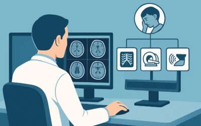
A 10-year-old with a known facial venous malformation presents to the emergency department with sudden-onset left-sided weakness and aphasia. The clinical team is concerned for an acute stroke, but the underlying vascular anomaly complicates the diagnostic picture. Is the stroke arterial or venous? Is it ischemic or hemorrhagic? Choosing the right initial imaging study is critical for guiding immediate management and preventing long-term disability. This article provides a detailed workflow for this specific scenario: a child with stroke symptoms and a known or suspected low-flow vascular anomaly. According to the American College of Radiology (ACR) Appropriateness Criteria, an MRI of the head without IV contrast is the ‘Usually appropriate’ first step in this complex presentation.
Who Fits This Clinical Scenario for Pediatric Stroke Imaging?
This guidance applies to a specific pediatric population: children, from infancy through adolescence, presenting with acute neurological deficits suggestive of a stroke, in the context of a known or suspected low-flow vascular anomaly. These anomalies include conditions like venous malformations, developmental venous anomalies (DVAs), or cavernous malformations. The key feature is the “low-flow” nature, which implies a distinct pathophysiology—primarily related to venous stasis, thrombosis, and congestion—compared to high-flow arterial lesions.
This workflow is not intended for all pediatric stroke presentations. It is crucial to distinguish this scenario from several related but distinct clinical situations that require different imaging pathways:
- Suspected High-Flow Arteriopathy: If the clinical suspicion is for a high-flow condition like moyamoya disease or an arteriovenous malformation (AVM), the imaging strategy shifts to prioritize arterial and hemodynamic assessment, often involving MRA or CTA.
- Confirmed Nontraumatic Hemorrhage: If a patient presents with a known intraparenchymal hemorrhage of unknown etiology, the next imaging study is aimed at identifying the underlying cause, which may require different sequences or modalities.
- Acute Intervention Candidate: For a child who is a candidate for emergent intervention (e.g., mechanical thrombectomy for a large vessel occlusion), the need for speed may elevate the role of CT and CTA to rapidly confirm the diagnosis and guide treatment.
Correctly identifying the patient’s specific clinical context ensures the most appropriate and highest-yield initial imaging study is selected.
What Diagnoses Are You Working Up in a Child with Stroke and a Low-Flow Anomaly?
In this scenario, the differential diagnosis extends beyond a typical arterial ischemic stroke. The presence of a low-flow vascular anomaly introduces unique pathophysiologic possibilities that the initial imaging must be able to differentiate.
The most urgent diagnosis to confirm or exclude is an acute ischemic stroke. In the setting of a low-flow malformation, this can occur via several mechanisms. Stagnant blood flow within the anomaly can lead to thrombus formation, which may then propagate or embolize, causing an arterial or venous infarction. The imaging must be highly sensitive to the earliest cellular changes of ischemia.
A closely related and critical consideration is venous infarction or congestion. Unlike a classic arterial stroke, this condition results from thrombosis within the low-flow anomaly or its draining veins. This impairs venous outflow, leading to parenchymal edema, ischemia, and potential hemorrhagic transformation. The imaging findings can be subtle and may not follow a typical arterial territory, making modality choice essential.
While less common as a primary event from a low-flow lesion compared to high-flow AVMs, hemorrhagic stroke or the hemorrhagic conversion of a venous infarct is a significant risk. The initial imaging study must be able to reliably detect acute hemorrhage, as its presence would fundamentally alter management, particularly regarding anticoagulation or antiplatelet therapy.
Finally, imaging helps differentiate true stroke from common stroke mimics in children. Conditions such as seizure with postictal paralysis (Todd’s paralysis), hemiplegic migraine, or metabolic encephalopathies can present with focal neurological deficits. While the diagnosis is often clinical, imaging is crucial for ruling out an underlying structural cause or acute vascular event.
Why Is MRI Head without IV Contrast the Recommended Initial Study?
For a child with suspected stroke in the setting of a low-flow vascular anomaly, MRI of the head without IV contrast is rated ‘Usually appropriate’ by the ACR because it provides the most comprehensive diagnostic information in a single, non-invasive study without ionizing radiation. Its superior soft-tissue contrast and specialized sequences are uniquely suited to address the key differential diagnoses in this scenario.
The cornerstone of this recommendation is Diffusion-Weighted Imaging (DWI), an MRI sequence that is exceptionally sensitive for detecting the cytotoxic edema of an acute ischemic stroke within minutes of onset. This is the most reliable way to confirm or exclude acute infarction. Furthermore, sequences like Susceptibility-Weighted Imaging (SWI) or Gradient-Recalled Echo (GRE) are highly sensitive for detecting blood products, making them excellent for identifying acute hemorrhage or the subtle microhemorrhages associated with cavernous malformations.
In contrast, other imaging modalities have significant limitations for this specific presentation:
- CT head without IV contrast, while also rated ‘Usually appropriate’ and often faster to obtain, is significantly less sensitive for early ischemic changes. An early CT can be falsely negative for infarction, delaying diagnosis. While excellent for detecting large acute hemorrhages, it is less sensitive than MRI’s SWI/GRE sequences for smaller bleeds or chronic blood products. Its use of ionizing radiation (pediatric RRL ☢☢☢ 0.3-3 mSv) is also a key consideration in children.
- CTA head with IV contrast is rated ‘May be appropriate (Disagreement)’. While it provides excellent arterial detail, it is less helpful for characterizing the venous pathology central to this scenario and offers limited information about the brain parenchyma compared to MRI. It also involves both IV contrast and a higher radiation dose (pediatric RRL ☢☢☢☢ 3-10 mSv).
The non-contrast MRI provides the critical parenchymal information needed to make an initial diagnosis. It directly visualizes infarction on DWI and hemorrhage on SWI/GRE, answering the most urgent clinical questions without the risks of radiation or contrast agents.
What’s Next After MRI Head without IV Contrast? Downstream Workflow
The results of the initial non-contrast head MRI will dictate the subsequent diagnostic and therapeutic pathway. The workflow branches based on whether the findings are positive, negative, or indeterminate.
If the MRI is positive for acute stroke: The findings guide the next steps. If DWI confirms an acute infarct, the pattern (arterial vs. venous territory) helps determine the likely mechanism. A venous-pattern infarct would prompt further investigation of the low-flow anomaly with dedicated vascular imaging, such as MR venography (MRV), to assess for thrombosis. An arterial-pattern infarct may suggest an embolic source, leading to a broader workup including cardiac and further cervicocranial vascular imaging. The presence of hemorrhage on SWI/GRE sequences would immediately influence decisions regarding anticoagulation.
If the MRI is negative for acute stroke or hemorrhage: This result makes an acute vascular event much less likely and increases the suspicion for a stroke mimic. Clinical re-evaluation for conditions like hemiplegic migraine, postictal state, or metabolic disturbance is warranted. If clinical suspicion for a vascular event remains high despite a negative initial MRI, repeating the MRI in 24-48 hours could be considered, as some changes may evolve.
If the MRI is indeterminate: Sometimes, findings can be ambiguous. For example, there may be non-specific T2/FLAIR signal abnormality without clear restricted diffusion, raising questions of subacute ischemia, inflammation, or postictal changes. In these cases, a follow-up MRI with IV contrast (‘May be appropriate’) can be valuable. Contrast enhancement can help characterize the age of an infarct, identify breakdown of the blood-brain barrier, or reveal underlying inflammation or neoplasm that could be mimicking a stroke.
Pitfalls to Avoid (and When to Get Help)
Navigating this clinical scenario requires careful attention to avoid common diagnostic traps.
- Over-reliance on CT: While fast, a negative non-contrast CT does not rule out an acute ischemic stroke. Delaying an MRI based on a normal CT in a child with high clinical suspicion for stroke is a significant pitfall.
- Ignoring the Venous System: In the context of a low-flow anomaly, the stroke mechanism is often venous. Ensure that the imaging and interpretation specifically evaluate for venous sinus or cortical vein thrombosis.
- Misinterpreting Stroke Mimics: Not all focal deficits are stroke. Be prepared to pivot the workup toward non-vascular causes if the high-sensitivity MRI is negative for an acute vascular event.
- Forgetting Sedation Needs: Young children often require sedation or anesthesia to obtain a high-quality, motion-free MRI. This requires logistical planning and should be anticipated to avoid imaging delays.
If the initial imaging is complex, non-diagnostic, or reveals an unexpected finding, immediate consultation with a pediatric neurologist and a neuroradiologist is essential to formulate the best plan for further investigation and management.
Related ACR Topics and Tools
For a comprehensive overview of imaging for pediatric cerebrovascular disease, including scenarios not covered here, please refer to the parent topic article. The following GigHz resources can also support clinical decision-making:
- For breadth across all scenarios in Cerebrovascular Disease-Child, see our parent guide: Cerebrovascular Disease-Child: ACR Appropriateness Decoded.
- For adjacent scenarios and alternative presentations, consult the ACR Appropriateness Criteria Lookup.
- For details on imaging techniques, explore the Imaging Protocol Library.
- To discuss cumulative radiation exposure with families, use the Radiation Dose Calculator.
Frequently Asked Questions
Why is non-contrast MRI preferred over a contrast-enhanced MRI as the initial study?
A non-contrast MRI is preferred initially because its core sequences—DWI for ischemia and SWI/GRE for hemorrhage—answer the most urgent clinical questions without the need for IV contrast. This avoids potential gadolinium exposure and associated risks, especially in children. A contrast-enhanced study is rated ‘May be appropriate’ and is typically reserved as a second step if the initial study is indeterminate or if there is a need to assess for inflammation or blood-brain barrier breakdown.
If my hospital’s MRI scanner is unavailable, is a non-contrast CT an acceptable first step?
Yes, a non-contrast CT of the head is also rated ‘Usually appropriate’ by the ACR and is a reasonable first step if MRI is not immediately available. It is fast and excellent for ruling out significant hemorrhage. However, it is crucial to remember that a negative CT does not rule out acute ischemic stroke. If clinical suspicion remains high, transfer or arrangement for an MRI as soon as possible is the necessary next step.
Does this guidance apply to a child with a known high-flow AVM presenting with a seizure?
No, this specific workflow is for suspected low-flow vascular anomalies. A child with a known high-flow arteriovenous malformation (AVM) falls under a different clinical scenario. The primary concern in that case is often hemorrhage, and the imaging workup is tailored to assess the AVM structure and potential bleeding, often involving CTA or MRA more emergently.
Should an MRA or MRV be included with the initial MRI?
The ACR rates MRA head without IV contrast as ‘May be appropriate (Disagreement)’. While it can be added, the primary goal of the initial scan is to evaluate the brain parenchyma for stroke or hemorrhage. Adding vascular sequences can increase scan time. Often, the most efficient workflow is to perform the non-contrast brain MRI first; if it confirms a stroke, tailored vascular imaging (MRA or MRV) can be performed as the immediate next step based on the parenchymal findings.
What if the low-flow anomaly is only ‘suspected’ and not confirmed?
This guidance still applies. The clinical presentation of stroke in a child should prompt consideration of an underlying vasculopathy. Even if an anomaly is only suspected (e.g., based on a cutaneous vascular birthmark associated with cerebrovascular anomalies), the MRI is the best initial test as it can both diagnose the stroke and characterize many types of underlying vascular malformations simultaneously.
Reviewed by Pouyan Golshani, MD, Interventional Radiologist — May 29, 2026