Why Order a Repeat Skeletal Survey for Suspected Child Abuse After a Negative Initial Workup?
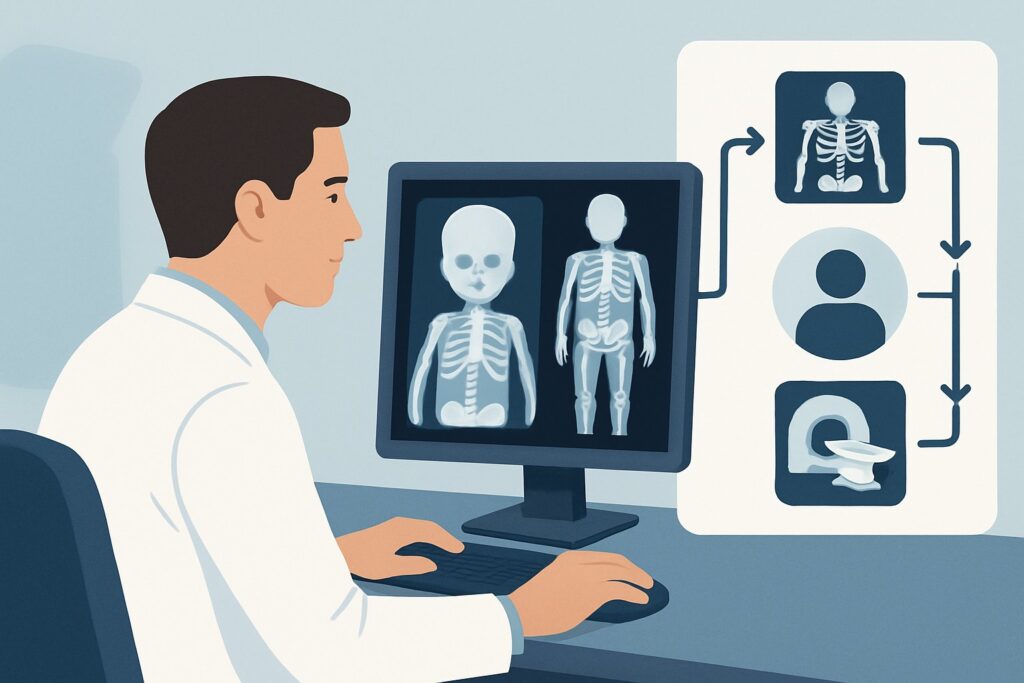
A one-year-old child is back for a follow-up visit two weeks after an emergency department evaluation for suspected physical abuse. The initial skeletal survey and head CT were interpreted as negative, but the clinical suspicion from the child protection team remains high. As the ordering physician, you face a critical decision: is further imaging warranted, and if so, what is the most appropriate study to perform now that time has passed? This scenario requires understanding the pathophysiology of healing bone to uncover injuries that may have been invisible on the initial studies.
For a child 24 months of age or younger with suspected physical abuse and a negative initial workup, the American College of Radiology (ACR) Appropriateness Criteria rate a follow-up Radiography skeletal survey as Usually Appropriate at the 10- to 14-day interval. This article details the clinical workflow and rationale for this specific recommendation.
Who Fits This Clinical Scenario for Follow-Up Abuse Imaging?
This guidance is narrowly focused on a specific follow-up situation. The recommendations apply to patients who meet all the following criteria:
- Age: 24 months or younger.
- Clinical Context: There is a continued, significant concern for non-accidental physical abuse based on the clinical presentation, history, or social situation.
- Prior Imaging: An initial, technically adequate skeletal survey and a non-contrast head CT were both performed and interpreted as negative for acute injury.
- Timing: The current clinical decision point is approximately 10 to 14 days after the initial evaluation.
It is crucial to distinguish this scenario from others that require a different imaging pathway. This article does not apply if:
- The patient is older than 24 months: Skeletal maturity, injury patterns, and the differential diagnosis change in older children, leading to different ACR recommendations.
- New or evolving neurological signs are present: If the child develops seizures, altered mental status, or new focal deficits, the workup shifts to a primary concern for Central Nervous System (CNS) injury, which triggers a different ACR variant.
- There is suspicion of visceral injury: Findings like abdominal bruising, tenderness, or abnormal liver function tests would necessitate an evaluation focused on abdominal organs, not a repeat skeletal survey.
- This is the initial workup: This guidance is exclusively for the planned follow-up imaging after a negative initial study, not for the first-line evaluation.
What Diagnoses Are You Working Up with Follow-Up Imaging?
The primary goal of the 10- to 14-day follow-up skeletal survey is to detect occult fractures that were radiographically invisible at the time of initial injury. The healing process itself makes these injuries detectable. The differential you are working up includes:
Occult Metaphyseal Fractures Also known as classic metaphyseal lesions (CMLs) or “corner fractures,” these are subtle fractures at the edges of the long bone metaphyses. They are caused by shearing or tensile forces from shaking or pulling on a limb and are highly specific for non-accidental trauma. Acutely, these tiny fracture lines can be nearly impossible to see. However, after 10-14 days, the body’s healing response creates visible callus and sclerosis, making the fracture line apparent.
Subtle Rib Fractures Posterior rib fractures, near the spine, are another hallmark of inflicted injury, often caused by forceful squeezing of the chest. Like CMLs, non-displaced rib fractures can be missed on initial radiographs. The development of healing callus at the fracture site makes them significantly more conspicuous on the follow-up study.
Healing Periosteal Reaction In young children, the periosteum (the membrane covering the bones) is loosely attached. Trauma can cause subperiosteal hemorrhage, which is not visible on an initial X-ray. As this blood clot organizes and calcifies over one to two weeks, it creates a layer of new bone formation along the shaft of the bone. This finding, known as periosteal reaction, is a key secondary sign of a healing fracture.
Why Is a Repeat Skeletal Survey the Recommended Follow-Up Study?
The ACR designates a repeat Radiography skeletal survey as Usually appropriate for this scenario because it directly leverages the biology of bone healing to maximize diagnostic yield for injuries highly specific to abuse.
The 10- to 14-day interval is a diagnostic sweet spot. By this time, the initial inflammatory response has subsided, and the reparative phase of healing has begun. This process involves the formation of callus and the calcification of subperiosteal hematomas, which act as natural contrast agents, highlighting previously occult injuries. The high spatial resolution of radiography is ideal for visualizing these subtle bony changes, such as the delicate pattern of a CML or the faint outline of a healing rib.
The pediatric radiation dose for a skeletal survey is categorized as ☢☢☢ (0.3-3 mSv). While any radiation exposure in children requires careful consideration, the critical importance of making an accurate diagnosis in cases of suspected abuse is deemed to justify this level of exposure.
Alternative studies are rated lower for specific reasons in this context:
- Bone scan whole body is rated May be appropriate. While highly sensitive for areas of increased bone turnover (including healing fractures), it is less specific. It cannot clearly define the fracture morphology, which is critical for distinguishing a CML from other causes of increased uptake. Furthermore, it typically involves a higher radiation dose (pediatric RRL ☢☢☢☢, 3-10 mSv) than a skeletal survey.
- MRI head without IV contrast is rated May be appropriate (Disagreement). This study is excellent for evaluating the brain for evolving or subtle injury that a CT might miss. However, the primary question in this specific ACR variant is skeletal, not neurological. Given the initial head CT was negative, a follow-up MRI of the head does not address the need to re-evaluate the skeleton for healing fractures. Its utility is debated and depends on institutional protocols and specific clinical concerns for brain injury.
Once you’ve decided on the follow-up skeletal survey, our protocol guide covers the specific views and technical considerations. You can find it here: Skeletal Survey.
What’s Next After a Repeat Skeletal Survey? Downstream Workflow
The results of the follow-up skeletal survey guide the subsequent steps in the child’s medical and protective management.
- If the study is POSITIVE: If the repeat survey reveals healing fractures (e.g., CMLs, posterior rib fractures, multiple fractures in different stages of healing), this finding is highly significant. It provides objective evidence of trauma that occurred around the time of the initial presentation. This information must be communicated immediately to the clinical team and the child protection services involved. It strengthens the case for non-accidental trauma and will guide decisions regarding the child’s safety.
- If the study is NEGATIVE: A second, technically adequate negative skeletal survey at 10-14 days significantly lowers the likelihood of occult bony injury. While abuse cannot be ruled out by imaging alone, this finding means that a different form of injury may be present, or the initial suspicion may have been incorrect. The focus would then return to the complete clinical picture, social evaluation, and a search for other medical conditions that could mimic abuse.
- If the study is INDETERMINATE: Occasionally, a finding may be ambiguous (e.g., a subtle periosteal reaction that could be physiologic). In these cases, consultation with a pediatric radiologist is paramount. Further imaging, such as dedicated cone-down views of the area in question or, in rare cases, a bone scan, might be considered to clarify the finding.
Pitfalls to Avoid (and When to Get Help)
Navigating these cases requires meticulous attention to detail. Common pitfalls to avoid include:
- Incorrect Timing: Performing the follow-up study too early (e.g., at 3-5 days) may be insufficient for healing changes to become visible. Performing it too late (e.g., after 4 weeks) can make it difficult to distinguish new from old injuries.
- Incomplete Study: A “babygram” (a single radiograph of the whole body) is unacceptable. A proper skeletal survey consists of a specific set of high-resolution images of individual body parts.
- Misinterpreting Normal Variants: Physiologic periostitis of the newborn or classic metaphyseal irregularities can sometimes mimic fractures. These cases demand expert interpretation.
- Over-reliance on a Negative Report: Remember that imaging is just one piece of the puzzle. A negative skeletal survey does not rule out abuse, especially other forms like neglect or abusive head trauma without skeletal manifestations.
If any high-specificity fractures are identified or suspected, immediate escalation to a pediatric radiologist and the institution’s child abuse specialist or team is mandatory.
Related ACR Topics and Tools
For a comprehensive overview of imaging in all scenarios related to this topic, see the parent guide. For tools to help with ordering, protocoling, and dose management, see the resources below.
- For breadth across all scenarios in Suspected Physical Abuse-Child, see our parent guide: Suspected Physical Abuse-Child: ACR Appropriateness Decoded.
- ACR Appropriateness Criteria Lookup: For adjacent or alternative clinical scenarios.
- Imaging Protocol Library: For technical details on the recommended study.
- Radiation Dose Calculator: For discussing and tracking cumulative radiation exposure.
Frequently Asked Questions
Why is the 10- to 14-day timeframe so important for the follow-up skeletal survey?
This interval is considered the optimal window to detect occult fractures. By 10-14 days, the body’s natural healing process has created visible signs like callus formation and periosteal reaction at fracture sites that were invisible on the initial X-rays. Imaging earlier may be too soon for these changes to appear, while imaging later can make it harder to date the injuries accurately.
If the initial head CT was negative, is there any role for follow-up brain imaging?
The ACR rates a follow-up MRI of the head as ‘May be appropriate (Disagreement)’. While an initial non-contrast head CT is excellent for acute hemorrhage, MRI is more sensitive for subtle signs of brain injury like non-hemorrhagic axonal injury or small subdural collections. The decision to obtain a follow-up MRI depends on institutional protocols and whether there is a high clinical concern for abusive head trauma despite the negative CT, but it does not replace the need for the follow-up skeletal survey to assess for bony injury.
Is a whole-body bone scan a reasonable alternative to a repeat skeletal survey?
A bone scan is rated ‘May be appropriate’ but is generally not the preferred study. While it is very sensitive for detecting fracture healing, it is less specific and provides poor anatomical detail. It cannot clearly define the morphology of a fracture, which is critical for identifying highly specific injuries like classic metaphyseal lesions (CMLs). The skeletal survey provides the necessary high-resolution detail, and typically with a lower radiation dose.
What if the initial skeletal survey was technically poor or incomplete?
If the initial study was inadequate (e.g., poor positioning, motion artifact, missing views), it should be repeated as soon as the deficiency is recognized, rather than waiting 10-14 days. The guidance in this article applies only when the initial survey was technically adequate and interpreted as negative.
Does a second negative skeletal survey definitively rule out physical abuse?
No. A negative follow-up skeletal survey significantly reduces the likelihood of occult bony injury but does not rule out abuse. Abuse can manifest in many ways, including soft tissue injuries, visceral injuries, abusive head trauma without fractures, or neglect, none of which would be detected on a skeletal survey. Imaging findings must always be interpreted in the context of the full clinical and social evaluation.
Reviewed by Pouyan Golshani, MD, Interventional Radiologist — May 30, 2026