What Is the Best Imaging Study for a Suspected Lung Abscess in a Child?
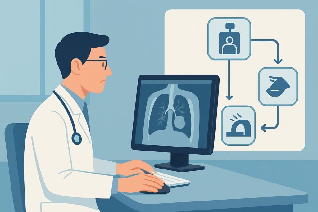
It’s late in the evening shift, and you are evaluating a 6-year-old with community-acquired pneumonia who is not improving as expected on oral antibiotics. The initial chest radiograph was concerning, showing a dense consolidation with a possible thick-walled cavity containing an air-fluid level. You suspect a complication, specifically a lung abscess, which would significantly alter management. The immediate clinical question is which advanced imaging study to order next to confirm the diagnosis and guide potential intervention. For this specific scenario, the American College of Radiology (ACR) Appropriateness Criteria rates CT chest with IV contrast as Usually Appropriate.
Who Fits This Clinical Scenario for a Suspected Lung Abscess?
This guidance applies to a specific patient population: an immunocompetent child who has been diagnosed with pneumonia, where the initial chest radiograph suggests a complication in the form of a lung abscess. Key radiographic findings prompting this workup include a cavitary lesion, often with a thick, irregular wall, and potentially an air-fluid level within the lung parenchyma.
This workflow is distinct from several similar-but-different clinical situations. It is crucial to ensure your patient fits this scenario and not a neighboring one, which would trigger a different diagnostic pathway.
Exclusion criteria for this specific workflow include:
- Simple Parapneumonic Effusion: If the chest radiograph primarily shows a moderate or large pleural effusion without clear signs of a parenchymal abscess, the workup is different. That scenario focuses on characterizing the pleural fluid, often starting with ultrasound.
- Uncomplicated Pneumonia: A child with typical community-acquired pneumonia who is clinically stable or improving as expected generally does not require advanced imaging beyond the initial radiograph.
- Immunocompromised Host: Children with known immunodeficiencies have a much broader differential for pulmonary infections and complications, requiring a distinct and often more aggressive imaging strategy.
- Suspected Bronchopleural Fistula: If the primary concern is an abnormal connection between the airway and pleural space, the imaging protocol may be tailored differently to assess for air leak.
What Diagnoses Are You Working Up in a Child with Suspected Lung Abscess?
When a chest radiograph suggests a lung abscess, the goal of advanced imaging is to confirm this finding and rule out critical mimics that require different management. The differential diagnosis is narrow but consequential.
Lung Abscess
This is the primary diagnosis of concern. A lung abscess is a localized, encapsulated collection of pus within the lung parenchyma, resulting from necrosis of lung tissue. Confirming its presence, size, and location is essential for determining whether management with antibiotics alone is sufficient or if percutaneous or surgical drainage is necessary.
Necrotizing Pneumonia
This is a severe complication of pneumonia characterized by liquefaction and destruction of lung tissue, often without a single, well-defined, encapsulated wall like a classic abscess. It may present with multiple small cavities. CT is highly effective at distinguishing a single drainable abscess from the more diffuse process of necrotizing pneumonia, which is typically managed medically.
Complicated Parapneumonic Effusion or Empyema
An empyema is a collection of pus within the pleural space, adjacent to the lung, rather than within the lung parenchyma itself. On a two-view chest radiograph, a loculated empyema can mimic an intraparenchymal abscess. Differentiating between these two is a critical step, as an empyema almost always requires drainage (thoracentesis, chest tube, or surgery), while many lung abscesses can be treated with antibiotics alone.
Infected Congenital Pulmonary Airway Malformation (CPAM)
In younger children, an underlying congenital lung lesion, such as a CPAM (formerly CCAM), can become secondarily infected and present identically to a primary lung abscess. Identifying a CPAM is important for long-term management, as surgical resection of the malformation is often recommended after the acute infection resolves to prevent recurrence.
Why Is CT Chest with IV Contrast the Recommended Study for a Suspected Lung Abscess?
The ACR designates CT chest with IV contrast as Usually Appropriate because it provides the most definitive anatomical information needed to manage this complex clinical problem. The rationale is based on its superior ability to characterize the abnormality seen on the initial radiograph.
The administration of intravenous contrast is the critical component. Contrast enhances the abscess wall, making it appear as a bright ring surrounding the non-enhancing central pus. This “ring enhancement” is the classic sign that confirms an abscess and clearly delineates its extent. Furthermore, contrast helps differentiate the abscess from surrounding consolidated or atelectatic lung and, crucially, distinguishes an intraparenchymal abscess from a pleural-based empyema. An empyema is identified by the “split pleura” sign, where the enhancing visceral and parietal pleura are separated by non-enhancing pus.
Alternative imaging modalities are rated lower for specific reasons in this context:
- US chest is rated May be appropriate. While excellent for evaluating pleural fluid and guiding drainage of peripheral lesions, it is limited by the acoustic window. It cannot reliably visualize central abscesses surrounded by aerated lung and is highly operator-dependent.
- MRI chest without and with IV contrast is also rated May be appropriate. It offers excellent soft tissue contrast without using ionizing radiation. However, it is more susceptible to motion artifact from breathing, requires longer scan times, and often necessitates sedation in young children, making it less practical in an acutely ill patient.
- CT chest without IV contrast is rated Usually not appropriate. Without contrast, it is extremely difficult to distinguish a fluid-filled abscess from surrounding consolidation or to differentiate an abscess from an empyema, defeating the primary purpose of the study.
While CT involves ionizing radiation (pediatric RRL ☢☢☢☢, 3-10 mSv), the diagnostic value in this specific scenario—where management may change from medical to procedural—justifies the exposure. Adherence to As Low As Reasonably Achievable (ALARA) principles with pediatric-specific, low-dose protocols is mandatory.
Once you’ve decided on CT chest with IV contrast, our protocol guide covers the technique, contrast, and reading principles: CT Chest/Abdomen/Pelvis with IV Contrast.
What’s Next After CT? Downstream Workflow
The results of the contrast-enhanced CT will directly guide the subsequent clinical pathway. The decision tree branches based on the definitive findings.
If the CT confirms a lung abscess:
The next steps depend on the size of the abscess and the child’s clinical status. For smaller abscesses in a clinically stable child, a prolonged course of intravenous antibiotics is the standard of care. For larger abscesses (often >6 cm) or in patients who are septic or failing to improve on antibiotics, consultation with interventional radiology for percutaneous catheter drainage or with pediatric surgery for surgical drainage is warranted.
If the CT demonstrates an empyema:
This finding almost always necessitates drainage. The next step is consultation with interventional radiology or pediatric surgery for placement of a chest tube or a video-assisted thoracoscopic surgery (VATS) procedure for decortication. Antibiotic therapy is continued as an adjunct to drainage.
If the CT shows necrotizing pneumonia without a drainable collection:
This is typically managed medically with broad-spectrum intravenous antibiotics. Drainage procedures are not effective for this diffuse process. The CT provides the confidence to continue with medical management and avoid an unnecessary procedure.
If the CT is negative or shows uncomplicated pneumonia:
If the CT rules out a complex collection and shows only simple consolidation, the initial suspicion from the radiograph was incorrect. Management can proceed along the standard pathway for uncomplicated or slowly-resolving pneumonia, which may involve adjusting antibiotic therapy and close clinical follow-up.
Pitfalls to Avoid (and When to Get Help)
Navigating the workup for a suspected pediatric lung abscess requires careful attention to detail to avoid common missteps.
A primary pitfall is ordering a CT of the chest without IV contrast. This provides limited additional information over the plain radiograph for this specific question and can lead to diagnostic uncertainty, potentially requiring a second, contrast-enhanced scan and doubling the radiation exposure. Another error is delaying advanced imaging in a child who is clinically deteriorating or failing to respond to appropriate therapy; timely diagnosis of a drainable collection is key to a good outcome. Finally, always ensure the CT protocol is tailored for pediatric patients to minimize radiation dose.
If the CT scan reveals a large, complex, or multiloculated collection, or if the child remains clinically unstable despite initial interventions, it is time to escalate. This involves a multidisciplinary discussion with pediatric infectious disease, interventional radiology, and pediatric surgery specialists to determine the optimal drainage strategy and medical management plan.
Related ACR Topics and Tools
This article focuses on one specific clinical variant. For a comprehensive overview of imaging for all common presentations of pediatric pneumonia, or to explore the tools used to make these decisions, the following resources are essential.
- For breadth across all scenarios in Pneumonia in the Immunocompetent Child, see our parent guide: Pneumonia in the Immunocompetent Child: ACR Appropriateness Decoded.
- To look up appropriateness ratings for other clinical scenarios, use the ACR Appropriateness Criteria Lookup.
- To review technical details for this and other studies, see the Imaging Protocol Library.
- To discuss radiation exposure with families, consult the Radiation Dose Calculator.
Frequently Asked Questions
Why is IV contrast so critical for a suspected pediatric lung abscess?
IV contrast is essential because it enhances the wall of the abscess, making it visible as a distinct ring. This allows the radiologist to confirm the diagnosis, measure its exact size, and, most importantly, differentiate it from a pleural empyema or surrounding consolidated lung, which require different treatments. A CT without contrast often cannot make these critical distinctions.
Is ultrasound a reasonable first step instead of CT to reduce radiation?
Ultrasound is rated as ‘May be appropriate’ by the ACR. It is an excellent radiation-free tool for evaluating abnormalities that are peripheral and touch the chest wall, including some abscesses and pleural effusions. However, its utility is severely limited for central lung lesions surrounded by air, which block the ultrasound waves. For a suspected abscess seen on a chest radiograph that is not clearly peripheral, CT with IV contrast is the more definitive next step to avoid a non-diagnostic ultrasound.
What if the CT shows an underlying congenital abnormality like a CPAM?
If the CT scan reveals that the abscess has formed within a congenital pulmonary airway malformation (CPAM), the immediate management is to treat the acute infection with antibiotics and drainage if necessary. However, this finding changes the long-term plan. The child will need follow-up with a pediatric surgeon to discuss elective surgical resection of the CPAM after the infection has completely resolved to prevent recurrent infections and other potential long-term complications.
Are there specific antibiotic considerations when a lung abscess is confirmed?
Yes. The bacteriology of lung abscesses often includes anaerobic organisms in addition to typical pneumonia pathogens like Streptococcus pneumoniae. Therefore, antibiotic coverage is typically broadened to include agents with strong anaerobic activity, such as clindamycin or a beta-lactam/beta-lactamase inhibitor combination. Treatment is also prolonged, often lasting 4-6 weeks or until imaging shows significant resolution. Consultation with a pediatric infectious disease specialist is recommended.
How do I explain the radiation risk of a CT scan to a child’s parents?
When discussing the need for a CT, it’s important to be transparent and frame the risk in the context of the benefit. Explain that while CT does use radiation, the diagnostic information it provides in this specific situation is critical for their child’s health and cannot be obtained reliably with other tests. Reassure them that the hospital uses special low-dose protocols specifically for children to minimize exposure. Emphasize that the risk of not diagnosing and properly treating a lung abscess is far greater than the small, long-term risk from the radiation of one necessary scan.
Reviewed by Pouyan Golshani, MD, Interventional Radiologist — May 29, 2026