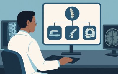
What Imaging Is Best for a Suspected Foot Foreign Body After Negative X-Rays?
A teenager presents to the urgent care clinic after stepping on an unknown object while running barefoot in the backyard. There is a small puncture wound on the plantar aspect of the foot, with localized tenderness and swelling. Standard two-view radiographs of the foot are performed and are negative for any radiopaque foreign body or fracture. The patient continues to experience significant pain, and you are concerned about a retained, non-visible object like a wood splinter or piece of plastic. This article details the appropriate next imaging step for this specific clinical scenario, based on the American College of Radiology (ACR) Appropriateness Criteria, which rates ultrasound of the foot as Usually appropriate.
Who Fits This Clinical Scenario?
This clinical workflow is designed for a specific patient presentation: an adult or child over the age of five who has sustained acute trauma to the foot with a suspected penetrating foreign body, and for whom initial radiographs are negative. The key inclusion criteria are a clear history or strong clinical suspicion of penetration (e.g., stepping on glass, a nail, or a wood splinter) and a normal initial X-ray.
This guidance does not apply to several similar-but-distinct clinical situations. If a radiopaque foreign body (like metal or most glass) is clearly visible on the initial radiograph, this workflow is unnecessary, and the clinical focus shifts to removal. This article also does not apply to patients whose primary clinical question is a fracture, where the Ottawa foot and ankle rules would guide the decision to obtain initial radiographs. Similarly, if the primary concern is a complex ligamentous injury, such as a Lisfranc injury, a different imaging pathway is indicated, as the search is for osseous or ligamentous disruption rather than a foreign object.
What Diagnoses Are You Working Up in This Scenario?
When initial radiographs are negative after a penetrating foot injury, the imaging workup is focused on identifying pathology that is invisible on X-ray. The differential diagnosis guides the choice of the next study.
The most common and pressing concern is a retained radiolucent foreign body. Many common materials that cause penetrating injuries, such as wood, thorns, plastic, and some types of glass, have low density and are not visible on standard radiographs. These objects can serve as a nidus for chronic inflammation and infection if not removed.
A secondary but critical diagnosis to consider is a developing abscess or deep soft tissue infection. A puncture wound introduces bacteria deep into the tissues, and a retained foreign body significantly increases this risk. Imaging can identify a localized, drainable fluid collection that requires intervention beyond simple antibiotics.
You must also consider direct injury to soft tissue structures. The penetrating object may have caused a tendon or ligament laceration. While less common than a simple retained object, a missed tendon injury can lead to long-term functional deficits. Imaging can help assess the integrity of these structures near the entry wound.
Finally, while typically a later complication, osteomyelitis can arise from direct inoculation of bacteria into the bone. Early imaging may show adjacent soft tissue inflammation or subtle periosteal reaction that could suggest an early bone infection, even before osseous changes are visible on radiographs.
Why Is US foot the Recommended Study for This Presentation?
For a suspected penetrating foreign body with negative radiographs, the ACR designates US foot as Usually appropriate. Ultrasound is the ideal first-line modality in this scenario due to its high sensitivity for detecting common radiolucent foreign bodies and its other diagnostic advantages.
Ultrasound excels at identifying objects like wood, plastic, and organic matter, which appear as hyperechoic structures, often with a characteristic posterior acoustic shadow. It provides real-time, dynamic evaluation, allowing the operator to correlate the point of maximal tenderness directly with the underlying anatomy. Furthermore, ultrasound is highly effective at identifying associated complications, such as a surrounding fluid collection, abscess formation, or inflammatory changes (cellulitis). It offers the significant benefit of having no ionizing radiation (0 mSv), making it particularly safe for all patients, including children.
Alternative studies are rated lower for specific reasons in this initial workup:
- MRI foot without IV contrast is rated as May be appropriate. While MRI offers superior soft tissue contrast and can detect foreign bodies and associated inflammation, it is more costly, less readily available, and more time-consuming than ultrasound. It is best reserved as a second-line study if ultrasound is negative or inconclusive but clinical suspicion remains high, especially for a deeply embedded object.
- CT foot without IV contrast is also rated as May be appropriate. CT is excellent for identifying materials that are only faintly radiopaque, like glass or gravel, which might be missed on plain films. However, it is significantly less sensitive than ultrasound for organic materials like wood and involves a small dose of ionizing radiation (Adult RRL: ☢ <0.1 mSv; Pediatric RRL: ☢☢ 0.03-0.3 mSv).
Studies with intravenous contrast, such as MRI foot without and with IV contrast or CT foot with IV contrast, are deemed Usually not appropriate for the initial search for a foreign body. Contrast does not typically improve the visualization of the foreign object itself and is generally reserved for cases where there is a specific concern for osteomyelitis or a vascular injury, which is not the primary question in this scenario.
What’s Next After US foot? Downstream Workflow
The results of the foot ultrasound will guide your next clinical steps. The workflow branches based on whether a foreign body, an abscess, or another finding is identified.
If the ultrasound is positive for a retained foreign body: The primary goal becomes removal. The ultrasound report should provide crucial details for the proceduralist, including the object’s size, depth, and precise location relative to nearby tendons, nerves, and vessels. This information determines whether a simple office-based removal is feasible or if a referral to surgery or interventional radiology for image-guided removal is necessary.
If the ultrasound is positive for an abscess (with or without a foreign body): The presence of a drainable fluid collection typically necessitates incision and drainage. The ultrasound can be used to guide the aspiration or drainage procedure, ensuring complete evacuation of the pus.
If the ultrasound is negative: If the study is negative and the patient’s symptoms are mild and improving, a course of conservative management with wound care and observation may be sufficient. However, if high clinical suspicion for a deep or missed foreign body persists despite a negative ultrasound (e.g., worsening pain, signs of infection), the next logical step is to consider the study rated as May be appropriate: MRI foot without IV contrast. MRI can detect deeply located objects or other soft tissue pathologies that may have been outside the scope or resolution of the ultrasound.
Pitfalls to Avoid (and When to Get Help)
Navigating the workup for a suspected retained foreign body requires avoiding several common pitfalls. First, do not assume a negative radiograph rules out a foreign body; this is the most frequent error and can lead to delayed diagnosis and complications. Second, recognize the limitations of ultrasound. The skill of the sonographer is paramount, as artifacts can mimic or obscure a foreign body. A very small or deeply embedded object may be missed. Third, do not delay imaging if the patient presents with systemic signs of infection or rapidly progressing cellulitis. In these cases, the search for a drainable abscess is urgent. If the ultrasound is equivocal or the clinical picture is worsening despite a negative initial workup, escalate care by obtaining an MRI or consulting with a surgical specialist.
Related ACR Topics and Tools
For a comprehensive overview of imaging for all foot trauma scenarios, this depth piece is best used alongside its parent topic article. For additional decision support, protocol details, or radiation dose information, the following resources are available.
- For breadth across all scenarios in Acute Trauma to the Foot, see our parent guide: Acute Trauma to the Foot: ACR Appropriateness Decoded.
- To explore other clinical presentations, consult the ACR Appropriateness Criteria Lookup tool.
- For technical details on performing imaging studies, see the Imaging Protocol Library.
- To discuss radiation exposure with patients, use the Radiation Dose Calculator.
Frequently Asked Questions
Why not just go straight to MRI if it’s better for soft tissue?
While MRI provides excellent soft tissue detail, ultrasound is rated higher as the first-line study in this scenario because it is faster, more accessible, less expensive, and highly effective for detecting the most common types of radiolucent foreign bodies (like wood and plastic) in superficial locations. MRI is best reserved as a problem-solving tool if ultrasound is negative or inconclusive but clinical suspicion remains high.
Does the type of suspected foreign body material change the imaging choice?
Yes, to some extent. Ultrasound is the best all-around initial test for unknown or organic materials. However, if you have a very high suspicion for a retained piece of glass, which can be difficult to see on X-ray but is dense enough to be seen on CT, a non-contrast CT (‘May be appropriate’) could be considered. For most cases where the material is unknown, ultrasound remains the best starting point.
Is a CT scan ever the best first choice after a negative X-ray?
CT is generally not the first choice but is rated as ‘May be appropriate’. It becomes a more compelling option if the primary suspicion is for a foreign body made of glass or gravel, or if there is a concern for an associated subtle fracture that was not apparent on the initial radiographs. However, for the general workup of a suspected radiolucent object, ultrasound’s lack of radiation and superior performance for organic materials make it the preferred initial modality.
What if the patient is a child under 5 years old?
This specific ACR variant applies to children older than 5 years. While the principles are similar for younger children, imaging decisions in very young patients often involve additional considerations, such as the need for sedation for longer studies like MRI and an even stronger emphasis on avoiding radiation. Ultrasound remains an excellent, radiation-free option and is frequently used in all pediatric age groups for this indication.
When should I order a study with IV contrast?
For the initial detection of a foreign body, IV contrast is ‘Usually not appropriate’ for both CT and MRI. Contrast does not make the foreign body itself more visible. Its use is reserved for secondary questions, such as evaluating for osteomyelitis, assessing the viability of surrounding tissue, or delineating the full extent of a complex abscess, which are typically downstream concerns rather than the initial diagnostic step.
Reviewed by Pouyan Golshani, MD, Interventional Radiologist — May 29, 2026