What Imaging Is Best for Altered Mental Status in a Patient with Known Brain Pathology?

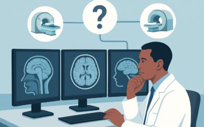
A 65-year-old male with a history of a resected glioblastoma, status post-chemoradiation, presents to the emergency department with two days of increasing confusion and lethargy. His family is concerned about tumor recurrence. The initial neurologic exam is non-focal, but his baseline has clearly changed. You need to determine if this new altered mental status (AMS) is related to his known intracranial pathology, a treatment complication, or an entirely new process. This article provides a clinical workflow for this specific scenario, guiding you through the American College of Radiology (ACR) Appropriateness Criteria to select the right initial imaging study. For this presentation, the ACR rates MRI head without and with IV contrast as Usually Appropriate.
Who Fits This Clinical Scenario?
This guidance applies specifically to adult patients presenting with a new or worsening alteration in mental status who have a known, pre-existing intracranial pathology. The key is “known”—the patient has a documented history of a condition like a primary brain tumor (e.g., glioma), metastasis, multiple sclerosis, a prior large-territory stroke, a treated aneurysm, an arteriovenous malformation (AVM), or post-surgical changes from a craniotomy.
This workflow is not intended for:
- Patients with suspected but not yet diagnosed intracranial pathology. If a patient presents with new focal neurologic deficits alongside AMS, suggesting a new stroke or mass, they fall into a different clinical variant.
- Patients where the primary suspicion is a toxic-metabolic cause. An elderly patient with a urinary tract infection and delirium, even with a remote history of a lacunar stroke, would first be managed under the toxic-metabolic pathway. This scenario is for when the known brain pathology itself is a primary suspect for the change in status.
- Patients with new-onset psychosis without a known structural brain lesion. This represents a distinct diagnostic challenge with its own imaging considerations.
Correctly identifying your patient’s scenario is crucial, as it directly influences the most appropriate initial imaging choice and avoids unnecessary or low-yield studies.
What Diagnoses Are You Working Up in This Scenario?
In a patient with known intracranial pathology, the differential diagnosis for altered mental status is focused and high-stakes. The primary goal of imaging is to differentiate between progression of the underlying disease, complications of its treatment, and new, unrelated events.
Progression or Recurrence of the Primary Pathology: This is often the chief concern. For a patient with a brain tumor, this means tumor growth or new metastatic lesions. In a patient with multiple sclerosis, it could represent a significant new wave of demyelinating plaque activity causing cognitive symptoms. Imaging is essential to confirm or exclude this possibility, as it directly dictates the next steps in management.
Treatment-Related Complications: Interventions for intracranial disease can cause delayed complications that mimic disease progression. Radiation necrosis, for example, can enhance with contrast and cause mass effect, looking very similar to tumor recurrence on basic imaging. Other possibilities include post-surgical infection (abscess), chemotherapy-induced leukoencephalopathy, or hydrocephalus due to a malfunctioning shunt or obstructed cerebrospinal fluid (CSF) pathways.
New Vascular Events: These patients are not immune to other common neurologic emergencies. A new ischemic or hemorrhagic stroke can occur independently of their known condition. Furthermore, some pathologies, particularly tumors like glioblastoma or metastases from melanoma or renal cell carcinoma, are prone to internal hemorrhage, which can present as an acute decline in mental status.
Seizure Activity: Structural brain lesions are a major risk factor for seizures. The patient’s altered mental status could be a non-convulsive seizure or a postictal state. While imaging may not show acute changes during a seizure, it is critical for evaluating the underlying structural focus that may be triggering the event.
Why Is MRI Head Without and With Contrast Usually Appropriate for This Presentation?
The American College of Radiology (ACR) designates MRI head without and with IV contrast as Usually Appropriate for this scenario because it provides the most comprehensive evaluation of the likely differential diagnoses. Its superior soft-tissue resolution is unmatched for assessing the complex brain parenchyma in a patient with pre-existing abnormalities.
The non-contrast sequences (like T1, T2, FLAIR, and DWI) are excellent for identifying edema, ischemia, non-acute hemorrhage, and cytotoxic edema from a new stroke. The addition of intravenous gadolinium-based contrast is the critical component. It highlights areas of blood-brain barrier disruption, which is fundamental for assessing tumor activity, inflammation from infection or demyelination, and certain post-treatment effects. This combined approach gives the clinical and radiology teams the best chance to distinguish active tumor from radiation necrosis, identify a developing abscess, or characterize new enhancing lesions.
Let’s compare this to other modalities rated by the ACR for this specific scenario:
- CT head without IV contrast: Also rated Usually Appropriate. This is a fast, accessible alternative, particularly in an unstable patient or if MRI is unavailable or contraindicated. Its primary role is to rapidly exclude acute, large-scale problems like significant hemorrhage, hydrocephalus, or a large mass causing herniation. However, it lacks the sensitivity of MRI for subtle tumor recurrence, small ischemic strokes, or differentiating treatment-related changes.
- CT head without and with IV contrast: Rated Usually Not Appropriate. If the clinical question is complex enough to warrant IV contrast, the diagnostic information gained from a contrast-enhanced MRI is substantially greater than that from a contrast-enhanced CT. Opting for the CT exposes the patient to ionizing radiation (ACR RRL=☢☢☢ 1-10 mSv) for a less definitive answer, often leading to a follow-up MRI anyway. It is generally better to proceed directly to MRI if contrast is deemed necessary and the patient is stable enough for the scan.
In summary, while a non-contrast CT has a role for initial safety screening, the definitive diagnostic study for a stable patient in this scenario is an MRI with and without contrast. It directly addresses the core clinical questions of disease progression versus treatment complication without the use of ionizing radiation (ACR RRL=O 0 mSv).
What’s Next After MRI? Downstream Workflow for Altered Mental Status
The results of the MRI will guide your next steps, branching into distinct clinical pathways. The goal is to move from diagnosis to a clear management plan.
If the MRI shows clear evidence of disease progression: For a patient with a known tumor, findings of a new, enlarging, or more avidly enhancing mass will typically prompt an urgent re-consultation with their neuro-oncology or neurosurgery team. This may lead to a change in systemic therapy, consideration for re-resection, or planning for further radiation.
If the MRI is equivocal for progression vs. treatment effect: This is a common and challenging situation, especially in post-radiation patients. Findings like new enhancement within a radiation field can represent either tumor recurrence or radiation necrosis. The next step often involves advanced imaging techniques to assess tissue metabolism and physiology. This may include MR spectroscopy, MR perfusion imaging, or a PET scan, which can help differentiate metabolically active tumor cells from non-neoplastic tissue changes.
If the MRI shows a new, unrelated pathology: A finding like an acute stroke, abscess, or subdural hematoma shifts the management focus entirely. This requires initiating the appropriate pathway for that specific diagnosis, such as stroke protocols, infectious disease consultation and potential surgical drainage for an abscess, or neurosurgical evaluation for a hematoma.
If the MRI is negative or stable compared to prior studies: A non-diagnostic MRI is a crucial finding. It strongly suggests the altered mental status is not due to a structural change in the known pathology. The workup should then pivot aggressively toward non-structural and systemic causes. This includes a thorough search for infection (including CSF analysis via lumbar puncture), metabolic derangements, medication side effects, or subclinical seizures (requiring an EEG). This patient’s workup now more closely resembles the scenario for AMS with a suspected medical or toxic-metabolic cause.
Pitfalls to Avoid (and When to Get Help)
Navigating this scenario requires careful consideration to avoid common diagnostic traps.
- Attribution Error: Do not automatically attribute the patient’s AMS to their known pathology. Always maintain a broad differential and rule out common reversible causes like infection, hypoglycemia, or medication effects.
- Relying on an old “baseline” scan: A “stable” MRI is only meaningful if the prior study is recent. Comparing to a scan from two years ago may miss subtle but significant interval progression.
- Ordering CT with contrast as a substitute for MRI: As noted, if the question requires contrast, MRI is almost always the superior test. Using CT with contrast can lead to indeterminate results and delay the definitive diagnosis.
- Forgetting MRI contraindications: Always screen for pacemakers/defibrillators, certain metallic implants, or severe claustrophobia before ordering an MRI. Have a plan for an alternative study (like non-contrast CT) if MRI is not feasible.
If the patient shows rapid neurologic decline, signs of impending herniation (e.g., pupillary changes, posturing), or is hemodynamically unstable, escalate immediately to your institution’s critical care and neurocritical care teams.
Related ACR Topics and Tools
This article focuses on one specific clinical variant. For a comprehensive overview of imaging for all related presentations, from coma to new-onset psychosis, please consult our parent guide. For further exploration of imaging criteria, protocols, and radiation safety, the following resources are available.
- For breadth across all scenarios in Altered Mental Status, Coma, Delirium, and Psychosis, see our parent guide: Altered Mental Status, Coma, Delirium, and Psychosis: ACR Appropriateness Decoded.
- ACR Appropriateness Criteria Lookup — for adjacent scenarios
- Imaging Protocol Library — for technique on the recommended study
- Radiation Dose Calculator — for cumulative dose conversations
Frequently Asked Questions
Why is MRI with contrast needed if the patient already had a non-contrast CT that was negative?
A non-contrast CT is excellent for ruling out acute large-scale events like major hemorrhage or hydrocephalus. However, it is insensitive to many of the key concerns in this scenario, such as subtle tumor recurrence, inflammatory changes, or small abscesses. Intravenous contrast is essential to visualize areas where the blood-brain barrier is compromised, which is a key feature of these pathologies. An MRI with contrast provides far more detail about these processes than a CT with contrast.
What if my patient has a contraindication to MRI, like a non-compatible pacemaker?
If MRI is absolutely contraindicated, the next best step depends on the primary suspicion. A non-contrast CT head remains a valuable first test to rule out hemorrhage or hydrocephalus. If a contrast-enhanced study is still felt to be necessary to look for a mass or abscess, a CT head with IV contrast may be appropriate, despite its lower sensitivity compared to MRI. This decision should be made in consultation with a radiologist.
The patient’s creatinine is elevated. Can I still order an MRI with gadolinium contrast?
This is an important consideration. For patients with severe renal dysfunction (e.g., eGFR < 30 mL/min/1.73m²), there is a risk of nephrogenic systemic fibrosis (NSF) with certain older gadolinium-based contrast agents. However, modern macrocyclic agents carry a much lower risk. The decision should be based on a risk-benefit analysis. Discuss the case with the radiology department; they can advise on the specific risk with the agents they use and whether the potential diagnostic benefit outweighs the risk.
How do I differentiate tumor progression from pseudoprogression on an MRI?
This is a classic diagnostic challenge. Pseudoprogression is a treatment-related effect, often seen after radiation, where an area can enhance and have surrounding edema, mimicking true tumor growth. Standard MRI sequences can be indeterminate. Differentiating often requires advanced imaging like MR perfusion (which assesses blood flow), MR spectroscopy (which assesses metabolites), or a nuclear medicine study like a PET scan. Often, the most practical approach is close interval follow-up imaging in 6-8 weeks to assess for change.
Should I order an MRI of the whole spine as well?
An MRI of the spine is generally not part of the initial workup for altered mental status unless there are specific clinical signs pointing to a spinal cord process (e.g., myelopathy, a sensory level, bowel/bladder dysfunction) or if the patient’s known pathology has a high propensity for ‘drop metastases’ to the spinal canal (e.g., ependymoma, medulloblastoma).
Reviewed by Pouyan Golshani, MD, Interventional Radiologist — May 29, 2026