What Imaging Is Best for Breast Cancer Screening After Autologous Reconstruction?
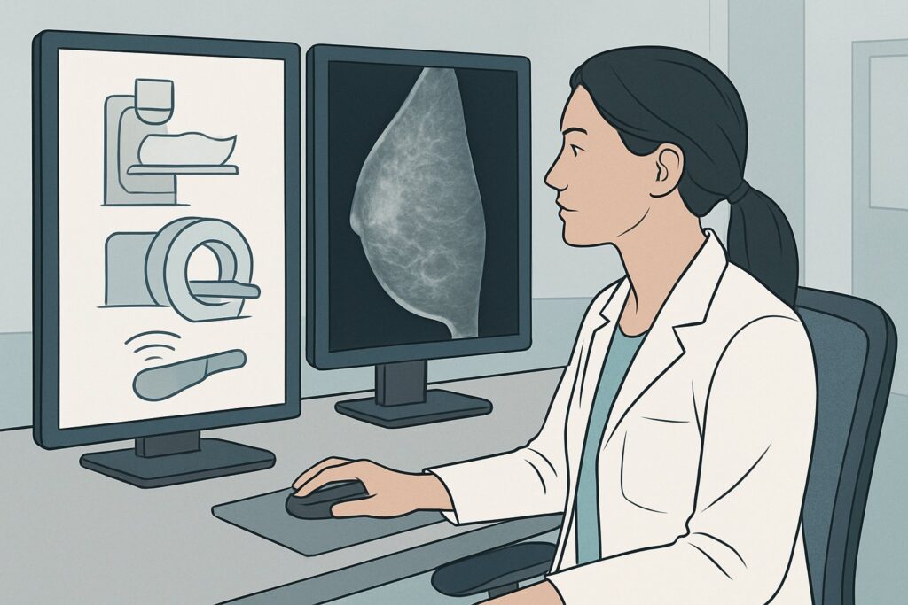
A 62-year-old female with a history of left-sided breast cancer, treated with mastectomy and a deep inferior epigastric perforator (DIEP) flap reconstruction six years ago, presents for her annual wellness visit. She is asymptomatic and feels well. As her primary care physician, you must decide on the appropriate imaging for her routine cancer screening. The reconstructed breast presents a different anatomical landscape than native breast tissue, raising questions about the utility and modality of surveillance. This article provides a detailed clinical workflow for this specific scenario, guiding you through the American College of Radiology (ACR) Appropriateness Criteria.
For this patient presentation, the ACR rates Digital breast tomosynthesis screening as May be appropriate, reflecting a nuanced decision-making process based on the balance of benefits and risks.
Who Fits This Clinical Scenario?
This guidance is specifically for asymptomatic females undergoing routine breast cancer screening who have a personal history of breast cancer and have undergone autologous breast reconstruction. Autologous reconstruction uses the patient’s own tissue—such as a transverse rectus abdominis myocutaneous (TRAM), deep inferior epigastric perforator (DIEP), or gluteal artery perforator (GAP) flap—to create a new breast mound. This scenario applies whether the reconstruction was performed with or without an underlying implant.
This workflow is not intended for:
- Patients with new clinical symptoms: If the patient presents with a new palpable lump, skin changes, or focal pain in the reconstructed breast or chest wall, this is a diagnostic workup, not a screening scenario. This requires a different imaging pathway, often starting with targeted ultrasound.
- Patients with implant-only reconstruction: Surveillance for nonautologous (implant-based) reconstruction involves different considerations, particularly regarding implant integrity, and is covered under a separate ACR variant.
- Patients with mastectomy and no reconstruction: Screening for this group focuses solely on the chest wall and remaining native tissue, which presents a distinct set of imaging challenges.
Correctly identifying your patient’s specific clinical context is the critical first step to ordering the most appropriate and valuable imaging study.
What Diagnoses Are You Working Up in This Scenario?
Even after a mastectomy, which removes the vast majority of breast glandular tissue, a small amount can remain. The primary goal of screening in this population is the early detection of locoregional recurrence or a new primary cancer. The differential diagnosis in this asymptomatic screening context includes both malignant and benign findings.
Locoregional Cancer Recurrence: This is the most consequential diagnosis to identify. Recurrence can occur in any residual breast tissue, within the skin or subcutaneous fat of the reconstructed mound, at the chest wall (involving the pectoralis muscle or ribs), or in regional lymph node basins (axillary, internal mammary, supraclavicular). While the overall risk is low, early detection significantly improves outcomes.
New Primary Breast Cancer: In patients with a contralateral native breast, screening aims to detect a new primary cancer on that side. For the reconstructed side, while rare, a new primary can theoretically arise from residual glandular tissue.
Benign Post-Surgical Changes: The most common imaging finding in an autologous flap is fat necrosis. This benign condition results from ischemia in the transferred adipose tissue and can present as oil cysts, calcifications, or spiculated masses that can closely mimic malignancy on imaging. Differentiating fat necrosis from recurrence is a primary task for the radiologist and a key reason that specialized imaging techniques are valuable.
Other Benign Findings: Scar tissue, seromas, and hematomas are also common post-surgically. While these typically resolve or stabilize over time, they can contribute to a complex imaging appearance, especially in the early years following reconstruction.
Why Is Digital Breast Tomosynthesis Screening Considered for This Presentation?
The ACR rates Digital breast tomosynthesis (DBT) screening as May be appropriate for screening a patient with autologous reconstruction. This rating acknowledges its value in specific contexts while recognizing that the evidence for routine screening of all such patients is still evolving. Standard Mammography screening also receives a May be appropriate rating.
The primary advantage of DBT over conventional 2D mammography is its ability to reduce the effect of overlapping tissue. By acquiring a series of low-dose images from different angles, DBT creates “slices” through the breast or reconstructed mound. This is particularly useful in the dense and architecturally complex environment of a reconstructed breast, where scar tissue and fat necrosis can obscure or mimic a true lesion on a flat 2D image. DBT can improve the characterization of findings, potentially reducing the need for additional workup of benign post-surgical changes.
In contrast, other modalities are rated lower for this specific screening scenario:
- Ultrasound (US) breast is rated Usually not appropriate for screening. While excellent for evaluating palpable lumps or mammographic abnormalities (i.e., in a diagnostic setting), its use as a primary screening tool for the entire breast is limited by lower specificity, operator dependence, and the lack of evidence demonstrating a mortality benefit in this role.
- Magnetic Resonance Imaging (MRI) breast without and with IV contrast is also rated Usually not appropriate for routine screening in this population, unless the patient meets separate high-risk criteria (e.g., genetic mutation, extensive family history). While MRI has very high sensitivity for detecting cancer, its specificity is lower, and it can lead to a higher rate of false positives and subsequent biopsies for benign findings, especially in the context of post-surgical inflammation and enhancement.
From a safety perspective, both DBT and standard mammography involve a low level of ionizing radiation (ACR RRL ☢☢, 0.1-1 mSv). This is considered a safe and acceptable dose for annual screening. For detailed technical guidance on image acquisition, refer to our comprehensive guide on the Screening Mammography (with DBT) protocol.
What’s Next After Digital Breast Tomosynthesis Screening? Downstream Workflow
The results of the screening DBT, reported using the Breast Imaging Reporting and Data System (BI-RADS), will guide your next steps.
- Negative or Benign Result (BI-RADS 1 or 2): If the examination shows no evidence of malignancy and only benign findings (like stable post-surgical scarring or classic oil cysts from fat necrosis), the patient can return to her routine annual screening schedule. No further immediate action is needed.
- Incomplete Assessment (BI-RADS 0): This indicates that the radiologist needs more information to make a final assessment. The patient will be called back for additional imaging, known as a diagnostic workup. This typically includes diagnostic mammographic views (e.g., spot compression, magnification) and/or a targeted ultrasound of the area in question.
- Probably Benign Result (BI-RADS 3): This is an uncommon outcome in a screening setting but may be used for a finding that has a very high likelihood of being benign (>98%). The standard recommendation is a short-interval follow-up imaging study, usually in six months, to ensure stability.
- Suspicious or Highly Suggestive of Malignancy (BI-RADS 4 or 5): If the DBT identifies a suspicious lesion, the definitive next step is a biopsy for histopathologic diagnosis. The imaging report will typically recommend an image-guided biopsy, most often using ultrasound guidance due to its real-time visualization and lack of radiation.
The key transition point is from a screening to a diagnostic pathway. A normal screening result reassures the patient and provider, while an abnormal result triggers a focused, problem-solving workup to establish a definitive diagnosis.
Pitfalls to Avoid (and When to Get Help)
Navigating imaging after autologous reconstruction requires careful consideration to avoid common pitfalls:
- Mistaking Fat Necrosis for Recurrence: This is the most frequent challenge. Rely on experienced breast radiologists who are familiar with the spectrum of post-surgical changes. Providing detailed operative history can be invaluable.
- Inadequate Field of View: Ensure the mammogram includes the entire reconstructed mound, the chest wall up to the clavicle, and the axillary tail region. Incomplete imaging can miss a peripheral recurrence.
- Ordering MRI for Routine Screening: Avoid ordering a screening breast MRI by default in this population unless the patient has separate, established high-risk indications. The higher false-positive rate can lead to unnecessary patient anxiety and invasive procedures.
- Ignoring the Contralateral Breast: Remember that the patient is also being screened for a new primary cancer in the contralateral, native breast, which requires standard, high-quality screening views.
If a patient develops a new, rapidly growing, or firm palpable mass, do not wait for a routine screening appointment. Escalate to an urgent diagnostic evaluation, which typically begins with diagnostic mammography and targeted ultrasound.
Related ACR Topics and Tools
For a comprehensive overview of all clinical variants related to imaging after mastectomy and reconstruction, and for tools to help with ordering decisions, please refer to the following resources:
- For breadth across all scenarios in Imaging after Mastectomy and Breast Reconstruction, see our parent guide: Imaging after Mastectomy and Breast Reconstruction: ACR Appropriateness Decoded.
- To look up other clinical scenarios, use the ACR Appropriateness Criteria Lookup tool.
- For detailed technical specifications on imaging studies, consult the Imaging Protocol Library.
- To discuss cumulative radiation exposure with your patients, the Radiation Dose Calculator can be a helpful aid.
Frequently Asked Questions
Why isn’t MRI the first choice for screening after autologous reconstruction?
While MRI is highly sensitive, it is rated ‘Usually not appropriate’ for routine screening in this scenario because of its lower specificity. Post-surgical changes, such as inflammation, scarring, and fat necrosis, can enhance with contrast, mimicking cancer. This leads to a higher rate of false positives, which can result in unnecessary anxiety, additional imaging, and biopsies. MRI is reserved for specific high-risk patients or for problem-solving in a diagnostic setting.
How does screening differ if the patient had an implant-only reconstruction instead of an autologous flap?
Screening after implant-only reconstruction is a different clinical scenario. While the goal of detecting recurrence is the same, imaging must also assess the integrity of the implant itself. Mammography requires specific techniques (Eklund displacement views) to visualize the surrounding tissue. This is covered in a separate ACR Appropriateness Criteria variant.
Is there still breast tissue left to screen after a mastectomy?
Yes, it is possible. A total mastectomy aims to remove all glandular breast tissue, but microscopic amounts can remain along the skin flaps or chest wall. It is this residual tissue that carries a small risk of locoregional recurrence, which is the primary target of screening in this population.
What is fat necrosis and how does it affect imaging?
Fat necrosis is a benign inflammatory process that occurs when part of the fatty tissue in the autologous flap loses its blood supply. On imaging, it can have a wide range of appearances, from benign-looking oil cysts to spiculated masses with calcifications that can be indistinguishable from cancer. Differentiating fat necrosis from a true recurrence is a primary challenge and a key reason why high-quality imaging and expert interpretation are crucial.
Should screening be performed annually for these patients?
The optimal frequency and duration of screening after mastectomy and reconstruction are areas of ongoing research, and guidelines can vary. However, many institutions and societies recommend annual mammographic screening of the reconstructed breast and chest wall, along with the contralateral breast, similar to standard screening protocols. The decision should be made in consultation with the patient’s oncology and surgical team.
Reviewed by Pouyan Golshani, MD, Interventional Radiologist — May 29, 2026