What Imaging Is Best for Nonspecific Dizziness Without Vertigo or Ataxia?
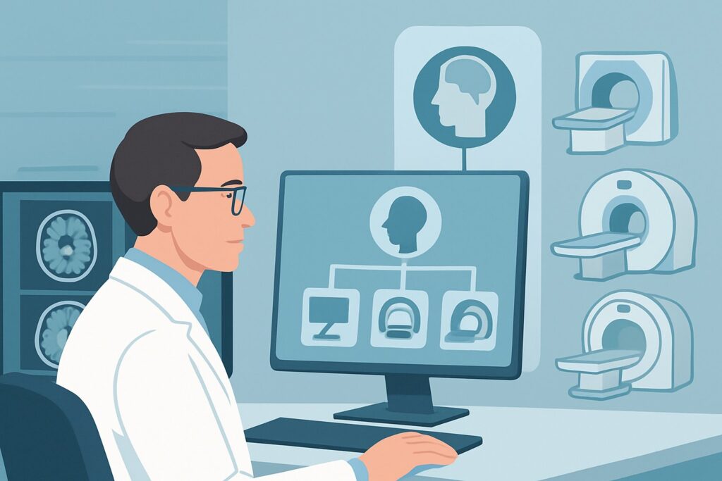
A 62-year-old patient reports several weeks of a vague “foggy” or “lightheaded” feeling in your primary care clinic. The sensation is intermittent and not true spinning. A detailed history and physical examination reveal no vertigo, no ataxia, and no other focal neurologic deficits. You’re now faced with a common clinical decision: is imaging necessary for this nonspecific presentation, and if so, which study provides the most diagnostic value with the least risk? This article provides a deep dive into the American College of Radiology (ACR) Appropriateness Criteria for this exact scenario, explaining why `MRI head without IV contrast` is rated as `May be appropriate` and how to navigate the diagnostic workflow.
Who Fits This Clinical Scenario?
This guidance applies specifically to adult patients presenting for initial imaging with a primary complaint of nonspecific dizziness. The key inclusion criteria are the absence of other localizing signs. The patient’s symptoms are vague—often described as lightheadedness, fogginess, or a feeling of being “off-balance” without objective ataxia.
Crucially, this workflow is NOT for patients with:
- True Vertigo: A distinct sensation of spinning or rotational movement. Patients with acute, persistent vertigo and an abnormal HINTS (Head-Impulse, Nystagmus, Test-of-Skew) examination, or those with chronic vertigo and unilateral hearing loss, fall into different diagnostic categories with distinct imaging recommendations.
- Objective Ataxia: Demonstrable problems with gait, coordination, or balance on physical examination. A patient with chronic disequilibrium and clear signs of cerebellar ataxia requires a different workup, as the pre-test probability of a posterior fossa lesion is significantly higher.
- Other Neurologic Deficits: Any associated weakness, numbness, vision changes, dysarthria, or other focal findings. Such symptoms raise immediate concern for an acute cerebrovascular event or other serious pathology, triggering a more urgent and specific imaging protocol.
This scenario represents a low-yield but common clinical presentation where the goal of imaging is primarily to rule out an occult structural cause after a thorough clinical evaluation has not revealed a clear etiology.
What Diagnoses Are You Working Up in This Scenario?
For nonspecific dizziness in an otherwise neurologically intact adult, the differential is broad, and many causes are non-structural (e.g., polypharmacy, orthostatic hypotension, metabolic disturbances, anxiety). When imaging is considered, it is to investigate the low-probability but high-consequence intracranial pathologies that could manifest with such vague symptoms.
Chronic Small Vessel Ischemic Disease: This is a common incidental finding but can be a contributor to generalized dizziness or imbalance, particularly in older adults with vascular risk factors. Extensive white matter disease can disrupt supratentorial and cerebellar connections, leading to vague symptoms long before focal deficits appear.
Slow-Growing Brain Tumor: While uncommon, a slow-growing extra-axial tumor like a meningioma or a low-grade glioma can present with nonspecific symptoms before causing mass effect or focal signs. The dizziness may result from subtle pressure on adjacent brain structures or early hydrocephalus.
Chronic Subdural Hematoma (cSDH): Particularly in elderly patients, those with a history of falls, or individuals on anticoagulation, a cSDH can present insidiously. Vague lightheadedness, mild headache, and subtle cognitive changes can be the only initial signs, without the classic focal deficits of an acute bleed.
Structural Abnormalities: Less common causes include occult hydrocephalus, which can present with vague dysequilibrium, or congenital anomalies like a Chiari I malformation that may become symptomatic in adulthood. While rare, these are important structural diagnoses that imaging can readily identify.
Why Is MRI Head Without IV Contrast the Recommended Study for This Presentation?
For an adult with nonspecific dizziness and a normal neurologic exam, the ACR rates MRI head without IV contrast as May be appropriate. This rating reflects the low diagnostic yield of imaging in this population but identifies MRI as the most suitable modality if a decision is made to proceed with an imaging workup.
The rationale is rooted in balancing diagnostic capability with risk. MRI without contrast provides excellent soft-tissue resolution, making it highly sensitive for detecting the key differential considerations: chronic ischemic changes, non-enhancing tumors, chronic blood products from a cSDH, and structural anomalies like hydrocephalus or Chiari malformations. It achieves this with no ionizing radiation (0 mSv), a critical safety consideration for a study that is often negative.
Alternative studies are rated lower for specific reasons in this context:
- CT head without IV contrast is rated Usually not appropriate. While fast and accessible, its lower contrast resolution may miss subtle ischemic disease, small non-calcified tumors, or an isodense chronic subdural hematoma. It also exposes the patient to ionizing radiation (☢☢☢ 1-10 mSv), which is difficult to justify given the low pre-test probability of a positive finding.
- MRI head with IV contrast is also rated Usually not appropriate. The addition of gadolinium-based contrast is not necessary for the primary differential. Most slow-growing tumors (like low-grade gliomas) or chronic ischemic changes will be visible on non-contrast sequences. Contrast administration adds cost, time, and the rare risk of systemic reactions or nephrogenic systemic fibrosis without providing significant additional diagnostic information for this specific initial presentation.
The choice of a non-contrast MRI acknowledges that while the likelihood of finding a serious underlying cause is low, if imaging is pursued, it should be the test that most effectively rules out the relevant pathologies with the least patient risk.
What’s Next After MRI Head Without IV Contrast? Downstream Workflow
The results of the MRI will guide the subsequent clinical pathway. Given the nonspecific nature of the presentation, a negative study is the most common outcome and is, in itself, a clinically valuable result.
- If the MRI is negative: This result effectively rules out a significant structural intracranial cause for the patient’s symptoms. The focus should pivot back to non-neurologic and systemic etiologies. The next steps include a thorough medication review (polypharmacy is a frequent culprit), assessment for orthostatic hypotension, cardiac evaluation (e.g., Holter monitor to rule out arrhythmia), and screening for anxiety or other psychiatric conditions. No further neuroimaging is typically warranted.
- If the MRI is positive for a specific finding: The downstream workflow is dictated by the abnormality. A finding of an unexpected meningioma or other tumor would prompt a referral to neurosurgery. The discovery of a chronic subdural hematoma would also require neurosurgical consultation. If the MRI reveals extensive chronic small vessel ischemic disease, the focus shifts to aggressive management of vascular risk factors: hypertension, diabetes, hyperlipidemia, and smoking cessation.
- If the MRI is indeterminate or shows incidental findings: The report may describe nonspecific white matter hyperintensities or age-related volume loss. These findings often do not fully explain the patient’s symptoms but should prompt a review of vascular risk factors. If a finding is truly equivocal, discussion with the interpreting radiologist can clarify the need for follow-up imaging or a different modality.
Pitfalls to Avoid (and When to Get Help)
Navigating the workup for nonspecific dizziness requires careful clinical judgment to avoid common missteps.
- Premature Imaging: The most significant pitfall is ordering imaging before a thorough history and physical examination. Many causes (orthostatic hypotension, medication side effects) can be identified at the bedside, obviating the need for a low-yield MRI.
- Ordering the Wrong Study: Defaulting to a head CT is a common error. For this elective, low-suspicion workup, the superior soft-tissue detail of MRI is necessary to confidently rule out the relevant pathology, and the lack of radiation is a key advantage.
- Over-interpreting Incidental Findings: A negative study is a good outcome. Be cautious about attributing a patient’s symptoms to minor, age-expected findings like mild white matter changes or cerebral atrophy, which could lead to diagnostic closure and delay the search for the true systemic cause.
If the patient’s symptoms evolve to include new focal neurologic deficits, true vertigo, or objective ataxia, this represents a change in clinical scenario. At that point, escalate care with an urgent neurology consultation and likely repeat, more specific imaging.
Related ACR Topics and Tools
For a comprehensive overview of all clinical variants related to dizziness and ataxia, and for tools to help in selecting the right imaging study, the following resources are available.
- For breadth across all scenarios in Dizziness and Ataxia, see our parent guide: Dizziness and Ataxia: ACR Appropriateness Decoded.
- To look up other clinical scenarios, visit the ACR Appropriateness Criteria Lookup tool.
- For details on imaging techniques, browse the Imaging Protocol Library.
- To discuss cumulative exposure with patients, use the Radiation Dose Calculator.
Frequently Asked Questions
Why is MRI rated ‘May be appropriate’ instead of ‘Usually appropriate’ for nonspecific dizziness?
The ‘May be appropriate’ rating reflects the low diagnostic yield of imaging in this specific patient population. Because nonspecific dizziness without other neurologic signs is often caused by systemic, metabolic, or psychiatric conditions, an MRI is frequently negative. The rating indicates that MRI is the best imaging choice if one is performed, but that imaging may not be necessary for every patient and the decision should be based on clinical judgment after a thorough initial evaluation.
If my patient is claustrophobic, is a CT head an acceptable alternative?
According to the ACR, a CT head without contrast is ‘Usually not appropriate’ for this indication due to its lower sensitivity for the key differential diagnoses (like chronic ischemia or small tumors) and its use of ionizing radiation. If a patient cannot tolerate a standard MRI, options include discussing sedation with the patient or using an open MRI, though the latter may have lower image quality. A CT is a significant compromise in diagnostic capability for this particular workup.
Should I order an MRA of the head and neck for nonspecific dizziness?
No, for this specific scenario, MRA of the head and neck (with or without contrast) is rated ‘Usually not appropriate.’ This presentation lacks specific symptoms suggestive of a vascular etiology, such as those seen in a transient ischemic attack (TIA) or stroke (e.g., focal weakness, aphasia, visual loss). Vascular imaging is reserved for cases with a higher suspicion of cerebrovascular disease.
Does this guidance apply if the patient is elderly and has a history of falls?
Yes, but with increased clinical suspicion for a chronic subdural hematoma (cSDH). While the scenario and recommendation remain the same, the pre-test probability for finding a cSDH is higher in this subgroup. An MRI head without contrast is still the ideal study, as it is highly sensitive for chronic blood products, which can sometimes be difficult to see on a non-contrast CT.
What if the dizziness started acutely after a minor head injury?
That presentation changes the clinical context significantly and would fall under the ACR Appropriateness Criteria for Head Trauma, not Dizziness and Ataxia. In cases of trauma, especially with any loss of consciousness or other concerning signs, a non-contrast head CT is often the first-line imaging modality to rapidly rule out acute intracranial hemorrhage.
Reviewed by Pouyan Golshani, MD, Interventional Radiologist — May 30, 2026