What Imaging Is Next for Chronic Knee Pain When Radiographs Are Negative?
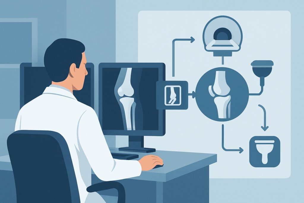
A 45-year-old patient presents with four months of insidious-onset right knee pain, localized medially and worse with squatting. There is no history of a single, acute traumatic event. You ordered initial weight-bearing radiographs, which have just returned with the read: “No acute fracture or dislocation. No significant degenerative change. Small joint effusion.” Now, with the patient’s symptoms persisting and the initial imaging unrevealing, you face the critical decision of what to order next to diagnose the underlying cause.
This article provides a focused workflow for this exact clinical scenario, guided by the American College of Radiology (ACR) Appropriateness Criteria. For a patient with chronic knee pain and negative or effusion-only initial radiographs, the ACR designates MRI knee without IV contrast as Usually appropriate for further evaluation. We will explore the rationale, differential diagnosis, and downstream pathways for this common clinical challenge.
Who Fits This Clinical Scenario?
This guidance is specifically for an adult or child aged 5 years or older presenting with chronic knee pain, where the initial diagnostic step—a knee radiograph—was either negative or demonstrated only a joint effusion. “Chronic” typically implies symptoms lasting for several weeks to months, distinguishing it from an acute injury.
Inclusion criteria for this workflow:
- Patient is an adult or a child at least 5 years old.
- Knee pain has been present for weeks or months (chronic).
- An initial knee radiograph has already been performed.
- The radiograph shows no specific pathology (e.g., fracture, significant osteoarthritis, loose body) other than a possible joint effusion.
Exclusion criteria (patients who should follow a different pathway):
- Acute, significant trauma: Patients with a recent, high-impact injury should be evaluated under acute knee trauma guidelines, as the pre-test probability for fracture is higher.
- Clear radiographic findings: If the initial radiograph shows definitive evidence of moderate-to-severe osteoarthritis, an osteochondral defect, or signs of inflammatory arthritis, you should consult the specific ACR variant for those findings. This workflow is for when the radiograph is unhelpful.
- Suspected infection or tumor: Patients with constitutional symptoms like fever, night sweats, weight loss, or a focal, palpable mass require a different, more urgent workup, often involving contrast-enhanced MRI and laboratory tests.
What Diagnoses Are You Working Up in This Scenario?
With significant bony pathology largely excluded by the negative radiograph, the differential diagnosis shifts toward internal derangement involving soft tissues and articular cartilage. The goal of the next imaging study is to visualize these radiographically occult structures.
Meniscal Tear
This is one of the most common causes of chronic knee pain and mechanical symptoms like locking, catching, or giving way. Degenerative tears can occur without a distinct traumatic event, especially in middle-aged and older adults. A negative radiograph is the expected finding.
Ligamentous Injury
While high-grade ligament tears (e.g., a complete Anterior Cruciate Ligament [ACL] rupture) often present acutely, chronic sprains, partial tears, or insufficiency of the cruciate or collateral ligaments can cause persistent pain and instability. These injuries are not visible on radiographs but are well-delineated on MRI.
Chondral or Osteochondral Lesions
Damage to the articular cartilage (chondral injury) or the underlying bone (osteochondral lesion) is a significant source of pain and can be a precursor to osteoarthritis. Radiographs are insensitive to all but the largest osteochondral defects. MRI is highly sensitive for detecting cartilage thinning, fissures, and associated subchondral bone marrow edema.
Patellofemoral Pain Syndrome or Chondromalacia Patellae
Anterior knee pain is frequently related to the patellofemoral joint. While often a clinical diagnosis, MRI can be valuable when symptoms are refractory to conservative treatment. It can reveal chondromalacia (cartilage softening and fibrillation), maltracking, and associated bone marrow edema patterns.
Occult Bony Injury
In some cases, a stress fracture or significant bone bruise (trabecular microfracture) may not be apparent on initial radiographs. If clinical suspicion is high (e.g., an endurance athlete with new, intense focal pain), MRI is the most sensitive modality for detecting bone marrow edema associated with these injuries.
Why Is MRI Knee without IV Contrast the Recommended Study for This Presentation?
The ACR rates MRI knee without IV contrast as “Usually appropriate” because it directly and non-invasively evaluates the primary differential diagnoses in this scenario with excellent diagnostic accuracy. Its superior soft-tissue contrast resolution is unmatched for visualizing the menisci, ligaments, cartilage, and bone marrow—the very structures suspected of pathology when radiographs are normal.
The key rationale includes:
- High Sensitivity and Specificity: MRI provides detailed anatomical information, allowing for the confident diagnosis or exclusion of meniscal tears, ligamentous injuries, and chondral defects. This directly guides subsequent management, whether it be physical therapy, injection, or surgical referral.
- No Ionizing Radiation: Unlike CT, MRI does not use ionizing radiation (adult and pediatric radiation relative level: O 0 mSv). This is a particularly important consideration in children and younger adults who may require imaging over their lifetime.
- Contrast is Unnecessary: For the vast majority of internal derangement cases, intravenous gadolinium-based contrast adds little diagnostic information, while increasing cost, scan time, and potential (though rare) risks. Contrast is typically reserved for evaluating suspected tumors, infection/abscess, or inflammatory arthropathies.
How do alternative studies compare?
The ACR rates other modalities lower for this specific clinical question. For example, US knee is rated “May be appropriate (Disagreement).” While ultrasound is excellent for evaluating superficial structures like tendons (patellar, quadriceps), bursae, and effusions, it is highly operator-dependent and cannot adequately visualize intra-articular structures like the cruciate ligaments or the posterior horns of the menisci. Its utility is limited to a narrow set of differential diagnoses not central to this scenario.
Similarly, CT knee without IV contrast is rated “May be appropriate.” CT provides exceptional detail of cortical and trabecular bone but offers poor soft-tissue contrast. It would not be the correct choice for assessing menisci or ligaments. It also involves ionizing radiation (adult RRL: ☢ <0.1 mSv; pediatric RRL: ☢☢ 0.03-0.3 mSv), making it less desirable than MRI when soft-tissue pathology is the primary concern.
Once you’ve decided on the top-rated procedure, our protocol guide covers the technique, sequences, and reading principles. For a deep dive, see: MRI Knee Without Contrast.
What’s Next After MRI Knee without IV Contrast? Downstream Workflow
The MRI result is a critical branch point that dictates the next steps in patient management. The goal is to translate the imaging finding into an effective treatment plan.
If the MRI is positive for a distinct structural cause (e.g., meniscal tear, ligament rupture, full-thickness chondral defect):
- The next step is typically a referral to Orthopedic Surgery or a sports medicine specialist. The imaging findings, combined with the patient’s age, activity level, and mechanical symptoms, will determine whether conservative management (physical therapy, activity modification, injections) or surgical intervention (e.g., arthroscopy) is most appropriate.
If the MRI is negative (no clear structural cause for pain):
- This is a valuable finding that rules out significant internal derangement. The focus should shift to non-structural or functional causes of pain. This often includes diagnoses like patellofemoral pain syndrome, IT band syndrome, or pes anserine bursitis, which are primarily managed with targeted physical therapy. Reassurance can be provided to the patient that no surgical target was identified.
If the MRI is indeterminate or shows findings of unclear clinical significance (e.g., low-grade meniscal signal, mild chondromalacia):
- These findings must be correlated closely with the patient’s clinical examination. A trial of conservative management is almost always the first step. If pain persists despite a robust course of physical therapy, a diagnostic/therapeutic injection (e.g., corticosteroid) may be considered. In some cases, an MR arthrogram (“May be appropriate”) could be considered to better evaluate for subtle labral or cartilage pathology, though this is less common.
Pitfalls to Avoid (and When to Get Help)
Navigating this workflow requires attention to a few common pitfalls to ensure timely and accurate diagnosis.
- Ordering Contrast by Default: Avoid ordering an “MRI knee with and without contrast” unless there is a specific concern for tumor, infection, or inflammatory synovitis. For standard internal derangement, it is “Usually not appropriate” and adds unnecessary cost and risk.
- Ignoring Referred Pain: Chronic knee pain, especially in the absence of clear intra-articular pathology on MRI, can be referred from the hip or lumbar spine. If the knee MRI is negative but symptoms persist, consider a clinical evaluation and potentially radiographs of the ipsilateral hip or lumbar spine (both rated “May be appropriate”).
- Over-interpreting Incidental Findings: Asymptomatic meniscal signal and mild cartilage changes are common, especially with increasing age. Always correlate imaging findings with the patient’s specific symptoms and physical exam findings to avoid attributing pain to an incidental finding.
If red flag symptoms such as unexplained fever, night sweats, or rapid symptom progression develop, escalate the workup immediately to evaluate for more sinister pathology like infection or malignancy.
Related ACR Topics and Tools
This article covers one specific scenario within the broader topic of chronic knee pain. For a comprehensive overview of all clinical variants and their recommended imaging pathways, or to explore the technical details of the recommended study, the following resources are available.
- For breadth across all scenarios in Chronic Knee Pain, see our parent guide: Chronic Knee Pain: ACR Appropriateness Decoded.
- To look up adjacent scenarios or other clinical questions, use the ACR Appropriateness Criteria Lookup.
- To review technical specifications for hundreds of studies, visit the Imaging Protocol Library.
- For discussions about radiation exposure with patients, consult the Radiation Dose Calculator.
Frequently Asked Questions
Is an MRI always necessary if the initial knee radiograph is negative?
Not always. If the patient’s symptoms are mild and improving with conservative measures like rest, ice, and physical therapy, a trial of non-operative management is reasonable before proceeding to advanced imaging. MRI is indicated when symptoms are persistent, severe, or associated with mechanical signs (locking, instability) that suggest an internal derangement requiring a specific diagnosis to guide further treatment.
Why is MRI without contrast preferred over MR arthrography for this scenario?
Standard non-contrast MRI has very high diagnostic accuracy for the most common causes of chronic knee pain, such as meniscal and ligamentous tears. MR arthrography, which involves injecting contrast directly into the joint, is an invasive procedure rated as ‘May be appropriate.’ It is typically reserved for specific, less common situations, such as evaluating for a recurrent meniscal tear in a post-operative knee or assessing subtle cartilage delamination.
What if my patient has a contraindication to MRI, like a non-compatible pacemaker?
If a patient cannot undergo an MRI, the next best test depends on the specific clinical question. For evaluating soft tissues, there is no perfect alternative. CT arthrography (‘May be appropriate’) can provide good detail of the menisci and cartilage surfaces but involves radiation and an invasive injection. Ultrasound (‘May be appropriate (Disagreement)’) can assess superficial tendons and ligaments but not deep intra-articular structures. A consultation with a radiologist and an orthopedic surgeon is recommended to choose the best alternative.
Does this guidance apply to a child under 5 years of age?
No. This specific ACR variant applies to children aged 5 and older. The differential diagnosis for chronic knee pain in very young children (under 5) is different and includes conditions like transient synovitis, septic arthritis, and developmental abnormalities. The imaging workup in that age group requires special consideration and often follows a different pathway.
Can I order a weight-bearing or upright MRI for this scenario?
While weight-bearing MRI is available in some centers, its role in routine clinical practice for chronic knee pain is not well-established, and it is not specifically addressed in the ACR Appropriateness Criteria for this scenario. A standard, high-quality supine MRI without contrast is the validated and recommended study for diagnosing the vast majority of internal knee derangements.
Reviewed by Pouyan Golshani, MD, Interventional Radiologist — June 27, 2026