What Imaging Is Next for Thoracic Back Pain with an Abnormal Radiograph?
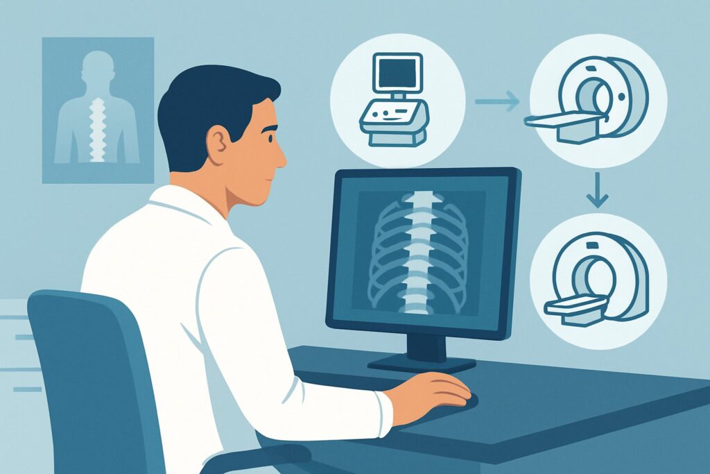
A 65-year-old patient presents with three months of worsening mid-back pain. Over the last week, they’ve developed bilateral leg weakness and unsteady gait. You obtain thoracic spine radiographs, which reveal a T8 vertebral body compression fracture and suspicious lucency in the pedicle. The initial imaging has confirmed a significant structural problem, but the underlying cause remains unclear. Is this a simple osteoporotic fracture, a pathologic fracture from malignancy, or an infectious process? The presence of myelopathy makes this an urgent clinical question.
This scenario is about choosing the definitive next imaging study when a plain film has already identified bone destruction, a fracture, or a significant deformity in the thoracic spine. According to the American College of Radiology (ACR) Appropriateness Criteria, the next step is clear: MRI of the thoracic spine without and with IV contrast is rated as Usually Appropriate. This article details the clinical workflow for this specific situation.
Who Fits This Clinical Scenario?
This guidance applies to a specific subset of adult patients with thoracic back pain. The critical inclusion criterion is a prior thoracic spine radiograph that has already demonstrated a significant structural abnormality. This includes findings such as:
- Vertebral body compression fracture
- Lytic or blastic bone lesions suggesting destruction
- Severe spinal deformity (e.g., kyphosis, scoliosis) that appears acute or progressive
The patient may or may not have associated neurologic symptoms, such as radiculopathy (nerve root compression) or myelopathy (spinal cord compression). The presence of these symptoms adds urgency but does not change the choice of the next imaging study.
This workflow is distinct from other common presentations. It does not apply to patients with initial, uncomplicated acute back pain where imaging may not be indicated at all. It also differs from the workup of a patient with clinical red flags (e.g., fever, history of cancer, unexplained weight loss) but a normal or not-yet-obtained radiograph. In those cases, the decision to proceed directly to advanced imaging follows a different rationale. This guidance is specifically for the “what’s next” question after an abnormal plain film has been obtained.
What Diagnoses Are You Working Up in This Scenario?
With an abnormal radiograph in hand, the differential diagnosis narrows significantly. The goal of the next imaging study is to characterize the known abnormality and identify its cause. The primary considerations include malignancy, infection, and complications of trauma or osteoporosis.
Malignancy (Metastatic Disease or Primary Bone Tumor) is a primary concern, especially in older adults or those with a known cancer history. Metastatic lesions from breast, lung, prostate, kidney, and thyroid cancer commonly spread to the spine. Radiographs may show lytic or blastic changes, but they cannot assess for epidural extension or spinal cord compression, which is a neurosurgical emergency. Primary bone tumors like multiple myeloma or lymphoma are also on the differential.
Spinal Infection (Osteomyelitis/Discitis) can cause bone destruction that mimics malignancy on plain films. An infection can lead to vertebral collapse, deformity, and the formation of an epidural abscess, which can rapidly cause irreversible spinal cord injury. Distinguishing infection from tumor is critical as their management is completely different.
Pathologic Fracture Characterization is another key goal. While many vertebral compression fractures are osteoporotic, a fracture in the setting of bone destruction on a radiograph raises suspicion for an underlying pathologic process. Advanced imaging is needed to assess the bone marrow for infiltration, evaluate the posterior elements for involvement (a red flag for malignancy), and determine the degree of spinal canal compromise and ligamentous stability.
Why Is MRI of the Thoracic Spine Without and With IV Contrast the Recommended Study?
When a radiograph shows a destructive bone process, fracture, or deformity, the clinical questions shift from detection to characterization and assessment of neurologic structures. Magnetic Resonance Imaging (MRI) is the superior modality for this purpose, leading to its Usually Appropriate rating from the ACR.
An MRI provides unparalleled detail of the bone marrow, intervertebral discs, spinal cord, nerve roots, and surrounding soft tissues. This allows it to directly address the key differential diagnoses:
- For Malignancy: MRI can detect abnormal marrow signal from tumor infiltration long before cortical bone destruction is advanced. The post-contrast sequences are crucial for identifying enhancing tumor tissue and delineating the extent of any epidural or paraspinal disease compressing the spinal cord.
- For Infection: MRI is highly sensitive for osteomyelitis and discitis. The combination of T2-weighted signal changes (edema) and characteristic enhancement patterns after IV contrast administration—often involving adjacent vertebral bodies and the intervening disc—is key to diagnosis. Contrast is essential for identifying and defining the extent of an epidural abscess.
Why are other studies rated lower?
A CT of the thoracic spine without IV contrast is also rated Usually Appropriate but serves a different, more limited role. It provides excellent bony detail and is superior for assessing fracture comminution and alignment. However, it offers poor visualization of the spinal cord, nerve roots, and bone marrow infiltration. It is often considered a complementary study or an alternative when MRI is contraindicated (e.g., incompatible implanted device, severe claustrophobia). A CT myelogram, rated May be appropriate, can delineate the spinal canal but is invasive and has been largely supplanted by MRI with contrast.
Studies like a whole-body bone scan are Usually not appropriate at this stage because while sensitive for metabolic bone activity, they are nonspecific. They cannot distinguish between fracture, infection, or tumor, nor can they evaluate the spinal cord.
The recommended study, MRI without and with contrast, involves no ionizing radiation (0 mSv). This is a significant advantage over CT-based methods, which carry a dose of 1-10 mSv (☢☢☢) or higher. Once you’ve decided on this study, our protocol guide covers key technical considerations for the non-contrast portion of the exam: MRI Thoracic Spine Without Contrast.
What’s Next After MRI? Downstream Workflow
The results of the contrast-enhanced thoracic MRI will guide the subsequent clinical pathway. The workflow branches based on whether the findings suggest tumor, infection, or a benign process.
If the MRI suggests malignancy: Findings like marrow-replacing lesions with epidural extension and preservation of the intervertebral disc point toward a tumor. The immediate next step is an urgent consultation with neurosurgery and/or radiation oncology, especially if there is spinal cord compression. A CT-guided biopsy is typically required to obtain tissue for a definitive pathologic diagnosis, which will guide systemic therapy decisions made by an oncologist.
If the MRI suggests infection: Findings of disc space destruction, adjacent vertebral endplate changes, and a phlegmon or fluid collection (abscess) are classic for osteomyelitis/discitis. This requires urgent consultation with infectious disease specialists and often neurosurgery. A CT-guided or open biopsy may be performed to obtain cultures and guide antibiotic therapy. If a significant epidural abscess is causing neurologic compromise, emergent surgical decompression and washout is necessary.
If the MRI confirms a benign osteoporotic fracture: If the marrow signal is normal (or shows simple edema) and there are no signs of an underlying destructive process, the diagnosis is likely a benign compression fracture. Management focuses on pain control, bracing, and medical treatment for osteoporosis. For patients with severe, persistent pain, procedures like kyphoplasty or vertebroplasty may be considered by interventional radiology or spine surgery.
Pitfalls to Avoid (and When to Get Help)
In this high-stakes clinical scenario, several pitfalls can delay diagnosis or lead to suboptimal outcomes. Be mindful of the following:
- Omitting IV Contrast: Ordering a non-contrast MRI when infection or tumor is suspected is a major error. Contrast is essential for differentiating an abscess from a phlegmon and for delineating the extent of an enhancing tumor.
- Misinterpreting Edema: Marrow edema can be seen in acute benign fractures, infection, and malignancy. Rely on the full constellation of MRI findings, including morphology and post-contrast enhancement, to differentiate these causes.
- Delaying Imaging in Myelopathy: The presence of myelopathy (spinal cord compression) is a neurologic emergency. Advanced imaging should be obtained emergently to guide intervention before deficits become permanent.
- Accepting a Non-diagnostic Biopsy: If a CT-guided biopsy is negative but clinical and imaging suspicion for malignancy or infection remains high, do not stop the workup. Escalate to an open biopsy or repeat the percutaneous procedure.
If a patient has rapidly progressing neurologic deficits, escalate immediately to your on-call neurosurgery or spine surgery service, often before the MRI is even completed.
Related ACR Topics and Tools
Navigating imaging decisions requires access to reliable, evidence-based resources. For a comprehensive overview of all clinical variants related to thoracic spine imaging, from uncomplicated pain to post-operative assessment, please consult our parent guide. For tools to help with ordering, protocoling, and patient communication, see the resources below.
- Parent Topic Hub: For breadth across all scenarios in Thoracic Back Pain, see our parent guide: Thoracic Back Pain: ACR Appropriateness Decoded.
- ACR Criteria Lookup: ACR Appropriateness Criteria Lookup
- Protocol Library: Imaging Protocol Library
- Dose Calculator: Radiation Dose Calculator
Frequently Asked Questions
Why is MRI with and without contrast recommended over MRI without contrast alone?
While a non-contrast MRI is excellent for showing bone marrow edema and spinal cord compression, the addition of IV gadolinium-based contrast is critical for differentiating the primary concerns in this scenario. Contrast helps delineate an epidural abscess in an infection, defines the vascularity and extent of a tumor, and can help distinguish a pathologic fracture from a benign one.
My patient has a pacemaker and cannot get an MRI. What is the best alternative?
If MRI is absolutely contraindicated, the next best study is a CT myelogram of the thoracic spine, which is rated ‘May be appropriate’ by the ACR. This involves injecting intrathecal contrast followed by a CT scan. It provides excellent visualization of the spinal canal and nerve roots to assess for compression, though it does not provide the bone marrow and soft tissue detail of an MRI and is an invasive procedure.
The radiograph showed a simple-appearing compression fracture. Do I still need an MRI?
If the patient has neurologic symptoms (myelopathy or radiculopathy), significant deformity, or clinical red flags for malignancy or infection (e.g., history of cancer, fever, IV drug use), an MRI is still strongly recommended to assess for canal compromise and rule out an underlying pathologic cause. For a classic osteoporotic-appearing fracture in a low-risk patient without neurologic deficits, a non-contrast MRI or even clinical follow-up may be sufficient, but that represents a different clinical scenario.
Should I order a whole-body bone scan to look for other lesions?
A whole-body bone scan is rated ‘Usually not appropriate’ as the *next* step after an abnormal radiograph. While it can detect other sites of metastatic disease, it is nonspecific and cannot evaluate the spinal cord or characterize the primary lesion. An MRI of the thoracic spine is the priority to address the immediate threat. A bone scan or PET/CT may be part of the subsequent systemic staging workup after the diagnosis is confirmed, but it should not delay the definitive spinal imaging.
Does the presence of myelopathy change the imaging recommendation from MRI to something faster like CT?
No, the presence of myelopathy makes the need for MRI *more* urgent, not less. While a CT is often faster to acquire, it cannot adequately visualize the spinal cord to determine the cause and severity of compression. The superior diagnostic information from an MRI is essential for appropriate surgical planning. In cases of suspected cord compression, the MRI should be ordered STAT.
Reviewed by Pouyan Golshani, MD, Interventional Radiologist — May 30, 2026