What Imaging Should You Order for a Child with Isolated Nystagmus?
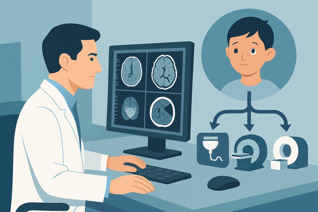
A three-year-old presents to your pediatric clinic for a well-child check. The parents mention they’ve recently noticed the child’s eyes “jiggling back and forth.” On examination, you confirm a conjugate, horizontal nystagmus. The rest of the neurologic and ophthalmologic exam is unremarkable, with no papilledema, proptosis, or focal deficits. You are now faced with the decision of how to appropriately work up this new, isolated finding. This article provides a focused clinical workflow for this exact scenario, guiding you through the American College of Radiology (ACR) recommendations. For a child with isolated nystagmus, the ACR rates MRI head without and with IV contrast as Usually Appropriate.
Who Fits This Clinical Scenario for Isolated Nystagmus?
This guidance is specifically for a child presenting with nystagmus as an isolated finding. The key inclusion criteria are:
- Patient: A child of any age.
- Primary Sign: Nystagmus (involuntary, rhythmic eye movements) that is the sole presenting neurologic or ophthalmologic abnormality.
- Clinical Context: The physical examination reveals no other concerning signs, such as proptosis, strabismus, papilledema, or focal neurologic deficits. There is no history of recent trauma.
It is critical to distinguish this presentation from similar but distinct clinical situations that require a different imaging approach. This workflow does not apply if the child also presents with:
- Papilledema or signs of increased intracranial pressure: This presentation shifts the primary concern to hydrocephalus or a mass causing elevated pressure, and it is covered under a different ACR variant.
- Known or suspected neurofibromatosis type 1 (NF1): In a child with NF1, nystagmus or vision loss raises high suspicion for an optic pathway tumor, a specific scenario with its own imaging considerations.
- Signs of orbital or periorbital infection: Findings like fever, eyelid swelling, pain with eye movements, or proptosis point toward an infectious etiology, which follows a separate diagnostic pathway.
What Diagnoses Are You Working Up with Imaging for Isolated Nystagmus?
While many cases of pediatric nystagmus are benign (e.g., congenital motor nystagmus), imaging is performed to exclude serious underlying structural causes. The differential diagnosis driving the need for neuroimaging is focused on the anterior and posterior visual pathways and the brainstem.
A primary concern, particularly in younger children, is an optic pathway glioma (OPG). These low-grade tumors can infiltrate the optic nerves, chiasm, or tracts, and nystagmus may be the earliest presenting sign, sometimes preceding clinically obvious vision loss. The nystagmus can be monocular or binocular and may resemble spasmus nutans.
Another critical consideration is a posterior fossa tumor. Masses such as a brainstem glioma, medulloblastoma, or cerebellar astrocytoma can disrupt the complex neural circuits that control eye movements, leading to various forms of nystagmus. These must be identified promptly due to their potential for rapid growth and hydrocephalus.
Less commonly, demyelinating diseases like acute disseminated encephalomyelitis (ADEM) or pediatric multiple sclerosis can present with nystagmus, although this is typically accompanied by other neurologic signs. Imaging helps identify characteristic inflammatory lesions.
Finally, imaging serves to rule out congenital structural abnormalities of the brain, such as a Chiari malformation or other hindbrain anomalies, which can cause nystagmus. If imaging is negative, it provides reassurance and supports a diagnosis of a benign condition like idiopathic congenital nystagmus, which is a diagnosis of exclusion.
Why Is MRI Head with and without Contrast Usually Appropriate for Isolated Nystagmus?
The ACR designates MRI head without and with IV contrast as Usually Appropriate because it provides the highest diagnostic yield for the key differential diagnoses in this scenario while avoiding ionizing radiation.
Magnetic Resonance Imaging (MRI) offers superior soft-tissue contrast compared to any other modality. This is essential for visualizing the fine details of the optic nerves, optic chiasm, brainstem, and cerebellum—the primary locations of concern. An MRI can clearly delineate tumor margins, identify subtle inflammation, and characterize structural anomalies that would be poorly visualized or entirely missed on a Computed Tomography (CT) scan.
The addition of intravenous gadolinium-based contrast is crucial. Many of the tumors on the differential, including optic pathway gliomas and posterior fossa masses, demonstrate enhancement after contrast administration. This enhancement increases their conspicuity, helps define their extent, and can differentiate them from non-enhancing lesions. Without contrast, a small or subtly infiltrating tumor could be missed.
Alternative studies are rated lower for specific reasons:
- CT head without and with IV contrast is rated Usually not appropriate. CT has significantly lower soft-tissue resolution, particularly in the posterior fossa, which is often obscured by beam-hardening artifact from the skull base. Most importantly, it exposes the child to ionizing radiation (pediatric dose ☢☢☢☢ 3-10 mSv), which should be avoided whenever a non-radiation alternative like MRI can answer the clinical question.
- MRI head without IV contrast is rated May be appropriate. While it still provides excellent anatomical detail and avoids radiation, omitting contrast reduces sensitivity for detecting enhancing tumors or inflammatory plaques. It is a reasonable choice only when there is a strong contraindication to gadolinium.
The recommended MRI study carries a pediatric radiation level of O (0 mSv), making it the safest and most effective initial imaging test for a child with isolated nystagmus.
What’s Next After the MRI? Downstream Workflow for Nystagmus
The results of the MRI will guide the subsequent clinical pathway. The downstream workflow depends on whether the findings are positive, negative, or indeterminate.
If the study is positive for a tumor (e.g., optic pathway glioma or posterior fossa mass): An urgent referral to pediatric neuro-oncology is the immediate next step. A pediatric neurosurgery consultation will also be required to discuss biopsy, resection, or cerebrospinal fluid diversion if hydrocephalus is present. Further staging imaging or specialized MRI sequences may be requested by the consulting teams.
If the study is negative: A normal MRI is a very reassuring finding. It effectively rules out the most serious structural causes of nystagmus. The patient should be referred to a pediatric ophthalmologist and, if not already involved, a pediatric neurologist for further characterization of the nystagmus (e.g., via electroretinography or video-oculography). The likely diagnosis becomes congenital motor nystagmus or another benign etiology, managed with observation and supportive care for any associated visual deficits.
If the study shows a structural anomaly (e.g., Chiari malformation): The next step is a referral to a pediatric neurosurgeon to evaluate the clinical significance of the finding and determine if surgical intervention is warranted.
If the study is indeterminate (e.g., nonspecific white matter signal abnormalities): This scenario requires close collaboration with a pediatric neurologist. These findings may prompt a broader workup for demyelinating, metabolic, or mitochondrial disorders, which could include a lumbar puncture, metabolic testing, and follow-up MRI.
Common Pitfalls to Avoid in the Nystagmus Workup
Navigating the workup for pediatric nystagmus requires careful attention to detail to avoid common missteps.
- Inadequate Sedation Plan: Young children often cannot remain still for the duration of an MRI. Motion artifact can render a study non-diagnostic, leading to repeat scans and delays. Always coordinate with the radiology department and, if necessary, pediatric anesthesiology to ensure an appropriate plan for sedation or general anesthesia.
- Defaulting to CT: In a non-emergent setting, ordering a CT scan is a significant pitfall. It provides a lower diagnostic yield for the primary concerns in the brain and posterior fossa and exposes the child to unnecessary radiation.
- Delaying the Workup: While isolated nystagmus is not typically a medical emergency, it warrants a prompt and orderly evaluation. Unreasonable delays in scheduling imaging can postpone the diagnosis of a potentially serious underlying condition like a brain tumor.
If the clinical picture evolves to include new neurologic deficits, seizures, or signs of increased intracranial pressure, escalate care immediately for urgent neuroimaging and inpatient evaluation.
Related ACR Topics and Tools
This article focuses on a single clinical variant. For a comprehensive overview of all scenarios related to pediatric orbital imaging and vision loss, or to explore the tools used to build these guidelines, please refer to the following resources.
- For breadth across all scenarios in Orbital Imaging and Vision Loss-Child, see our parent guide: Orbital Imaging and Vision Loss-Child: ACR Appropriateness Decoded.
- ACR Appropriateness Criteria Lookup — for adjacent scenarios
- Imaging Protocol Library — for technique on the recommended study
- Radiation Dose Calculator — for cumulative dose conversations
Frequently Asked Questions
Does every child with nystagmus need an MRI?
Not necessarily, but new-onset nystagmus without a clear benign cause (like congenital nystagmus diagnosed in early infancy with a normal exam) generally warrants neuroimaging to rule out a structural lesion. The ACR guidelines recommend imaging for the initial workup of isolated nystagmus to exclude serious underlying pathology like a brain tumor.
Why is an MRI of the head recommended instead of just the orbits?
While an optic pathway glioma is a key concern, the differential also includes pathology in the posterior fossa (cerebellum and brainstem). An MRI of the orbits alone would miss these crucial areas. An MRI of the head provides comprehensive coverage of the entire brain, including the visual pathways and posterior fossa structures that control eye movements.
Is an MRA or MRV necessary for the initial workup of isolated nystagmus?
No. For this specific scenario, both MRA (Magnetic Resonance Angiography) and MRV (Magnetic Resonance Venography) are rated as *Usually not appropriate*. The primary concerns are parenchymal tumors or structural anomalies, not vascular pathology. Adding these sequences is not indicated unless a specific vascular lesion is suspected based on other findings.
What if my patient has a contraindication to MRI, such as a non-compatible implanted device?
This is a rare situation in young children but requires careful consideration. If MRI is absolutely contraindicated, a contrast-enhanced CT of the head and orbits would be the next best test, despite its limitations with radiation and lower soft-tissue resolution. This decision should be made in consultation with a pediatric radiologist to weigh the risks and benefits.
If the initial MRI is negative, is follow-up imaging ever needed?
Generally, if the initial high-quality MRI is negative and the clinical exam is stable, routine follow-up imaging is not required. The diagnosis shifts to a likely benign or idiopathic cause. However, if the child develops new symptoms, such as vision loss, headache, or other neurologic deficits, repeat imaging would be warranted.
Reviewed by Pouyan Golshani, MD, Interventional Radiologist — May 30, 2026